Intra-individual comparison of two-dimensional shear wave elastography techniques using plane wave imaging and the multi-beam technique: are they interchangeable in measuring liver fibrosis?
IF 2.5 3区 医学Q2 RADIOLOGY, NUCLEAR MEDICINE & MEDICAL IMAGING
Jae Hyun Kim, Jeong Hee Yoon, Ijin Joo, Jeong Min Lee
{"title":"Intra-individual comparison of two-dimensional shear wave elastography techniques using plane wave imaging and the multi-beam technique: are they interchangeable in measuring liver fibrosis?","authors":"Jae Hyun Kim, Jeong Hee Yoon, Ijin Joo, Jeong Min Lee","doi":"10.14366/usg.22135","DOIUrl":null,"url":null,"abstract":"<p><strong>Purpose: </strong>This study compared two different two-dimensional shear wave elastography techniques-plane wave imaging (PWI) and multi-beam (MB) imaging-from the same vendor to evaluate liver fibrosis.</p><p><strong>Methods: </strong>In this prospective study, 42 patients with chronic liver disease who had recently undergone magnetic resonance elastography (<3 months) were enrolled, and their liver stiffness (LS) values were measured using PWI or MB. The LS values (kPa) were compared using the Wilcoxon rank-sum test. Inter-technique reproducibility and intra-observer repeatability were assessed using Bland-Altman analysis with 95% limits of agreement (LOA) and coefficients of variation (CVs). The cutoff values for predicting severe fibrosis (≥F3) were estimated using receiver operating characteristic curve (ROC) analysis, with magnetic resonance elastography as the reference standard.</p><p><strong>Results: </strong>PWI exhibited technical failure in four patients. Therefore, 38 patients underwent both examinations. The LS values showed moderate agreement between PWI and MB (CV, 22.5%) and 95% LOA of -3.71 to 7.44 kPa. The MB technique showed good intra-observer agreement (CV, 8.1%), while PWI showed moderate agreement (CV, 11.0%). The cutoff values of PWI and MB for diagnosing ≥F3 were 12.3 kPa and 13.8 kPa, respectively, with areas under the ROC curve of 0.89 and 0.95 (sensitivity, 100% and 100%; specificity, 65.6% and 85.7%).</p><p><strong>Conclusion: </strong>The LS values significantly differed between PWI and MB, hindering their interchangeable use in longitudinal follow-up. Considering its low technical failure rate and better repeatability, the MB technique may be preferable for evaluating liver fibrosis in chronic liver disease patients.</p>","PeriodicalId":54227,"journal":{"name":"Ultrasonography","volume":"42 2","pages":"265-274"},"PeriodicalIF":2.5000,"publicationDate":"2023-04-01","publicationTypes":"Journal Article","fieldsOfStudy":null,"isOpenAccess":false,"openAccessPdf":"https://ftp.ncbi.nlm.nih.gov/pub/pmc/oa_pdf/24/c5/usg-22135.PMC10071060.pdf","citationCount":"0","resultStr":null,"platform":"Semanticscholar","paperid":null,"PeriodicalName":"Ultrasonography","FirstCategoryId":"3","ListUrlMain":"https://doi.org/10.14366/usg.22135","RegionNum":3,"RegionCategory":"医学","ArticlePicture":[],"TitleCN":null,"AbstractTextCN":null,"PMCID":null,"EPubDate":"","PubModel":"","JCR":"Q2","JCRName":"RADIOLOGY, NUCLEAR MEDICINE & MEDICAL IMAGING","Score":null,"Total":0}
引用次数: 0
Abstract
Purpose: This study compared two different two-dimensional shear wave elastography techniques-plane wave imaging (PWI) and multi-beam (MB) imaging-from the same vendor to evaluate liver fibrosis.
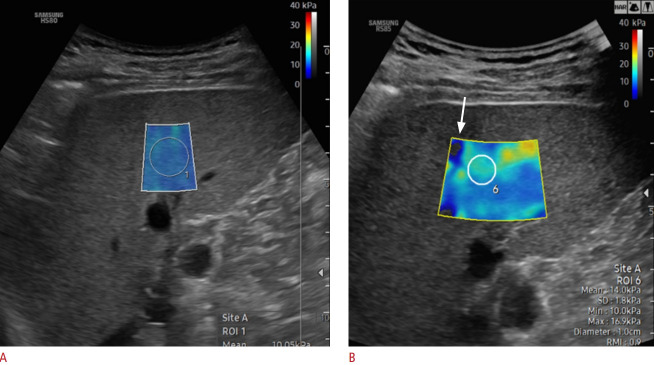
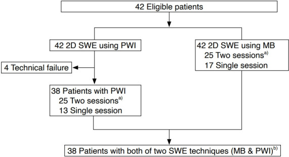
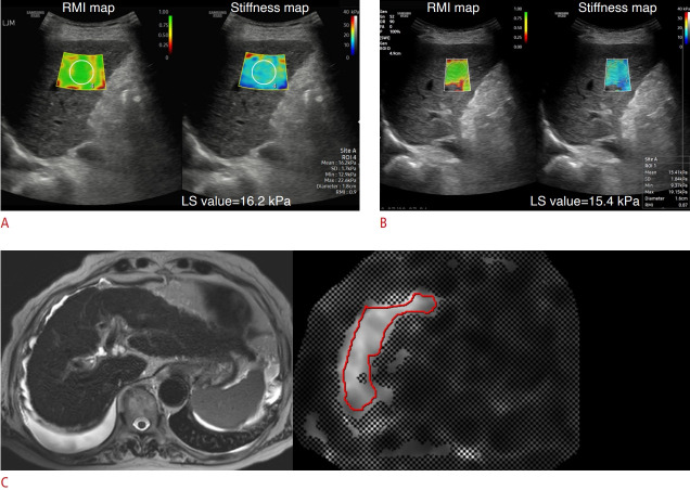
Methods: In this prospective study, 42 patients with chronic liver disease who had recently undergone magnetic resonance elastography (<3 months) were enrolled, and their liver stiffness (LS) values were measured using PWI or MB. The LS values (kPa) were compared using the Wilcoxon rank-sum test. Inter-technique reproducibility and intra-observer repeatability were assessed using Bland-Altman analysis with 95% limits of agreement (LOA) and coefficients of variation (CVs). The cutoff values for predicting severe fibrosis (≥F3) were estimated using receiver operating characteristic curve (ROC) analysis, with magnetic resonance elastography as the reference standard.
Results: PWI exhibited technical failure in four patients. Therefore, 38 patients underwent both examinations. The LS values showed moderate agreement between PWI and MB (CV, 22.5%) and 95% LOA of -3.71 to 7.44 kPa. The MB technique showed good intra-observer agreement (CV, 8.1%), while PWI showed moderate agreement (CV, 11.0%). The cutoff values of PWI and MB for diagnosing ≥F3 were 12.3 kPa and 13.8 kPa, respectively, with areas under the ROC curve of 0.89 and 0.95 (sensitivity, 100% and 100%; specificity, 65.6% and 85.7%).
Conclusion: The LS values significantly differed between PWI and MB, hindering their interchangeable use in longitudinal follow-up. Considering its low technical failure rate and better repeatability, the MB technique may be preferable for evaluating liver fibrosis in chronic liver disease patients.
UltrasonographyMedicine-Radiology, Nuclear Medicine and Imaging
CiteScore
5.10
自引率
6.50%
发文量
78
审稿时长
15 weeks
期刊介绍:
Ultrasonography, the official English-language journal of the Korean Society of Ultrasound in Medicine (KSUM), is an international peer-reviewed academic journal dedicated to practice, research, technology, and education dealing with medical ultrasound. It is renamed from the Journal of Korean Society of Ultrasound in Medicine in January 2014, and published four times per year: January 1, April 1, July 1, and October 1. Original articles, technical notes, topical reviews, perspectives, pictorial essays, and timely editorial materials are published in Ultrasonography covering state-of-the-art content.
Ultrasonography aims to provide updated information on new diagnostic concepts and technical developments, including experimental animal studies using new equipment in addition to well-designed reviews of contemporary issues in patient care. Along with running KSUM Open, the annual international congress of KSUM, Ultrasonography also serves as a medium for cooperation among physicians and specialists from around the world who are focusing on various ultrasound technology and disease problems and relevant basic science.
 求助内容:
求助内容: 应助结果提醒方式:


