Ivana Mravicic, Selma Lukacevic, Alma Biscevic, Melisa Ahmedbegovic Pjano, Nina Ziga, Mateja Tusek
{"title":"A Treatment Approach in Congenital Fibrosis of Extraocular Muscles.","authors":"Ivana Mravicic, Selma Lukacevic, Alma Biscevic, Melisa Ahmedbegovic Pjano, Nina Ziga, Mateja Tusek","doi":"10.5455/medarh.2023.77.137-141","DOIUrl":null,"url":null,"abstract":"<p><strong>Background: </strong>Congenital fibrosis of extraocular muscles ( CFEOM) is a group of genetically defined eye-moving disorders. The syndrome is clinically characterized by congenital non-progressive ophthalmoplegia caused by dysinervation of the cranial nerves with or without ptosis. As a main sign of a CFEOM, extraocular muscles get shrunken and fibrotic, which makes surgery more technically demanding and the result more unpredictable, which makes the treatment challenging and highly customized. Our paper presents variations of the clinical picture and treatment cases of CFEOM1.</p><p><strong>Objective: </strong>To outline the importance of the clinical examination with the exact measurement of deviations for the patients with ocular fibrosis and passive duction test under general anesthesia, establishing them as the main criteria for treatment.</p><p><strong>Methods: </strong>We treated seven patients (14 eyes) with CFEOM1. The decision of the treatment was based on the measurement of the eye position in the primary position (PP), the severity of compensatory head position (CHP), restriction of motility, and passive motility test performed before surgery in general anesthesia. In 3 cases, patients were treated conservatively with the treatment of refractive error and amblyopia. However, in 4 patients, CHP and position of the eyes in PP were not acceptable, motility was severely impaired, and patients underwent surgery. The first surgery was performed on eye muscles: recession of inferior rectus muscle (IRM), anteposition, and resection of superior rectus muscle (SRM). As a second step procedure, ptosis surgery was performed. When the muscle was too tight, and it wasn't possible to have a satisfying result with conventional surgery, we used a tissue expander to improve the position and motility of the affected eyes.</p><p><strong>Results: </strong>In all operated cases, CHP has significantly improved and the position of the eyes in PP.</p><p><strong>Conclusion: </strong>Exact eye and head position measurements and a passive motility test during general anesthesia should guide the surgery. In the case when conventional surgery is not possible, implantation of a bovine pericard is a safe and effective method.</p>","PeriodicalId":18421,"journal":{"name":"Medicinski arhiv","volume":"77 2","pages":"137-141"},"PeriodicalIF":0.0000,"publicationDate":"2023-04-01","publicationTypes":"Journal Article","fieldsOfStudy":null,"isOpenAccess":false,"openAccessPdf":"https://ftp.ncbi.nlm.nih.gov/pub/pmc/oa_pdf/74/a5/medarch-77-137.PMC10227850.pdf","citationCount":"0","resultStr":null,"platform":"Semanticscholar","paperid":null,"PeriodicalName":"Medicinski arhiv","FirstCategoryId":"1085","ListUrlMain":"https://doi.org/10.5455/medarh.2023.77.137-141","RegionNum":0,"RegionCategory":null,"ArticlePicture":[],"TitleCN":null,"AbstractTextCN":null,"PMCID":null,"EPubDate":"","PubModel":"","JCR":"Q2","JCRName":"Medicine","Score":null,"Total":0}
引用次数: 0
Abstract
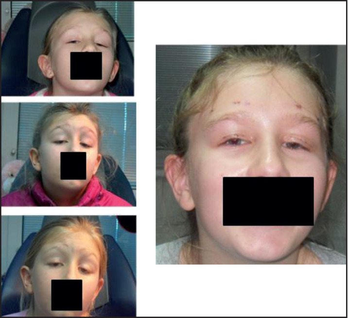
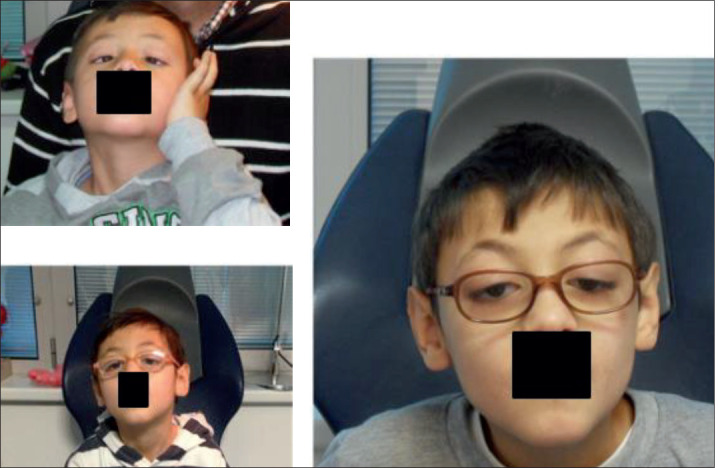
Background: Congenital fibrosis of extraocular muscles ( CFEOM) is a group of genetically defined eye-moving disorders. The syndrome is clinically characterized by congenital non-progressive ophthalmoplegia caused by dysinervation of the cranial nerves with or without ptosis. As a main sign of a CFEOM, extraocular muscles get shrunken and fibrotic, which makes surgery more technically demanding and the result more unpredictable, which makes the treatment challenging and highly customized. Our paper presents variations of the clinical picture and treatment cases of CFEOM1.
Objective: To outline the importance of the clinical examination with the exact measurement of deviations for the patients with ocular fibrosis and passive duction test under general anesthesia, establishing them as the main criteria for treatment.
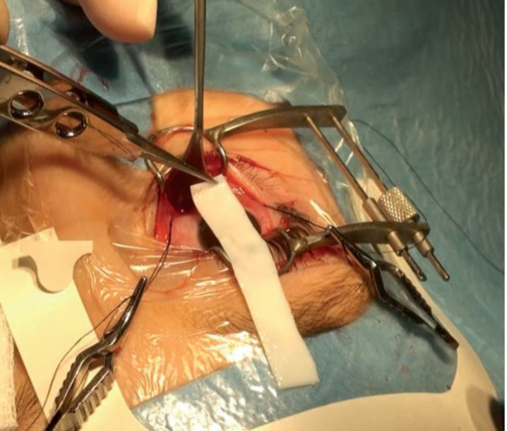
Methods: We treated seven patients (14 eyes) with CFEOM1. The decision of the treatment was based on the measurement of the eye position in the primary position (PP), the severity of compensatory head position (CHP), restriction of motility, and passive motility test performed before surgery in general anesthesia. In 3 cases, patients were treated conservatively with the treatment of refractive error and amblyopia. However, in 4 patients, CHP and position of the eyes in PP were not acceptable, motility was severely impaired, and patients underwent surgery. The first surgery was performed on eye muscles: recession of inferior rectus muscle (IRM), anteposition, and resection of superior rectus muscle (SRM). As a second step procedure, ptosis surgery was performed. When the muscle was too tight, and it wasn't possible to have a satisfying result with conventional surgery, we used a tissue expander to improve the position and motility of the affected eyes.
Results: In all operated cases, CHP has significantly improved and the position of the eyes in PP.
Conclusion: Exact eye and head position measurements and a passive motility test during general anesthesia should guide the surgery. In the case when conventional surgery is not possible, implantation of a bovine pericard is a safe and effective method.

 求助内容:
求助内容: 应助结果提醒方式:


