Benjamin J Lee, Audrey Grossen, Helen Shi, Sara Abu Mehsen, Zhongxin Yu, Kar-Ming A Fung, Khairuddin Memon, Joanna E Gernsback
{"title":"Review of Pediatric Extraosseous Chordomas with a Unique, Illustrative Case.","authors":"Benjamin J Lee, Audrey Grossen, Helen Shi, Sara Abu Mehsen, Zhongxin Yu, Kar-Ming A Fung, Khairuddin Memon, Joanna E Gernsback","doi":"10.1159/000528761","DOIUrl":null,"url":null,"abstract":"<p><strong>Introduction: </strong>Chordoma is a rare, aggressive tumor that is believed to originate from notochord remnants. It can occur anywhere from the clivus to the sacrum and often recurs even after resection and radiotherapy. We present a unique case that initially suggested a different pathology based on imaging and presentation but was found to be a chordoma on gross and pathological analysis.</p><p><strong>Case presentation: </strong>An 11-year-old girl presented outpatient for scoliosis evaluation and was found to have what appeared to be a right L4 peripheral nerve sheath tumor on MRI, causing dextroconvex scoliosis. She underwent a gross total resection via a retroperitoneal approach and was found to have what appeared to be an extraosseous, extradural, extra-spinal canal lumbar chordoma. Immunohistochemical features on surgical pathology were consistent with chordoma. The patient was referred to radiation oncology for adjuvant radiotherapy and pediatric hematology/oncology for recurrence monitoring.</p><p><strong>Discussion: </strong>Our case is the first to present in such a manner, was shown to be external to the spinal canal, encasing the nerve root, and was the first such case in a pediatric patient. We reviewed the growing body of literature on spinal extraosseous chordomas and their characteristics within the pediatric patient population. We also reviewed chordoma pathogenesis theories as well as current and future treatment options.</p>","PeriodicalId":54631,"journal":{"name":"Pediatric Neurosurgery","volume":"58 1","pages":"29-37"},"PeriodicalIF":0.9000,"publicationDate":"2023-01-01","publicationTypes":"Journal Article","fieldsOfStudy":null,"isOpenAccess":false,"openAccessPdf":"https://www.ncbi.nlm.nih.gov/pmc/articles/PMC10064394/pdf/","citationCount":"0","resultStr":null,"platform":"Semanticscholar","paperid":null,"PeriodicalName":"Pediatric Neurosurgery","FirstCategoryId":"3","ListUrlMain":"https://doi.org/10.1159/000528761","RegionNum":4,"RegionCategory":"医学","ArticlePicture":[],"TitleCN":null,"AbstractTextCN":null,"PMCID":null,"EPubDate":"","PubModel":"","JCR":"Q4","JCRName":"CLINICAL NEUROLOGY","Score":null,"Total":0}
引用次数: 0
Abstract
Introduction: Chordoma is a rare, aggressive tumor that is believed to originate from notochord remnants. It can occur anywhere from the clivus to the sacrum and often recurs even after resection and radiotherapy. We present a unique case that initially suggested a different pathology based on imaging and presentation but was found to be a chordoma on gross and pathological analysis.
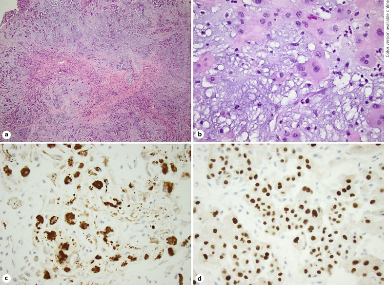
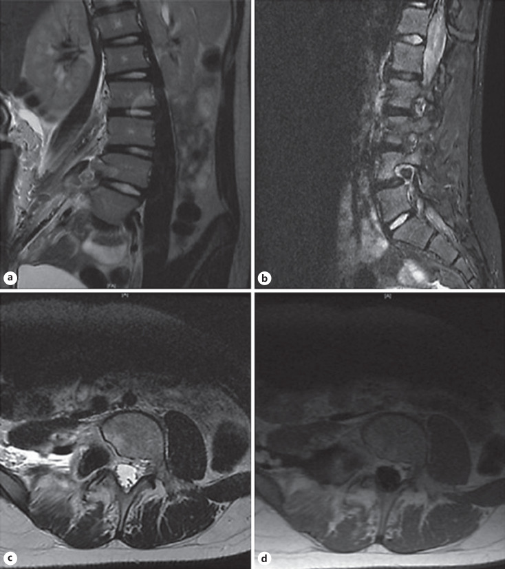
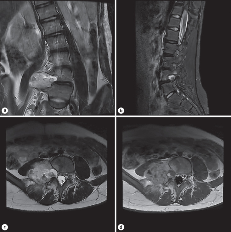
Case presentation: An 11-year-old girl presented outpatient for scoliosis evaluation and was found to have what appeared to be a right L4 peripheral nerve sheath tumor on MRI, causing dextroconvex scoliosis. She underwent a gross total resection via a retroperitoneal approach and was found to have what appeared to be an extraosseous, extradural, extra-spinal canal lumbar chordoma. Immunohistochemical features on surgical pathology were consistent with chordoma. The patient was referred to radiation oncology for adjuvant radiotherapy and pediatric hematology/oncology for recurrence monitoring.
Discussion: Our case is the first to present in such a manner, was shown to be external to the spinal canal, encasing the nerve root, and was the first such case in a pediatric patient. We reviewed the growing body of literature on spinal extraosseous chordomas and their characteristics within the pediatric patient population. We also reviewed chordoma pathogenesis theories as well as current and future treatment options.
期刊介绍:
Articles in ''Pediatric Neurosurgery'' strives to publish new information and observations in pediatric neurosurgery and the allied fields of neurology, neuroradiology and neuropathology as they relate to the etiology of neurologic diseases and the operative care of affected patients. In addition to experimental and clinical studies, the journal presents critical reviews which provide the reader with an update on selected topics as well as case histories and reports on advances in methodology and technique. This thought-provoking focus encourages dissemination of information from neurosurgeons and neuroscientists around the world that will be of interest to clinicians and researchers concerned with pediatric, congenital, and developmental diseases of the nervous system.
 求助内容:
求助内容: 应助结果提醒方式:


