Mao Vásquez, Luis J Saavedra, Hector H García, Evelyn Vela, Jorge E Medina, Miguel Lozano, Carlos Hoyos, William W Lines-Aguilar
{"title":"Trigeminal neuralgia secondary to vascular compression and neurocysticercosis: illustrative case.","authors":"Mao Vásquez, Luis J Saavedra, Hector H García, Evelyn Vela, Jorge E Medina, Miguel Lozano, Carlos Hoyos, William W Lines-Aguilar","doi":"10.3171/CASE23127","DOIUrl":null,"url":null,"abstract":"<p><strong>Background: </strong>Trigeminal neuralgia (TN) is a frequent neurosurgical problem negatively influencing the quality of life of patients. The standard surgical treatment is microvascular decompression for primary cases and decompression of the mass effect, mainly tumors, for secondary cases. Neurocysticercosis (NCC) in the cerebellopontine angle is a rare etiology of TN. The authors report a case in which NCC cysts around the trigeminal nerve coexisted with a vascular loop, which compressed the exit of the trigeminal nerve from the pons.</p><p><strong>Observations: </strong>A 78-year-old woman presented with a 3-year history of persistent severe pain in the left side of her face, refractory to medical treatment. On gadolinium-enhanced magnetic resonance imaging, cystic lesions were observed around the left trigeminal nerve and a vascular loop was also present and in contact with the nerve. A retrosigmoid approach for cyst excision plus microvascular decompression of the trigeminal nerve was successfully performed. There were no complications. The patient was discharged without facial pain.</p><p><strong>Lessons: </strong>Albeit rare, TN secondary to NCC cysts should be considered in the differential diagnosis in NCC-endemic regions. In this case, the cause of the neuralgia was probably both problems, because when both were treated, the patient improved.</p>","PeriodicalId":16554,"journal":{"name":"Journal of Neurosurgery: Case Lessons","volume":"5 21","pages":""},"PeriodicalIF":0.0000,"publicationDate":"2023-05-22","publicationTypes":"Journal Article","fieldsOfStudy":null,"isOpenAccess":false,"openAccessPdf":"https://ftp.ncbi.nlm.nih.gov/pub/pmc/oa_pdf/f1/37/CASE23127.PMC10550649.pdf","citationCount":"1","resultStr":null,"platform":"Semanticscholar","paperid":null,"PeriodicalName":"Journal of Neurosurgery: Case Lessons","FirstCategoryId":"1085","ListUrlMain":"https://doi.org/10.3171/CASE23127","RegionNum":0,"RegionCategory":null,"ArticlePicture":[],"TitleCN":null,"AbstractTextCN":null,"PMCID":null,"EPubDate":"","PubModel":"","JCR":"","JCRName":"","Score":null,"Total":0}
引用次数: 1
Abstract
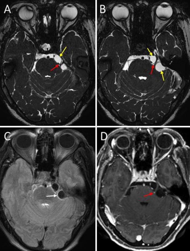
Background: Trigeminal neuralgia (TN) is a frequent neurosurgical problem negatively influencing the quality of life of patients. The standard surgical treatment is microvascular decompression for primary cases and decompression of the mass effect, mainly tumors, for secondary cases. Neurocysticercosis (NCC) in the cerebellopontine angle is a rare etiology of TN. The authors report a case in which NCC cysts around the trigeminal nerve coexisted with a vascular loop, which compressed the exit of the trigeminal nerve from the pons.
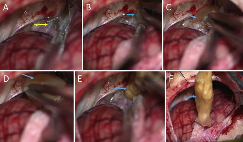
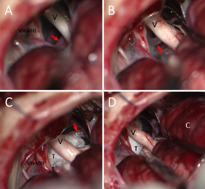
Observations: A 78-year-old woman presented with a 3-year history of persistent severe pain in the left side of her face, refractory to medical treatment. On gadolinium-enhanced magnetic resonance imaging, cystic lesions were observed around the left trigeminal nerve and a vascular loop was also present and in contact with the nerve. A retrosigmoid approach for cyst excision plus microvascular decompression of the trigeminal nerve was successfully performed. There were no complications. The patient was discharged without facial pain.
Lessons: Albeit rare, TN secondary to NCC cysts should be considered in the differential diagnosis in NCC-endemic regions. In this case, the cause of the neuralgia was probably both problems, because when both were treated, the patient improved.

 求助内容:
求助内容: 应助结果提醒方式:


