Mohammad Bilal Alsavaf, Kyle C Wu, Guilherme Finger, Eman H Salem, Maria Jose Castello Ruiz, Saniya S Godil, Luma Ghalib, Ricardo L Carrau, Daniel M Prevedello
{"title":"A silent corticotroph adenoma: making the case for a pars intermedia origin. Illustrative case.","authors":"Mohammad Bilal Alsavaf, Kyle C Wu, Guilherme Finger, Eman H Salem, Maria Jose Castello Ruiz, Saniya S Godil, Luma Ghalib, Ricardo L Carrau, Daniel M Prevedello","doi":"10.3171/CASE2350","DOIUrl":null,"url":null,"abstract":"<p><strong>Background: </strong>Silent corticotroph adenomas (SCAs) are the only pituitary adenomas thought to originate from the pars intermedia. This case report presents the rare finding of a multimicrocystic corticotroph macroadenoma displacing the anterior and posterior lobes of the pituitary gland on magnetic resonance imaging (MRI). This finding supports the hypothesis that silent corticotroph adenomas may originate from the pars intermedia and should be considered in the differential for tumors arising from this location.</p><p><strong>Observations: </strong>A 55-year-old man presented with an episode of confusion and blurred vision. MRI demonstrated separation of the anterior and posterior glands by a solid-cystic lesion located within the pars intermedia that superiorly displaced the optic chiasm. Endocrinologic evaluation was unremarkable. The differential diagnosis included pituitary adenoma, Rathke cleft cyst, and craniopharyngioma. The tumor was confirmed to be an SCA on pathology and was completely removed through the endoscopic endonasal transsphenoidal approach.</p><p><strong>Lessons: </strong>The case highlights the importance of preoperative screening for subclinical hypercortisolism for tumors arising from this location. Knowledge of a patient's preoperative functional status is critical and dictates their postoperative biochemical assessment to determine remission. The case also illustrates surgical strategies for resecting pars intermedia lesions without injuring the gland.</p>","PeriodicalId":16554,"journal":{"name":"Journal of Neurosurgery: Case Lessons","volume":"5 20","pages":""},"PeriodicalIF":0.0000,"publicationDate":"2023-05-15","publicationTypes":"Journal Article","fieldsOfStudy":null,"isOpenAccess":false,"openAccessPdf":"https://ftp.ncbi.nlm.nih.gov/pub/pmc/oa_pdf/e2/41/CASE2350.PMC10550526.pdf","citationCount":"0","resultStr":null,"platform":"Semanticscholar","paperid":null,"PeriodicalName":"Journal of Neurosurgery: Case Lessons","FirstCategoryId":"1085","ListUrlMain":"https://doi.org/10.3171/CASE2350","RegionNum":0,"RegionCategory":null,"ArticlePicture":[],"TitleCN":null,"AbstractTextCN":null,"PMCID":null,"EPubDate":"","PubModel":"","JCR":"","JCRName":"","Score":null,"Total":0}
引用次数: 0
Abstract
Background: Silent corticotroph adenomas (SCAs) are the only pituitary adenomas thought to originate from the pars intermedia. This case report presents the rare finding of a multimicrocystic corticotroph macroadenoma displacing the anterior and posterior lobes of the pituitary gland on magnetic resonance imaging (MRI). This finding supports the hypothesis that silent corticotroph adenomas may originate from the pars intermedia and should be considered in the differential for tumors arising from this location.
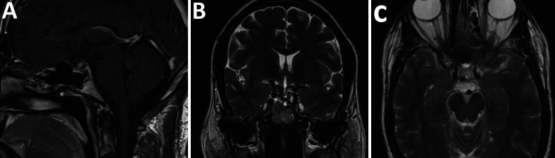
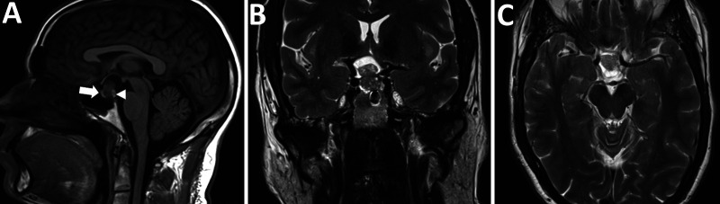
Observations: A 55-year-old man presented with an episode of confusion and blurred vision. MRI demonstrated separation of the anterior and posterior glands by a solid-cystic lesion located within the pars intermedia that superiorly displaced the optic chiasm. Endocrinologic evaluation was unremarkable. The differential diagnosis included pituitary adenoma, Rathke cleft cyst, and craniopharyngioma. The tumor was confirmed to be an SCA on pathology and was completely removed through the endoscopic endonasal transsphenoidal approach.
Lessons: The case highlights the importance of preoperative screening for subclinical hypercortisolism for tumors arising from this location. Knowledge of a patient's preoperative functional status is critical and dictates their postoperative biochemical assessment to determine remission. The case also illustrates surgical strategies for resecting pars intermedia lesions without injuring the gland.

 求助内容:
求助内容: 应助结果提醒方式:


