Alexander S. Barrett , Ori Maller , Michael W. Pickup , Valerie M. Weaver , Kirk C. Hansen
{"title":"Compartment resolved proteomics reveals a dynamic matrisome in a biomechanically driven model of pancreatic ductal adenocarcinoma","authors":"Alexander S. Barrett , Ori Maller , Michael W. Pickup , Valerie M. Weaver , Kirk C. Hansen","doi":"10.1016/j.regen.2018.03.002","DOIUrl":null,"url":null,"abstract":"<div><p><span>Pancreatic ductal adenocarcinoma<span><span><span> (PDAC) is characterized by a severe fibrotic component that compromises treatment, alters the </span>immune cell<span><span> profile and contributes to patient mortality. It has been shown that early on in this process, dynamic changes in tissue biomechanics play an integral role in supporting </span>pancreatic cancer development and progression. Despite the acknowledgement of its importance, a granular view of how stromal composition changes during the course of PDAC progression remains largely unknown. To mimic the quasi-mesenchymal phenotype and pronounced desmoplastic response observed clinically, we utilized a </span></span>genetically engineered mouse model of PDAC that is driven by a Kras</span></span><sup>G12D</sup><span><span> mutation and loss of Tgfbr2 expression. Application of compartment resolved proteomics<span><span> revealed that PDAC progression in this KTC model is associated with dynamic stromal alterations that are indicative of a wound healing program. We identified an early provisional matricellular fibrosis that was accompanied by markers of macrophage activation and </span>infiltration<span>, consistent with the inflammatory phase of wound healing. At 20 weeks a proliferative phenotype was observed with increased fibroblast markers, further collagen deposition and loss of </span></span></span>basement membrane and native cell markers.</span></p></div>","PeriodicalId":94333,"journal":{"name":"Journal of immunology and regenerative medicine","volume":"1 ","pages":"Pages 67-75"},"PeriodicalIF":0.0000,"publicationDate":"2018-03-01","publicationTypes":"Journal Article","fieldsOfStudy":null,"isOpenAccess":false,"openAccessPdf":"https://sci-hub-pdf.com/10.1016/j.regen.2018.03.002","citationCount":"11","resultStr":null,"platform":"Semanticscholar","paperid":null,"PeriodicalName":"Journal of immunology and regenerative medicine","FirstCategoryId":"1085","ListUrlMain":"https://www.sciencedirect.com/science/article/pii/S2468498818300027","RegionNum":0,"RegionCategory":null,"ArticlePicture":[],"TitleCN":null,"AbstractTextCN":null,"PMCID":null,"EPubDate":"","PubModel":"","JCR":"","JCRName":"","Score":null,"Total":0}
引用次数: 11
Abstract
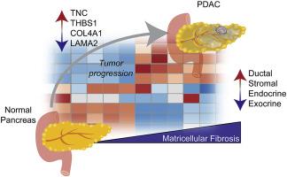
Pancreatic ductal adenocarcinoma (PDAC) is characterized by a severe fibrotic component that compromises treatment, alters the immune cell profile and contributes to patient mortality. It has been shown that early on in this process, dynamic changes in tissue biomechanics play an integral role in supporting pancreatic cancer development and progression. Despite the acknowledgement of its importance, a granular view of how stromal composition changes during the course of PDAC progression remains largely unknown. To mimic the quasi-mesenchymal phenotype and pronounced desmoplastic response observed clinically, we utilized a genetically engineered mouse model of PDAC that is driven by a KrasG12D mutation and loss of Tgfbr2 expression. Application of compartment resolved proteomics revealed that PDAC progression in this KTC model is associated with dynamic stromal alterations that are indicative of a wound healing program. We identified an early provisional matricellular fibrosis that was accompanied by markers of macrophage activation and infiltration, consistent with the inflammatory phase of wound healing. At 20 weeks a proliferative phenotype was observed with increased fibroblast markers, further collagen deposition and loss of basement membrane and native cell markers.

 求助内容:
求助内容: 应助结果提醒方式:


