Yuan Chen, Yixiang Zhou, Xue Zhu, Ge Yan, Donghui Pan, Lizhen Wang, Min Yang, Ke Wang
{"title":"PET imaging of retinal inflammation in mice exposed to blue light using [<sup>18</sup>F]-DPA-714.","authors":"Yuan Chen, Yixiang Zhou, Xue Zhu, Ge Yan, Donghui Pan, Lizhen Wang, Min Yang, Ke Wang","doi":"","DOIUrl":null,"url":null,"abstract":"<p><strong>Purpose: </strong>Positron emission tomography (PET) is widely used in high-precision imaging, which may provide a simple and noninvasive method for the detection of pathology and therapeutic effects. [<sup>18</sup>F]-DPA-714 is a second-generation translocator protein (TSPO) positron emission tomography radiotracer that shows great promise in a model of neuroinflammation. In this study, [<sup>18</sup>F]-DPA-714 micro-PET imaging was used to evaluate retinal inflammation in mice exposed to blue light, a well-established model of age-related macular degeneration (AMD) for molecular mechanism research and drug screening.</p><p><strong>Methods: </strong>C57BL/6J melanized mice were subjected to 10,000, 15,000, and 20,000 lux blue light for 5 days (8 h/day) to develop the retinal injury model, and the structure and function of the retina were assessed using hematoxylin-eosin (HE) staining, electroretinography (ERG), and terminal-deoxynucleotidyl transferase (TdT)-mediated nick-end labeling (TUNEL) immunostaining. Then, [<sup>18</sup>F]-DPA-714 was injected approximately 100 μCi through each tail vein, and static imaging was performed 1 h after injection. Finally, the mice eyeballs were collected for biodistribution and immune analysis.</p><p><strong>Results: </strong>The blue light exposure significantly destroyed the structure and function of the retina, and the uptake of [<sup>18</sup>F]-DPA-714 in the retinas of the mice exposed to blue light were the most significantly upregulated, which was consistent with the biodistribution data. In addition, the immunohistochemical, western blot, and immunofluorescence data showed an increase in microglial TSPO expression.</p><p><strong>Conclusions: </strong>[<sup>18</sup>F]-DPA-714 micro-PET imaging might be a good method for evaluating early inflammatory status during retinal pathology.</p>","PeriodicalId":18866,"journal":{"name":"Molecular Vision","volume":"28 ","pages":"507-515"},"PeriodicalIF":1.4000,"publicationDate":"2022-01-01","publicationTypes":"Journal Article","fieldsOfStudy":null,"isOpenAccess":false,"openAccessPdf":"https://ftp.ncbi.nlm.nih.gov/pub/pmc/oa_pdf/43/d4/mv-v28-507.PMC10115360.pdf","citationCount":"0","resultStr":null,"platform":"Semanticscholar","paperid":null,"PeriodicalName":"Molecular Vision","FirstCategoryId":"3","ListUrlMain":"","RegionNum":3,"RegionCategory":"医学","ArticlePicture":[],"TitleCN":null,"AbstractTextCN":null,"PMCID":null,"EPubDate":"","PubModel":"","JCR":"Q4","JCRName":"BIOCHEMISTRY & MOLECULAR BIOLOGY","Score":null,"Total":0}
引用次数: 0
Abstract
Purpose: Positron emission tomography (PET) is widely used in high-precision imaging, which may provide a simple and noninvasive method for the detection of pathology and therapeutic effects. [18F]-DPA-714 is a second-generation translocator protein (TSPO) positron emission tomography radiotracer that shows great promise in a model of neuroinflammation. In this study, [18F]-DPA-714 micro-PET imaging was used to evaluate retinal inflammation in mice exposed to blue light, a well-established model of age-related macular degeneration (AMD) for molecular mechanism research and drug screening.
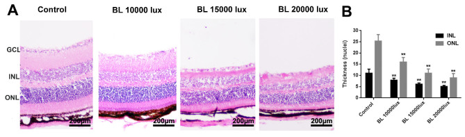
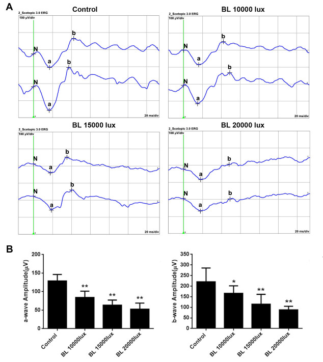
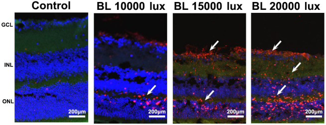
Methods: C57BL/6J melanized mice were subjected to 10,000, 15,000, and 20,000 lux blue light for 5 days (8 h/day) to develop the retinal injury model, and the structure and function of the retina were assessed using hematoxylin-eosin (HE) staining, electroretinography (ERG), and terminal-deoxynucleotidyl transferase (TdT)-mediated nick-end labeling (TUNEL) immunostaining. Then, [18F]-DPA-714 was injected approximately 100 μCi through each tail vein, and static imaging was performed 1 h after injection. Finally, the mice eyeballs were collected for biodistribution and immune analysis.
Results: The blue light exposure significantly destroyed the structure and function of the retina, and the uptake of [18F]-DPA-714 in the retinas of the mice exposed to blue light were the most significantly upregulated, which was consistent with the biodistribution data. In addition, the immunohistochemical, western blot, and immunofluorescence data showed an increase in microglial TSPO expression.
Conclusions: [18F]-DPA-714 micro-PET imaging might be a good method for evaluating early inflammatory status during retinal pathology.
期刊介绍:
Molecular Vision is a peer-reviewed journal dedicated to the dissemination of research results in molecular biology, cell biology, and the genetics of the visual system (ocular and cortical).
Molecular Vision publishes articles presenting original research that has not previously been published and comprehensive articles reviewing the current status of a particular field or topic. Submissions to Molecular Vision are subjected to rigorous peer review. Molecular Vision does NOT publish preprints.
For authors, Molecular Vision provides a rapid means of communicating important results. Access to Molecular Vision is free and unrestricted, allowing the widest possible audience for your article. Digital publishing allows you to use color images freely (and without fees). Additionally, you may publish animations, sounds, or other supplementary information that clarifies or supports your article. Each of the authors of an article may also list an electronic mail address (which will be updated upon request) to give interested readers easy access to authors.
 求助内容:
求助内容: 应助结果提醒方式:


