{"title":"Characterization of myelomonocytoid progenitor cells with mesenchymal differentiation potential obtained by outgrowth from pancreas explants.","authors":"Marc-Estienne Roehrich, Giuseppe Vassalli","doi":"10.1155/2012/429868","DOIUrl":null,"url":null,"abstract":"<p><p>Progenitor cells can be obtained by outgrowth from tissue explants during primary ex vivo tissue culture. We have isolated and characterized cells outgrown from neonatal mouse pancreatic explants. A relatively uniform population of cells showing a distinctive morphology emerged over time in culture. This population expressed monocyte/macrophage and hematopoietic markers (CD11b(+) and CD45(+)), and some stromal-related markers (CD44(+) and CD29(+)), but not mesenchymal stem cell (MSC)-defining markers (CD90(-) and CD105(-)) nor endothelial (CD31(-)) or stem cell-associated markers (CD133(-) and stem cell antigen-1; Sca-1(-)). Cells could be maintained in culture as a plastic-adherent monolayer in culture medium (MesenCult MSC) for more than 1 year. Cells spontaneously formed sphere clusters \"pancreatospheres\" which, however, were nonclonal. When cultured in appropriate media, cells differentiated into multiple mesenchymal lineages (fat, cartilage, and bone). Positive dithizone staining suggested that a subset of cells differentiated into insulin-producing cells. However, further studies are needed to characterize the endocrine potential of these cells. These findings indicate that a myelomonocytoid population from pancreatic explant outgrowths has mesenchymal differentiation potential. These results are in line with recent data onmonocyte-derivedmesenchymal progenitors (MOMPs).</p>","PeriodicalId":9268,"journal":{"name":"Biotechnology Research International","volume":"2012 ","pages":"429868"},"PeriodicalIF":0.0000,"publicationDate":"2012-01-01","publicationTypes":"Journal Article","fieldsOfStudy":null,"isOpenAccess":false,"openAccessPdf":"https://www.ncbi.nlm.nih.gov/pmc/articles/PMC3431127/pdf/","citationCount":"2","resultStr":null,"platform":"Semanticscholar","paperid":null,"PeriodicalName":"Biotechnology Research International","FirstCategoryId":"1085","ListUrlMain":"https://doi.org/10.1155/2012/429868","RegionNum":0,"RegionCategory":null,"ArticlePicture":[],"TitleCN":null,"AbstractTextCN":null,"PMCID":null,"EPubDate":"","PubModel":"","JCR":"","JCRName":"","Score":null,"Total":0}
引用次数: 2
Abstract
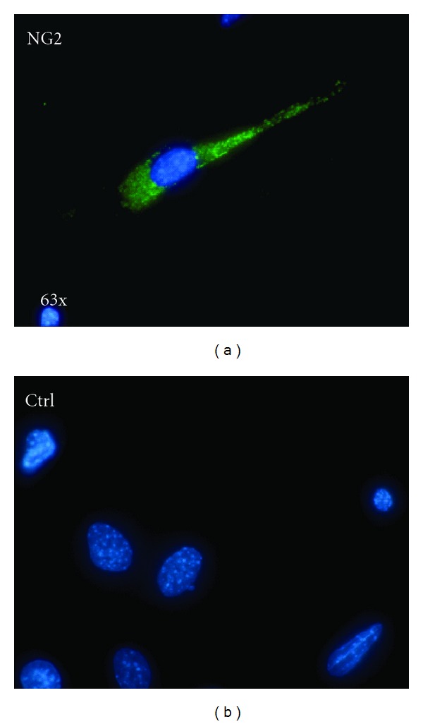
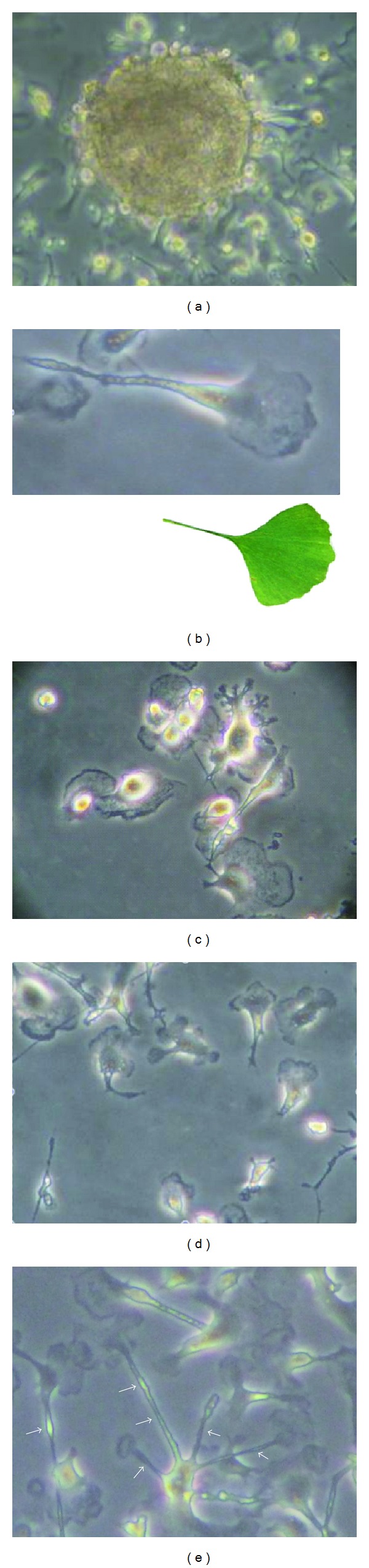
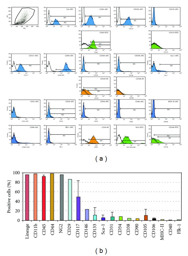
Progenitor cells can be obtained by outgrowth from tissue explants during primary ex vivo tissue culture. We have isolated and characterized cells outgrown from neonatal mouse pancreatic explants. A relatively uniform population of cells showing a distinctive morphology emerged over time in culture. This population expressed monocyte/macrophage and hematopoietic markers (CD11b(+) and CD45(+)), and some stromal-related markers (CD44(+) and CD29(+)), but not mesenchymal stem cell (MSC)-defining markers (CD90(-) and CD105(-)) nor endothelial (CD31(-)) or stem cell-associated markers (CD133(-) and stem cell antigen-1; Sca-1(-)). Cells could be maintained in culture as a plastic-adherent monolayer in culture medium (MesenCult MSC) for more than 1 year. Cells spontaneously formed sphere clusters "pancreatospheres" which, however, were nonclonal. When cultured in appropriate media, cells differentiated into multiple mesenchymal lineages (fat, cartilage, and bone). Positive dithizone staining suggested that a subset of cells differentiated into insulin-producing cells. However, further studies are needed to characterize the endocrine potential of these cells. These findings indicate that a myelomonocytoid population from pancreatic explant outgrowths has mesenchymal differentiation potential. These results are in line with recent data onmonocyte-derivedmesenchymal progenitors (MOMPs).

 求助内容:
求助内容: 应助结果提醒方式:


