Biochemical and immuno-histochemical localization of type IIA procollagen in annulus fibrosus of mature bovine intervertebral disc

Q1 Medicine
Audrey McAlinden , David M. Hudson , Aysel A. Fernandes , Soumya Ravindran , Russell J. Fernandes
{"title":"Biochemical and immuno-histochemical localization of type IIA procollagen in annulus fibrosus of mature bovine intervertebral disc","authors":"Audrey McAlinden ,&nbsp;David M. Hudson ,&nbsp;Aysel A. Fernandes ,&nbsp;Soumya Ravindran ,&nbsp;Russell J. Fernandes","doi":"10.1016/j.mbplus.2021.100077","DOIUrl":null,"url":null,"abstract":"<div><p>For next generation tissue-engineered constructs and regenerative medicine to succeed clinically, the basic biology and extracellular matrix composition of tissues that these repair techniques seek to restore have to be fully determined. Using the latest reagents coupled with tried and tested methodologies, we continue to uncover previously undetected structural proteins in mature intervertebral disc. In this study we show that the “embryonic” type IIA procollagen isoform (containing a cysteine-rich amino propeptide) was biochemically detectable in the annulus fibrosus of both calf and mature steer caudal intervertebral discs, but not in the nucleus pulposus where the type IIB isoform was predominantly localized. Specifically, the triple-helical type IIA procollagen isoform immunolocalized in the outer margins of the inner annulus fibrosus. Triple helical processed type II collagen exclusively localized within the inter-lamellae regions and with type IIA procollagen in the intra-lamellae regions. Mass spectrometry of the α1(II) collagen chains from the region where type IIA procollagen localized showed high 3-hydroxylation of Proline-944, a post-translational modification that is correlated with thin collagen fibrils as in the nucleus pulposus. The findings implicate small diameter fibrils of type IIA procollagen in select regions of the annulus fibrosus where it likely contributes to the organization of collagen bundles and structural properties within the type I-type II collagen transition zone.</p></div>","PeriodicalId":52317,"journal":{"name":"Matrix Biology Plus","volume":"12 ","pages":"Article 100077"},"PeriodicalIF":0.0000,"publicationDate":"2021-12-01","publicationTypes":"Journal Article","fieldsOfStudy":null,"isOpenAccess":false,"openAccessPdf":"https://sci-hub-pdf.com/10.1016/j.mbplus.2021.100077","citationCount":"1","resultStr":null,"platform":"Semanticscholar","paperid":null,"PeriodicalName":"Matrix Biology Plus","FirstCategoryId":"1085","ListUrlMain":"https://www.sciencedirect.com/science/article/pii/S2590028521000211","RegionNum":0,"RegionCategory":null,"ArticlePicture":[],"TitleCN":null,"AbstractTextCN":null,"PMCID":null,"EPubDate":"","PubModel":"","JCR":"Q1","JCRName":"Medicine","Score":null,"Total":0}
引用次数: 1

Abstract

For next generation tissue-engineered constructs and regenerative medicine to succeed clinically, the basic biology and extracellular matrix composition of tissues that these repair techniques seek to restore have to be fully determined. Using the latest reagents coupled with tried and tested methodologies, we continue to uncover previously undetected structural proteins in mature intervertebral disc. In this study we show that the “embryonic” type IIA procollagen isoform (containing a cysteine-rich amino propeptide) was biochemically detectable in the annulus fibrosus of both calf and mature steer caudal intervertebral discs, but not in the nucleus pulposus where the type IIB isoform was predominantly localized. Specifically, the triple-helical type IIA procollagen isoform immunolocalized in the outer margins of the inner annulus fibrosus. Triple helical processed type II collagen exclusively localized within the inter-lamellae regions and with type IIA procollagen in the intra-lamellae regions. Mass spectrometry of the α1(II) collagen chains from the region where type IIA procollagen localized showed high 3-hydroxylation of Proline-944, a post-translational modification that is correlated with thin collagen fibrils as in the nucleus pulposus. The findings implicate small diameter fibrils of type IIA procollagen in select regions of the annulus fibrosus where it likely contributes to the organization of collagen bundles and structural properties within the type I-type II collagen transition zone.
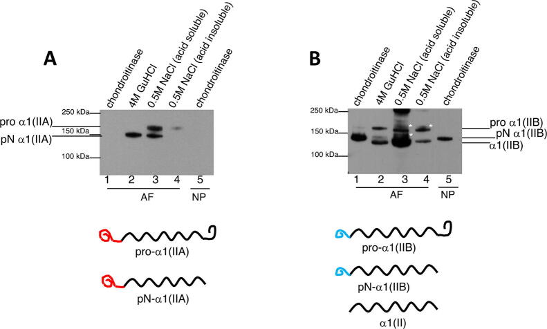
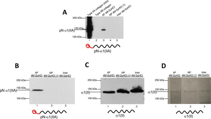
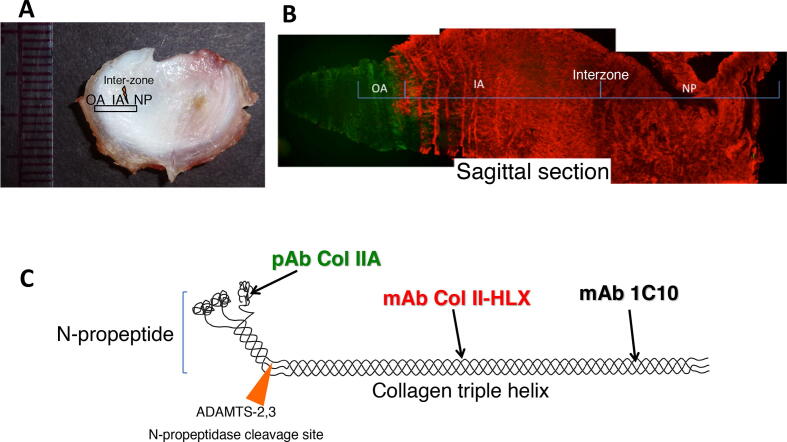
Abstract Image

Abstract Image

Abstract Image

成熟牛椎间盘纤维环IIA型前胶原的生化及免疫组织化学定位
为了使下一代组织工程构建和再生医学在临床上取得成功,必须充分确定这些修复技术所寻求恢复的组织的基本生物学和细胞外基质组成。使用最新的试剂加上久经考验的方法,我们继续在成熟的椎间盘中发现以前未检测到的结构蛋白。在这项研究中,我们发现“胚胎”型IIA型前胶原异构体(含有富含半胱氨酸的氨基前肽)在小牛和成熟的牛尾椎间盘的纤维环中都可以生化检测到,但在IIB型异构体主要集中的髓核中却没有检测到。具体来说,三螺旋型IIA前胶原异构体免疫定位于纤维内环的外边缘。三螺旋加工的II型胶原仅局限于片层间区域,而IIA型前胶原则位于片层内区域。IIA型前胶原定位区域α1(II)胶原链的质谱分析显示脯氨酸-944的3-羟基化程度高,这是一种翻译后修饰,与髓核中薄的胶原原纤维相关。研究结果表明,在纤维环的特定区域,IIA型前胶原的小直径原纤维可能有助于胶原束的组织和i - II型胶原过渡区内的结构特性。
本文章由计算机程序翻译,如有差异,请以英文原文为准。
求助全文
约1分钟内获得全文 求助全文
来源期刊
Matrix Biology Plus
Matrix Biology Plus Medicine-Histology
CiteScore
9.00
自引率
0.00%
发文量
25
审稿时长
105 days
×
引用
GB/T 7714-2015
复制
MLA
复制
APA
复制
导出至
BibTeX EndNote RefMan NoteFirst NoteExpress
×
提示
您的信息不完整,为了账户安全,请先补充。
现在去补充
×
提示
您因"违规操作"
具体请查看互助需知
我知道了
×
提示
确定
请完成安全验证×
copy
已复制链接
快去分享给好友吧!
我知道了
右上角分享
点击右上角分享
0
联系我们:info@booksci.cn Book学术提供免费学术资源搜索服务,方便国内外学者检索中英文文献。致力于提供最便捷和优质的服务体验。 Copyright © 2023 布克学术 All rights reserved.
京ICP备2023020795号-1
ghs 京公网安备 11010802042870号
Book学术文献互助
Book学术文献互助群
群 号:481959085
Book学术官方微信