Hong Seok Choi, Yun-Hyeon Kim, Won Gi Jeong, Jong Eun Lee, Hye Mi Park
{"title":"Clinicoradiological Features of Pulmonary Cryptococcosis in Immunocompetent Patients.","authors":"Hong Seok Choi, Yun-Hyeon Kim, Won Gi Jeong, Jong Eun Lee, Hye Mi Park","doi":"10.3348/jksr.2022.0008","DOIUrl":null,"url":null,"abstract":"<p><strong>Purpose: </strong>To assess the clinicoradiological features of pulmonary cryptococcosis in immunocompetent patients.</p><p><strong>Materials and methods: </strong>This retrospective study included immunocompetent patients who had been diagnosed with pulmonary cryptococcosis on the histopathologic exam and underwent chest CT between January 2008 and November 2019. Imaging features were divided into major imaging patterns, distributions, and ancillary imaging findings. Univariable analysis was performed to evaluate clinicoradiological features according to the presence of serum cryptococcal antigen.</p><p><strong>Results: </strong>Thirty-one patients were evaluated (mean age: 60 years, range: 19-78 years). A single nodular lesion confined to a single lobe was the most common imaging pattern (14/31, 45.2%). Serum cryptococcal antigen tests were performed in 19 patients (19/31, 61.3%). The presence of serum cryptococcal antigen was observed in six patients (6/19, 31.6%), all of whom showed a consolidation-dominant pattern. The presence of serum cryptococcal antigen was significantly associated with the consolidation-dominant pattern compared to those associated with a nodule-dominant pattern (<i>p</i> = 0.011).</p><p><strong>Conclusion: </strong>A combination of CT findings of consolidation and a positive serum cryptococcal antigen test may be helpful for diagnosing pulmonary cryptococcosis in immunocompetent patients.</p>","PeriodicalId":17455,"journal":{"name":"Journal of the Korean Society of Radiology","volume":"84 1","pages":"253-262"},"PeriodicalIF":0.0000,"publicationDate":"2023-01-01","publicationTypes":"Journal Article","fieldsOfStudy":null,"isOpenAccess":false,"openAccessPdf":"https://ftp.ncbi.nlm.nih.gov/pub/pmc/oa_pdf/9c/2f/jksr-84-253.PMC9935965.pdf","citationCount":"0","resultStr":null,"platform":"Semanticscholar","paperid":null,"PeriodicalName":"Journal of the Korean Society of Radiology","FirstCategoryId":"1085","ListUrlMain":"https://doi.org/10.3348/jksr.2022.0008","RegionNum":0,"RegionCategory":null,"ArticlePicture":[],"TitleCN":null,"AbstractTextCN":null,"PMCID":null,"EPubDate":"","PubModel":"","JCR":"Q4","JCRName":"Medicine","Score":null,"Total":0}
引用次数: 0
Abstract
Purpose: To assess the clinicoradiological features of pulmonary cryptococcosis in immunocompetent patients.
Materials and methods: This retrospective study included immunocompetent patients who had been diagnosed with pulmonary cryptococcosis on the histopathologic exam and underwent chest CT between January 2008 and November 2019. Imaging features were divided into major imaging patterns, distributions, and ancillary imaging findings. Univariable analysis was performed to evaluate clinicoradiological features according to the presence of serum cryptococcal antigen.
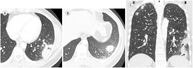
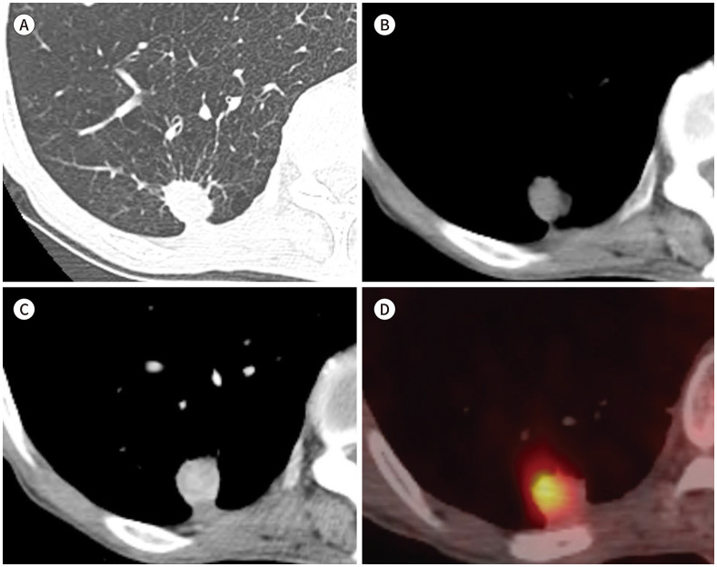
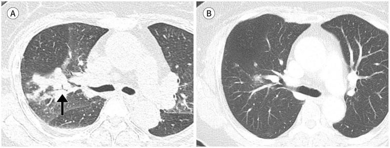
Results: Thirty-one patients were evaluated (mean age: 60 years, range: 19-78 years). A single nodular lesion confined to a single lobe was the most common imaging pattern (14/31, 45.2%). Serum cryptococcal antigen tests were performed in 19 patients (19/31, 61.3%). The presence of serum cryptococcal antigen was observed in six patients (6/19, 31.6%), all of whom showed a consolidation-dominant pattern. The presence of serum cryptococcal antigen was significantly associated with the consolidation-dominant pattern compared to those associated with a nodule-dominant pattern (p = 0.011).
Conclusion: A combination of CT findings of consolidation and a positive serum cryptococcal antigen test may be helpful for diagnosing pulmonary cryptococcosis in immunocompetent patients.

 求助内容:
求助内容: 应助结果提醒方式:


