Evaluation of Success of Arterial Cannulation Employing the Dorsalis Pedis Artery Versus Posterior Tibial Artery: A Clinical Comparative Study.
IF 0.6
Q3 ANESTHESIOLOGY
Turkish journal of anaesthesiology and reanimation
Pub Date : 2023-02-01
DOI:10.5152/TJAR.2023.22826
引用次数: 0
Abstract
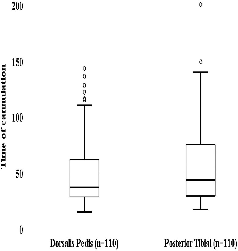
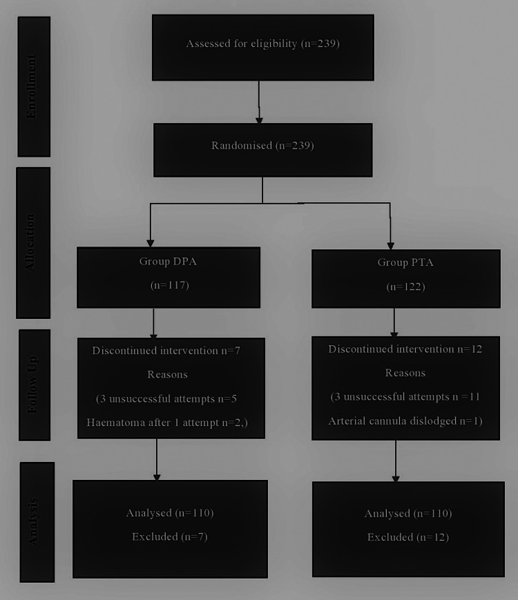
Objective: The dorsalis pedis artery and posterior tibial artery are recognised sites for arterial cannulation. This study aimed to compare the first-attempt success rates of cannulation along with other cannulation characteristics of these 2 arteries in adult patients undergoing surgery under general anaesthesia using the conventional palpatory method. Methods: Two hundred twenty adults were allocated randomly into 2 groups. The dorsalis pedis artery and posterior tibial artery were attempted for cannulation in the dorsalis pedis artery and posterior tibial artery group, respectively. First-attempt success rates, cannulation times, number of attempts, ease of cannulation, and complications were recorded. Results: Demographic characteristics, pulse characteristics, single-attempt success rates, ease of cannulation, reasons for failure, and complications were similar. Single-attempt success rates were similar (64.5% and 61.8%, P = .675) with equal median attempt. Easy cannulation (Visual Analogue Scale score ≤4) was the same in both groups, whereas percentages of difficult cannulation (Visual Analogue Scale scores ≥4) were 16.4% and 19.1% in the dorsalis pedis artery and posterior tibial artery groups, respectively. Cannulation time was lower in the dorsalis pedis artery group [median time in seconds: 37 (28, 63) seconds vs. 44 (29, 75) seconds, P = .027]. Single-attempt success rates were lower in the feeble pulse group as compared to the strong pulse group (48.61% vs. 70.27%, P = .002). Likewise, a higher Visual Analogue Scale of ease of cannulation (>4 score) was seen in the feeble pulse group compared to the strong pulse group (26.39% vs. 13.51%, P = .019). Conclusions: The single-attempt success rate was similar for both dorsalis pedis artery and posterior tibial artery. However, the time taken for cannulating the posterior tibial artery is significantly higher than that for dorsalis pedis artery.

足背动脉与胫后动脉插管成功率的临床比较研究。
目的:足背动脉和胫后动脉是动脉插管的识别部位。本研究旨在比较在全身麻醉下采用常规触诊方法进行手术的成人患者第一次插管成功率以及这两条动脉的其他插管特点。方法:将220名成人随机分为2组。在足背动脉组和胫后动脉组分别尝试足背动脉和胫后动脉插管。记录首次尝试成功率、插管次数、插管次数、插管难易程度及并发症。结果:人口学特征、脉搏特征、单次插管成功率、插管难易程度、失败原因及并发症相似。单次尝试成功率相似(64.5%和61.8%,P = 0.675)。两组容易插管(视觉模拟评分≤4分)的比例相同,而足背动脉组和胫后动脉组插管困难(视觉模拟评分≥4分)的比例分别为16.4%和19.1%。足背动脉组插管时间较短[中位时间秒:37(28,63)秒比44(29,75)秒,P = 0.027]。脉搏微弱组的单次尝试成功率低于脉搏强烈组(48.61%比70.27%,P = 0.002)。同样,弱脉组插管易度视觉模拟量表(>4分)高于强脉组(26.39% vs. 13.51%, P = 0.019)。结论:足背动脉和胫后动脉单次手术成功率相近。胫后动脉插管时间明显高于足背动脉插管时间。
本文章由计算机程序翻译,如有差异,请以英文原文为准。
求助全文
约1分钟内获得全文
求助全文
来源期刊

Turkish journal of anaesthesiology and reanimation
Medicine-Emergency Medicine
CiteScore
1.10
自引率
0.00%
发文量
0
 求助内容:
求助内容: 应助结果提醒方式:


