Nicole Hindley, Anna Sanchez Avila, Christopher Henstridge
{"title":"Bringing synapses into focus: Recent advances in synaptic imaging and mass-spectrometry for studying synaptopathy.","authors":"Nicole Hindley, Anna Sanchez Avila, Christopher Henstridge","doi":"10.3389/fnsyn.2023.1130198","DOIUrl":null,"url":null,"abstract":"<p><p>Synapses are integral for healthy brain function and are becoming increasingly recognized as key structures in the early stages of brain disease. Understanding the pathological processes driving synaptic dysfunction will unlock new therapeutic opportunities for some of the most devastating diseases of our time. To achieve this we need a solid repertoire of imaging and molecular tools to interrogate synaptic biology at greater resolution. Synapses have historically been examined in small numbers, using highly technical imaging modalities, or in bulk, using crude molecular approaches. However, recent advances in imaging techniques are allowing us to analyze large numbers of synapses, at single-synapse resolution. Furthermore, multiplexing is now achievable with some of these approaches, meaning we can examine multiple proteins at individual synapses in intact tissue. New molecular techniques now allow accurate quantification of proteins from isolated synapses. The development of increasingly sensitive mass-spectrometry equipment means we can now scan the synaptic molecular landscape almost in totality and see how this changes in disease. As we embrace these new technical developments, synapses will be viewed with clearer focus, and the field of synaptopathy will become richer with insightful and high-quality data. Here, we will discuss some of the ways in which synaptic interrogation is being facilitated by methodological advances, focusing on imaging, and mass spectrometry.</p>","PeriodicalId":12650,"journal":{"name":"Frontiers in Synaptic Neuroscience","volume":"15 ","pages":"1130198"},"PeriodicalIF":4.1000,"publicationDate":"2023-03-15","publicationTypes":"Journal Article","fieldsOfStudy":null,"isOpenAccess":false,"openAccessPdf":"https://www.ncbi.nlm.nih.gov/pmc/articles/PMC10050382/pdf/","citationCount":"0","resultStr":null,"platform":"Semanticscholar","paperid":null,"PeriodicalName":"Frontiers in Synaptic Neuroscience","FirstCategoryId":"3","ListUrlMain":"https://doi.org/10.3389/fnsyn.2023.1130198","RegionNum":4,"RegionCategory":"医学","ArticlePicture":[],"TitleCN":null,"AbstractTextCN":null,"PMCID":null,"EPubDate":"2023/1/1 0:00:00","PubModel":"eCollection","JCR":"Q2","JCRName":"NEUROSCIENCES","Score":null,"Total":0}
引用次数: 0
Abstract
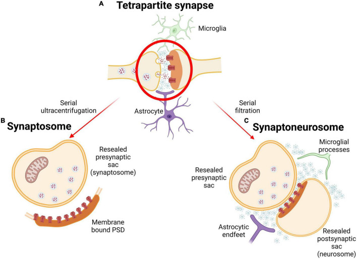
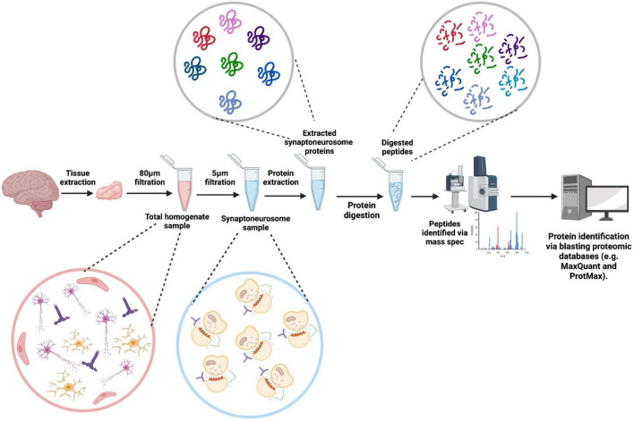
Synapses are integral for healthy brain function and are becoming increasingly recognized as key structures in the early stages of brain disease. Understanding the pathological processes driving synaptic dysfunction will unlock new therapeutic opportunities for some of the most devastating diseases of our time. To achieve this we need a solid repertoire of imaging and molecular tools to interrogate synaptic biology at greater resolution. Synapses have historically been examined in small numbers, using highly technical imaging modalities, or in bulk, using crude molecular approaches. However, recent advances in imaging techniques are allowing us to analyze large numbers of synapses, at single-synapse resolution. Furthermore, multiplexing is now achievable with some of these approaches, meaning we can examine multiple proteins at individual synapses in intact tissue. New molecular techniques now allow accurate quantification of proteins from isolated synapses. The development of increasingly sensitive mass-spectrometry equipment means we can now scan the synaptic molecular landscape almost in totality and see how this changes in disease. As we embrace these new technical developments, synapses will be viewed with clearer focus, and the field of synaptopathy will become richer with insightful and high-quality data. Here, we will discuss some of the ways in which synaptic interrogation is being facilitated by methodological advances, focusing on imaging, and mass spectrometry.

 求助内容:
求助内容: 应助结果提醒方式:


