Yeomyeong Kim, Cho-Rong Bae, Dongyeop Kim, Hyejeong Kim, Sunghye Lee, Haiying Zhang, Minyoung Noh, Young-Myeong Kim, Naoki Mochizuki, Young-Guen Kwon
{"title":"Efficacy of CU06-1004 via regulation of inflammation and endothelial permeability in LPS-induced acute lung injury.","authors":"Yeomyeong Kim, Cho-Rong Bae, Dongyeop Kim, Hyejeong Kim, Sunghye Lee, Haiying Zhang, Minyoung Noh, Young-Myeong Kim, Naoki Mochizuki, Young-Guen Kwon","doi":"10.1186/s12950-023-00338-x","DOIUrl":null,"url":null,"abstract":"<p><strong>Background: </strong>Acute lung injury (ALI) is a life-threatening condition that fundamentally results from inflammation and edema in the lung. There are no effective treatments available for clinical use. Previously, we found that as a leakage blocker CU06-1004 prevents endothelial barrier disruption and enhances endothelial cell survival under inflammatory conditions. In this study, we aimed to elucidate the effect of CU06-1004 in terms of prevention of inflammation and endothelial dysfunction in an ALI mouse model.</p><p><strong>Methods: </strong>An ALI model was established that included intraperitoneal administration of LPS. Following LPS administration, survival rates and lung wet/dry ratios were assessed. Histological analysis was performed using hematoxylin and eosin staining. Scanning electron microscopy was used to examine alveolar and capillary morphology. Cytokines such as IL-1β, IL-6, and TNF-α were analyzed using an ELISA assay of bronchoalveolar lavage fluid (BALF) and serum. Neutrophil infiltration was observed in BALF using Wright-Giemsa staining, and myeloperoxidase (MPO) activity was assessed. Pulmonary vascular leakage was confirmed using Evans-blue dye, and the expression of junctional proteins was evaluated using immunofluorescent staining. Expression of adhesion molecules was observed using immunofluorescence staining. NF-κB activation was determined using immunohistochemistry and western blot analysis.</p><p><strong>Results: </strong>Survival rates and pulmonary edema were ameliorated with CU06-1004 treatment. Administration of CU06-1004 normalized histopathological changes induced by LPS, and alveolar-capillary wall thickening was reduced. Compared with the LPS-challenged group, after CU06-1004 treatment, the infiltration of immune cells was decreased in the BALF, and MPO activity in lung tissue was reduced. Similarly, in the CU06-1004 treatment group, pro-inflammatory cytokines were significantly inhibited in both BALF and serum. Evans-blue leakage was reduced, and the expression of junctional proteins was recovered in the CU06-1004 group. Adhesion molecules were downregulated and NF-κB activation was inhibited after CU06-1004 treatment.</p><p><strong>Conclusions: </strong>These results suggested that CU06-1004 had a therapeutic effect against LPS-induced ALI via alleviation of the inflammatory response and protection of vascular integrity.</p>","PeriodicalId":56120,"journal":{"name":"Journal of Inflammation-London","volume":"20 1","pages":"13"},"PeriodicalIF":4.4000,"publicationDate":"2023-04-06","publicationTypes":"Journal Article","fieldsOfStudy":null,"isOpenAccess":false,"openAccessPdf":"https://www.ncbi.nlm.nih.gov/pmc/articles/PMC10078077/pdf/","citationCount":"2","resultStr":null,"platform":"Semanticscholar","paperid":null,"PeriodicalName":"Journal of Inflammation-London","FirstCategoryId":"3","ListUrlMain":"https://doi.org/10.1186/s12950-023-00338-x","RegionNum":3,"RegionCategory":"医学","ArticlePicture":[],"TitleCN":null,"AbstractTextCN":null,"PMCID":null,"EPubDate":"","PubModel":"","JCR":"Q2","JCRName":"IMMUNOLOGY","Score":null,"Total":0}
引用次数: 2
Abstract
Background: Acute lung injury (ALI) is a life-threatening condition that fundamentally results from inflammation and edema in the lung. There are no effective treatments available for clinical use. Previously, we found that as a leakage blocker CU06-1004 prevents endothelial barrier disruption and enhances endothelial cell survival under inflammatory conditions. In this study, we aimed to elucidate the effect of CU06-1004 in terms of prevention of inflammation and endothelial dysfunction in an ALI mouse model.
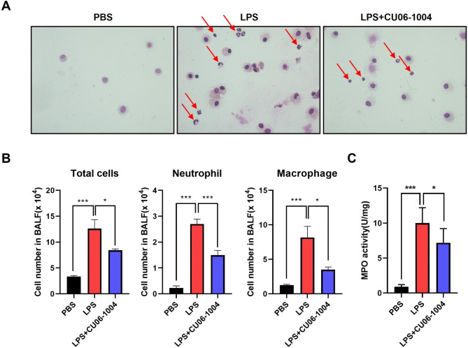
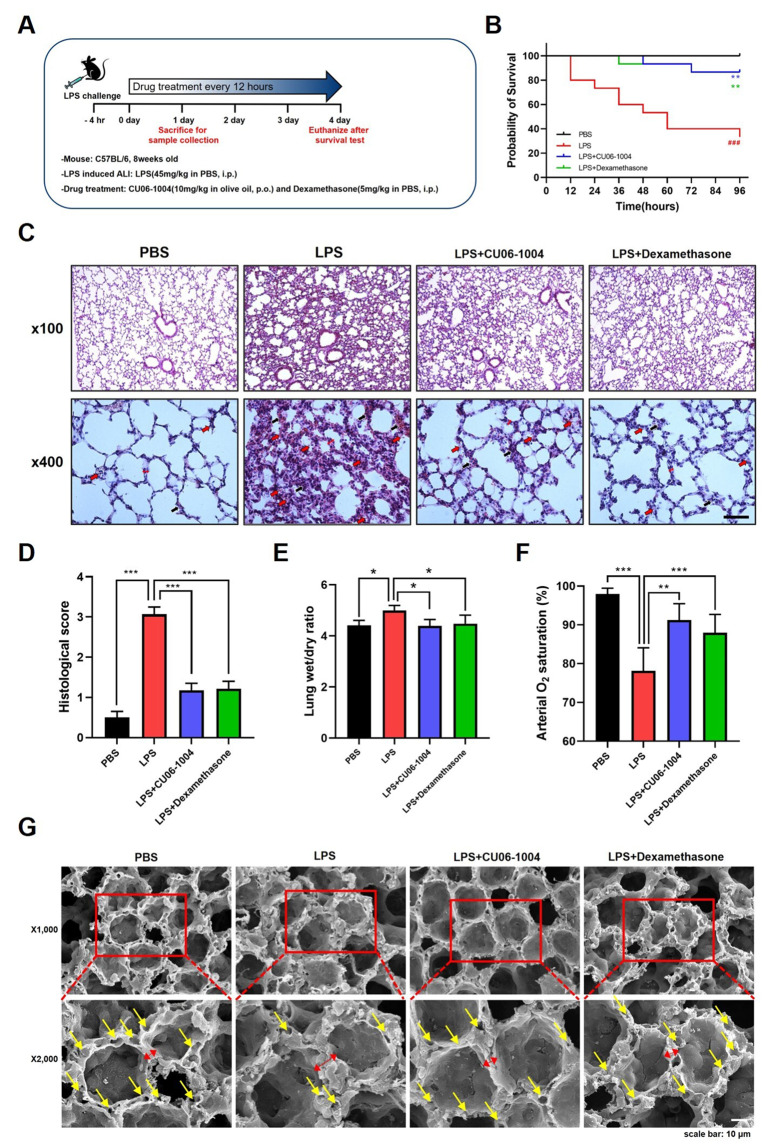
Methods: An ALI model was established that included intraperitoneal administration of LPS. Following LPS administration, survival rates and lung wet/dry ratios were assessed. Histological analysis was performed using hematoxylin and eosin staining. Scanning electron microscopy was used to examine alveolar and capillary morphology. Cytokines such as IL-1β, IL-6, and TNF-α were analyzed using an ELISA assay of bronchoalveolar lavage fluid (BALF) and serum. Neutrophil infiltration was observed in BALF using Wright-Giemsa staining, and myeloperoxidase (MPO) activity was assessed. Pulmonary vascular leakage was confirmed using Evans-blue dye, and the expression of junctional proteins was evaluated using immunofluorescent staining. Expression of adhesion molecules was observed using immunofluorescence staining. NF-κB activation was determined using immunohistochemistry and western blot analysis.
Results: Survival rates and pulmonary edema were ameliorated with CU06-1004 treatment. Administration of CU06-1004 normalized histopathological changes induced by LPS, and alveolar-capillary wall thickening was reduced. Compared with the LPS-challenged group, after CU06-1004 treatment, the infiltration of immune cells was decreased in the BALF, and MPO activity in lung tissue was reduced. Similarly, in the CU06-1004 treatment group, pro-inflammatory cytokines were significantly inhibited in both BALF and serum. Evans-blue leakage was reduced, and the expression of junctional proteins was recovered in the CU06-1004 group. Adhesion molecules were downregulated and NF-κB activation was inhibited after CU06-1004 treatment.
Conclusions: These results suggested that CU06-1004 had a therapeutic effect against LPS-induced ALI via alleviation of the inflammatory response and protection of vascular integrity.
期刊介绍:
Journal of Inflammation welcomes research submissions on all aspects of inflammation.
The five classical symptoms of inflammation, namely redness (rubor), swelling (tumour), heat (calor), pain (dolor) and loss of function (functio laesa), are only part of the story. The term inflammation is taken to include the full range of underlying cellular and molecular mechanisms involved, not only in the production of the inflammatory responses but, more importantly in clinical terms, in the healing process as well. Thus the journal covers molecular, cellular, animal and clinical studies, and related aspects of pharmacology, such as anti-inflammatory drug development, trials and therapeutic developments. It also considers publication of negative findings.
Journal of Inflammation aims to become the leading online journal on inflammation and, as online journals replace printed ones over the next decade, the main open access inflammation journal. Open access guarantees a larger audience, and thus impact, than any restricted access equivalent, and increasingly so, as the escalating costs of printed journals puts them outside University budgets. The unrestricted access to research findings in inflammation aids in promoting dynamic and productive dialogue between industrial and academic members of the inflammation research community, which plays such an important part in the development of future generations of anti-inflammatory therapies.

 求助内容:
求助内容: 应助结果提醒方式:


