Overall patterns of eye-specific retino-geniculo-cortical projections to layers III, IV, and VI in primary visual cortex of the greater galago (Otolemur crassicudatus), and correlation with cytochrome oxidase blobs.
Jaime F Olavarria, Huixin Qi, Toru Takahata, Jon H Kaas
{"title":"Overall patterns of eye-specific retino-geniculo-cortical projections to layers III, IV, and VI in primary visual cortex of the greater galago (<i>Otolemur crassicudatus</i>), and correlation with cytochrome oxidase blobs.","authors":"Jaime F Olavarria, Huixin Qi, Toru Takahata, Jon H Kaas","doi":"10.1017/S0952523822000062","DOIUrl":null,"url":null,"abstract":"<p><p>Studies in the greater galago have not provided a comprehensive description of the organization of eye-specific retino-geniculate-cortical projections to the recipient layers in V1. Here we demonstrate the overall patterns of ocular dominance domains in layers III, IV, and VI revealed following a monocular injection of the transneuronal tracer wheat germ agglutinin conjugated with horseradish peroxidase (WGA-HRP). We also correlate these patterns with the array of cytochrome oxidase (CO) blobs in tangential sections through the unfolded and flattened cortex. In layer IV, we observed for the first time that eye-specific domains form an interconnected pattern of bands 200-250 μm wide arranged such that they do not show orientation bias and do not meet the V1 border at right angles, as is the case in macaques. We also observed distinct WGA-HRP labeled patches in layers III and VI. The patches in layer III, likely corresponding to patches of K lateral geniculate nucleus (LGN) input, align with layer IV ocular dominance columns (ODCs) of the same eye dominance and overlap partially with virtually all CO blobs in both hemispheres, implying that CO blobs receive K LGN input from both eyes. We further found that CO blobs straddle the border between layer IV ODCs, such that the distribution of CO staining is approximately equal over ipsilateral and contralateral ODCs. These results, together with studies showing that a high percentage of cells in CO blobs are monocular, suggest that CO blobs consist of ipsilateral and contralateral subregions that are in register with underlying layer IV ODCs of the same eye dominance. In macaques and humans, CO blobs are centered on ODCs in layer IV. Our finding that CO blobs in galago straddle the border of neighboring layer IV ODCs suggests that this novel feature may represent an alternative way by which visual information is processed by eye-specific modular architecture in mammalian V1.</p>","PeriodicalId":23556,"journal":{"name":"Visual Neuroscience","volume":"39 ","pages":"E007"},"PeriodicalIF":2.3000,"publicationDate":"2022-11-02","publicationTypes":"Journal Article","fieldsOfStudy":null,"isOpenAccess":false,"openAccessPdf":"https://www.ncbi.nlm.nih.gov/pmc/articles/PMC9634673/pdf/","citationCount":"0","resultStr":null,"platform":"Semanticscholar","paperid":null,"PeriodicalName":"Visual Neuroscience","FirstCategoryId":"3","ListUrlMain":"https://doi.org/10.1017/S0952523822000062","RegionNum":4,"RegionCategory":"医学","ArticlePicture":[],"TitleCN":null,"AbstractTextCN":null,"PMCID":null,"EPubDate":"","PubModel":"","JCR":"Q4","JCRName":"NEUROSCIENCES","Score":null,"Total":0}
引用次数: 0
Abstract
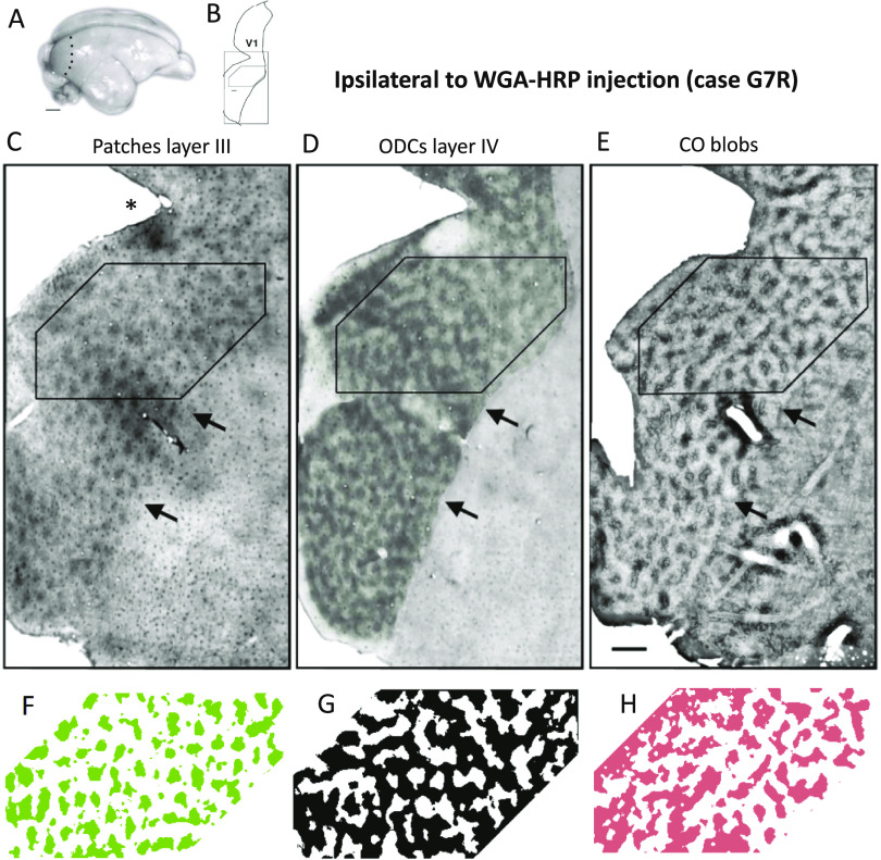
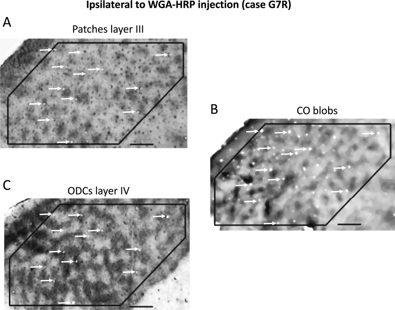
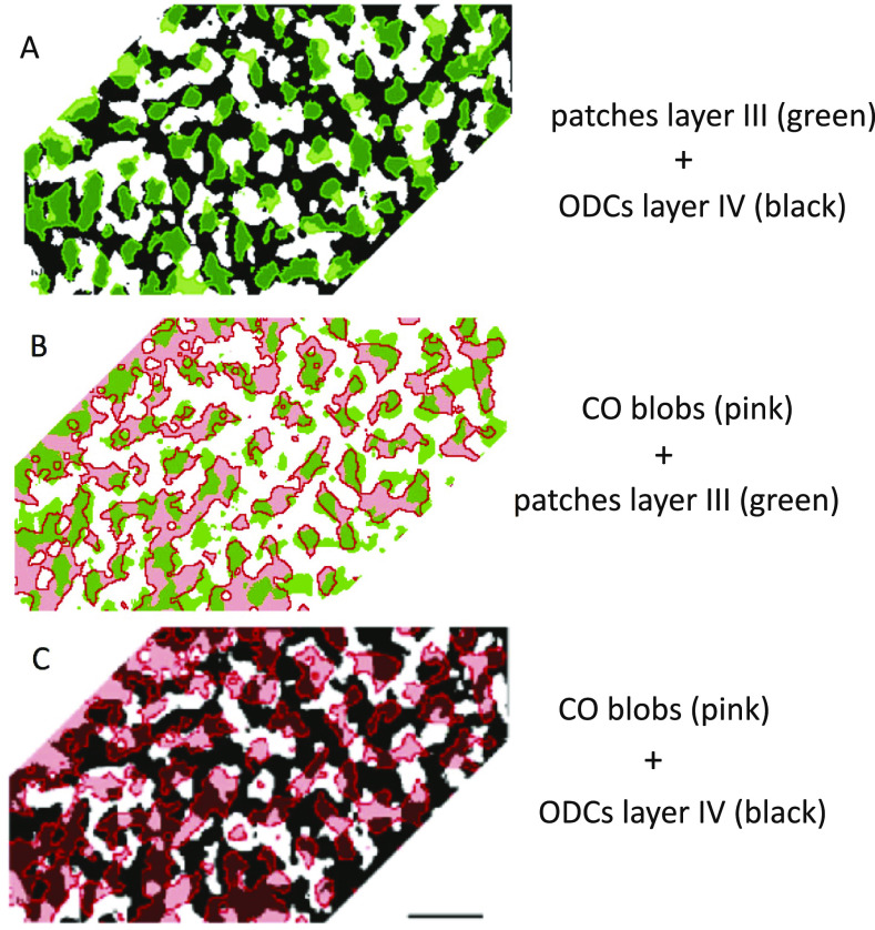
Studies in the greater galago have not provided a comprehensive description of the organization of eye-specific retino-geniculate-cortical projections to the recipient layers in V1. Here we demonstrate the overall patterns of ocular dominance domains in layers III, IV, and VI revealed following a monocular injection of the transneuronal tracer wheat germ agglutinin conjugated with horseradish peroxidase (WGA-HRP). We also correlate these patterns with the array of cytochrome oxidase (CO) blobs in tangential sections through the unfolded and flattened cortex. In layer IV, we observed for the first time that eye-specific domains form an interconnected pattern of bands 200-250 μm wide arranged such that they do not show orientation bias and do not meet the V1 border at right angles, as is the case in macaques. We also observed distinct WGA-HRP labeled patches in layers III and VI. The patches in layer III, likely corresponding to patches of K lateral geniculate nucleus (LGN) input, align with layer IV ocular dominance columns (ODCs) of the same eye dominance and overlap partially with virtually all CO blobs in both hemispheres, implying that CO blobs receive K LGN input from both eyes. We further found that CO blobs straddle the border between layer IV ODCs, such that the distribution of CO staining is approximately equal over ipsilateral and contralateral ODCs. These results, together with studies showing that a high percentage of cells in CO blobs are monocular, suggest that CO blobs consist of ipsilateral and contralateral subregions that are in register with underlying layer IV ODCs of the same eye dominance. In macaques and humans, CO blobs are centered on ODCs in layer IV. Our finding that CO blobs in galago straddle the border of neighboring layer IV ODCs suggests that this novel feature may represent an alternative way by which visual information is processed by eye-specific modular architecture in mammalian V1.
大鸮(Otolemur crassicudatus)初级视觉皮层第 III、IV 和 VI 层眼球特异性视网膜-皮质-神经投射的总体模式,以及与细胞色素氧化酶斑点的相关性。
对大galago的研究尚未全面描述V1受体层的眼特异性视网膜-皮质投射组织。在这里,我们展示了单眼注射跨神经元示踪剂--辣根过氧化物酶共轭的小麦胚芽凝集素(WGA-HRP)后,第三、第四和第六层眼支配域的整体模式。我们还将这些模式与通过展开和扁平皮层的切向切片中的细胞色素氧化酶(CO)球阵列联系起来。在第四层,我们首次观察到眼球特异性结构域形成了宽 200-250 μm 的带状相互连接模式,这些带状排列不显示方向偏差,也不像猕猴那样与 V1 边界成直角相交。我们还在第三层和第六层观察到不同的 WGA-HRP 标记斑块。第 III 层的斑块可能对应于 K 侧膝状核(LGN)输入的斑块,与第 IV 层的同眼优势列(ODC)对齐,并与两个半球的几乎所有 CO blobs 部分重叠,这意味着 CO blobs 接受来自双眼的 K LGN 输入。我们还发现,CO Blobs 横跨第四层 ODC 之间的边界,因此同侧和对侧 ODC 上的 CO 染色分布大致相等。这些结果,再加上研究显示 CO 信号团中有很大比例的细胞是单眼细胞,表明 CO 信号团由同侧和对侧亚区组成,这些亚区与底层第四层 ODC 的同眼优势一致。在猕猴和人类中,CO 信号团以第四层的 ODC 为中心。我们发现,galago 的 CO blobs 跨过了相邻的第四层 ODC 的边界,这表明这一新颖特征可能代表了哺乳动物 V1 中眼睛特异性模块结构处理视觉信息的另一种方式。
期刊介绍:
Visual Neuroscience is an international journal devoted to the publication of experimental and theoretical research on biological mechanisms of vision. A major goal of publication is to bring together in one journal a broad range of studies that reflect the diversity and originality of all aspects of neuroscience research relating to the visual system. Contributions may address molecular, cellular or systems-level processes in either vertebrate or invertebrate species. The journal publishes work based on a wide range of technical approaches, including molecular genetics, anatomy, physiology, psychophysics and imaging, and utilizing comparative, developmental, theoretical or computational approaches to understand the biology of vision and visuo-motor control. The journal also publishes research seeking to understand disorders of the visual system and strategies for restoring vision. Studies based exclusively on clinical, psychophysiological or behavioral data are welcomed, provided that they address questions concerning neural mechanisms of vision or provide insight into visual dysfunction.
 求助内容:
求助内容: 应助结果提醒方式:


