{"title":"Issue Information TOC","authors":"","doi":"10.1002/cpph.51","DOIUrl":null,"url":null,"abstract":"<p><b>Cover</b>: In Marchelletta et al. (https://doi.org/10.1002/cpph.54), the image show human colonic organoids retain in vivo morphology and cellular composition. Representative images of hematoxylin and eosin (H&E) staining of sections from paraffin-embedded organoids reveal a closed unit of differentiated columnar cells that resemble cells along the crypt axis in human colonic histologic sections from explant tissue. (<b>A</b>, <b>B</b>) Nuclei (dark purple) rest on the basal side in both explant tissue and organoids. (<b>C-F</b>) Organoids retain the cellular composition, frequency, and spatial orientation of tissue-resident colonic epithelial cells, as revealed by immunohistochemistry staining (arrows and brown staining) for the goblet-cell marker mucin 2 (MUC2) (<b>C</b>, <b>D</b>) and the enteroendocrine marker chromogranin A (<b>E</b>, <b>F</b>). \n\n <figure>\n <div><picture>\n <source></source></picture><p></p>\n </div>\n </figure></p>","PeriodicalId":10871,"journal":{"name":"Current Protocols in Pharmacology","volume":"85 1","pages":""},"PeriodicalIF":0.0000,"publicationDate":"2019-06-18","publicationTypes":"Journal Article","fieldsOfStudy":null,"isOpenAccess":false,"openAccessPdf":"https://sci-hub-pdf.com/10.1002/cpph.51","citationCount":"0","resultStr":null,"platform":"Semanticscholar","paperid":null,"PeriodicalName":"Current Protocols in Pharmacology","FirstCategoryId":"1085","ListUrlMain":"https://onlinelibrary.wiley.com/doi/10.1002/cpph.51","RegionNum":0,"RegionCategory":null,"ArticlePicture":[],"TitleCN":null,"AbstractTextCN":null,"PMCID":null,"EPubDate":"","PubModel":"","JCR":"Q2","JCRName":"Pharmacology, Toxicology and Pharmaceutics","Score":null,"Total":0}
引用次数: 0
Abstract
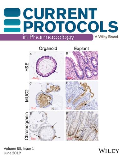
Cover: In Marchelletta et al. (https://doi.org/10.1002/cpph.54), the image show human colonic organoids retain in vivo morphology and cellular composition. Representative images of hematoxylin and eosin (H&E) staining of sections from paraffin-embedded organoids reveal a closed unit of differentiated columnar cells that resemble cells along the crypt axis in human colonic histologic sections from explant tissue. (A, B) Nuclei (dark purple) rest on the basal side in both explant tissue and organoids. (C-F) Organoids retain the cellular composition, frequency, and spatial orientation of tissue-resident colonic epithelial cells, as revealed by immunohistochemistry staining (arrows and brown staining) for the goblet-cell marker mucin 2 (MUC2) (C, D) and the enteroendocrine marker chromogranin A (E, F).

 求助内容:
求助内容: 应助结果提醒方式:


