{"title":"MiR-146b-5p/SEMA3G regulates epithelial-mesenchymal transition in clear cell renal cell carcinoma.","authors":"Mengxi Tang, Tao Xiong","doi":"10.1186/s13008-023-00083-w","DOIUrl":null,"url":null,"abstract":"<p><strong>Objective: </strong>The primary purpose was to unveil how the miR-146b-5p/SEMA3G axis works in clear cell renal cell carcinoma (ccRCC).</p><p><strong>Methods: </strong>ccRCC dataset was acquired from TCGA database, and target miRNA to be studied was further analyzed using survival analysis. We performed miRNA target gene prediction through the database, and those predicted miRNAs were intersected with differential mRNAs. After calculating the correlation between miRNAs and mRNAs, we completed the GSEA pathway enrichment analysis on mRNAs. MiRNA and mRNA expression was examined by qRT-PCR. Western blot was introduced to detect SEMA3G, MMP2, MMP9 expression, epithelial-mesenchymal transition (EMT) marker proteins, and Notch/TGF-β signaling pathway-related proteins. Targeted relationship between miRNA and mRNA was validated using a dual-luciferase test. Transwell assay was employed to assess cell migration and invasion. Wound healing assay was adopted for evaluation of migration ability. The effect of different treatments on cell morphology was observed by a microscope.</p><p><strong>Results: </strong>In ccRCC cells, miR-146b-5p was remarkably overexpressed, yet SEMA3G was markedly less expressed. MiR-146b-5p was capable of stimulating ccRCC cell invasion, migration and EMT, and promoting the transformation of ccRCC cell morphology to mesenchymal state. SEMA3G was targeted and inhibited via miR-146b-5p. MiR-146b-5p facilitated ccRCC cell migration, invasion, morphology transforming to mesenchymal state and EMT process by targeting SEMA3G and regulating Notch and TGF-β signaling pathways.</p><p><strong>Conclusion: </strong>MiR-146b-5p regulated Notch and TGF-β signaling pathway by suppressing SEMA3G expression, thus promoting the growth of ccRCC cells, which provides a possible target for ccRCC therapy and prognosis prediction.</p>","PeriodicalId":49263,"journal":{"name":"Cell Division","volume":"18 1","pages":"4"},"PeriodicalIF":2.8000,"publicationDate":"2023-03-07","publicationTypes":"Journal Article","fieldsOfStudy":null,"isOpenAccess":false,"openAccessPdf":"https://www.ncbi.nlm.nih.gov/pmc/articles/PMC9993666/pdf/","citationCount":"3","resultStr":null,"platform":"Semanticscholar","paperid":null,"PeriodicalName":"Cell Division","FirstCategoryId":"99","ListUrlMain":"https://doi.org/10.1186/s13008-023-00083-w","RegionNum":4,"RegionCategory":"生物学","ArticlePicture":[],"TitleCN":null,"AbstractTextCN":null,"PMCID":null,"EPubDate":"","PubModel":"","JCR":"Q3","JCRName":"CELL BIOLOGY","Score":null,"Total":0}
引用次数: 3
Abstract
Objective: The primary purpose was to unveil how the miR-146b-5p/SEMA3G axis works in clear cell renal cell carcinoma (ccRCC).
Methods: ccRCC dataset was acquired from TCGA database, and target miRNA to be studied was further analyzed using survival analysis. We performed miRNA target gene prediction through the database, and those predicted miRNAs were intersected with differential mRNAs. After calculating the correlation between miRNAs and mRNAs, we completed the GSEA pathway enrichment analysis on mRNAs. MiRNA and mRNA expression was examined by qRT-PCR. Western blot was introduced to detect SEMA3G, MMP2, MMP9 expression, epithelial-mesenchymal transition (EMT) marker proteins, and Notch/TGF-β signaling pathway-related proteins. Targeted relationship between miRNA and mRNA was validated using a dual-luciferase test. Transwell assay was employed to assess cell migration and invasion. Wound healing assay was adopted for evaluation of migration ability. The effect of different treatments on cell morphology was observed by a microscope.
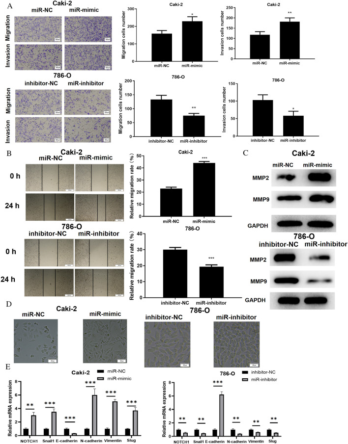
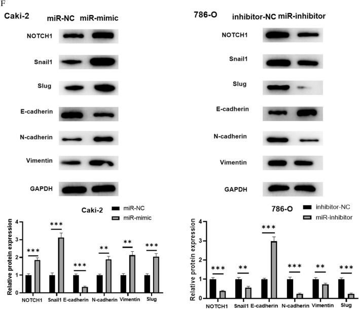
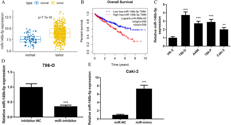
Results: In ccRCC cells, miR-146b-5p was remarkably overexpressed, yet SEMA3G was markedly less expressed. MiR-146b-5p was capable of stimulating ccRCC cell invasion, migration and EMT, and promoting the transformation of ccRCC cell morphology to mesenchymal state. SEMA3G was targeted and inhibited via miR-146b-5p. MiR-146b-5p facilitated ccRCC cell migration, invasion, morphology transforming to mesenchymal state and EMT process by targeting SEMA3G and regulating Notch and TGF-β signaling pathways.
Conclusion: MiR-146b-5p regulated Notch and TGF-β signaling pathway by suppressing SEMA3G expression, thus promoting the growth of ccRCC cells, which provides a possible target for ccRCC therapy and prognosis prediction.
期刊介绍:
Cell Division is an open access, peer-reviewed journal that encompasses all the molecular aspects of cell cycle control and cancer, cell growth, proliferation, survival, differentiation, signalling, gene transcription, protein synthesis, genome integrity, chromosome stability, centrosome duplication, DNA damage and DNA repair.
Cell Division provides an online forum for the cell-cycle community that aims to publish articles on all exciting aspects of cell-cycle research and to bridge the gap between models of cell cycle regulation, development, and cancer biology. This forum is driven by specialized and timely research articles, reviews and commentaries focused on this fast moving field, providing an invaluable tool for cell-cycle biologists.
Cell Division publishes articles in areas which includes, but not limited to:
DNA replication, cell fate decisions, cell cycle & development
Cell proliferation, mitosis, spindle assembly checkpoint, ubiquitin mediated degradation
DNA damage & repair
Apoptosis & cell death
 求助内容:
求助内容: 应助结果提醒方式:


