Vinayak Mishra, Alain Cuna, Rachana Singh, Daniel M Schwartz, Sherwin Chan, Akhil Maheshwari
{"title":"Imaging for Diagnosis and Assessment of Necrotizing Enterocolitis.","authors":"Vinayak Mishra, Alain Cuna, Rachana Singh, Daniel M Schwartz, Sherwin Chan, Akhil Maheshwari","doi":"10.5005/jp-journals-11002-0002","DOIUrl":null,"url":null,"abstract":"<p><p>Necrotizing enterocolitis (NEC) is inflammatory bowel necrosis of preterm and critically ill infants. The disease is seen in 6-10% of preterm infants who weigh less than 1500 g at birth and carries considerable morbidity, mortality, and healthcare cost burden. Efforts focused on timely mitigation remain restricted due to challenges in early diagnosis as clinical features, and available laboratory tests remain nonspecific until late in the disease. There is renewed interest in the radiological and sonographic assessment of intestinal diseases due to technological advances making them safe, cost-efficient, and supporting Web-based transmission of images, thereby reducing time to diagnosis by disease experts. Most of our experience has been with plain abdominal radiography, which shows characteristic features such as pneumatosis intestinalis in up to 50-60% of patients. Many patients with advanced disease may also show features such as portal venous gas and pneumoperitoneum. Unfortunately, these features are not seen consistently in patients with early, treatable conditions, and hence, there has been an unfulfilled need for additional imaging modalities. In recent years, abdominal ultrasound (AUS) has emerged as a readily available, noninvasive imaging tool that may be a valuable adjunct to plain radiographs for evaluating NEC. AUS can allow real-time assessment of vascular perfusion, bowel wall thickness, with higher sensitivity in detecting pneumatosis, altered peristalsis, and characteristics of the peritoneal fluid. Several other modalities, such as contrast-enhanced ultrasound (CEUS), magnetic resonance imaging (MRI), and near-infrared spectroscopy (NIRS), are also emerging. In this article, we have reviewed the available imaging options for NEC evaluation.</p>","PeriodicalId":74306,"journal":{"name":"Newborn (Clarksville, Md.)","volume":"1 1","pages":"182-189"},"PeriodicalIF":0.0000,"publicationDate":"2022-01-01","publicationTypes":"Journal Article","fieldsOfStudy":null,"isOpenAccess":false,"openAccessPdf":"https://ftp.ncbi.nlm.nih.gov/pub/pmc/oa_pdf/52/bc/nihms-1870282.PMC9976546.pdf","citationCount":"3","resultStr":null,"platform":"Semanticscholar","paperid":null,"PeriodicalName":"Newborn (Clarksville, Md.)","FirstCategoryId":"1085","ListUrlMain":"https://doi.org/10.5005/jp-journals-11002-0002","RegionNum":0,"RegionCategory":null,"ArticlePicture":[],"TitleCN":null,"AbstractTextCN":null,"PMCID":null,"EPubDate":"2022/3/31 0:00:00","PubModel":"Epub","JCR":"","JCRName":"","Score":null,"Total":0}
引用次数: 3
Abstract
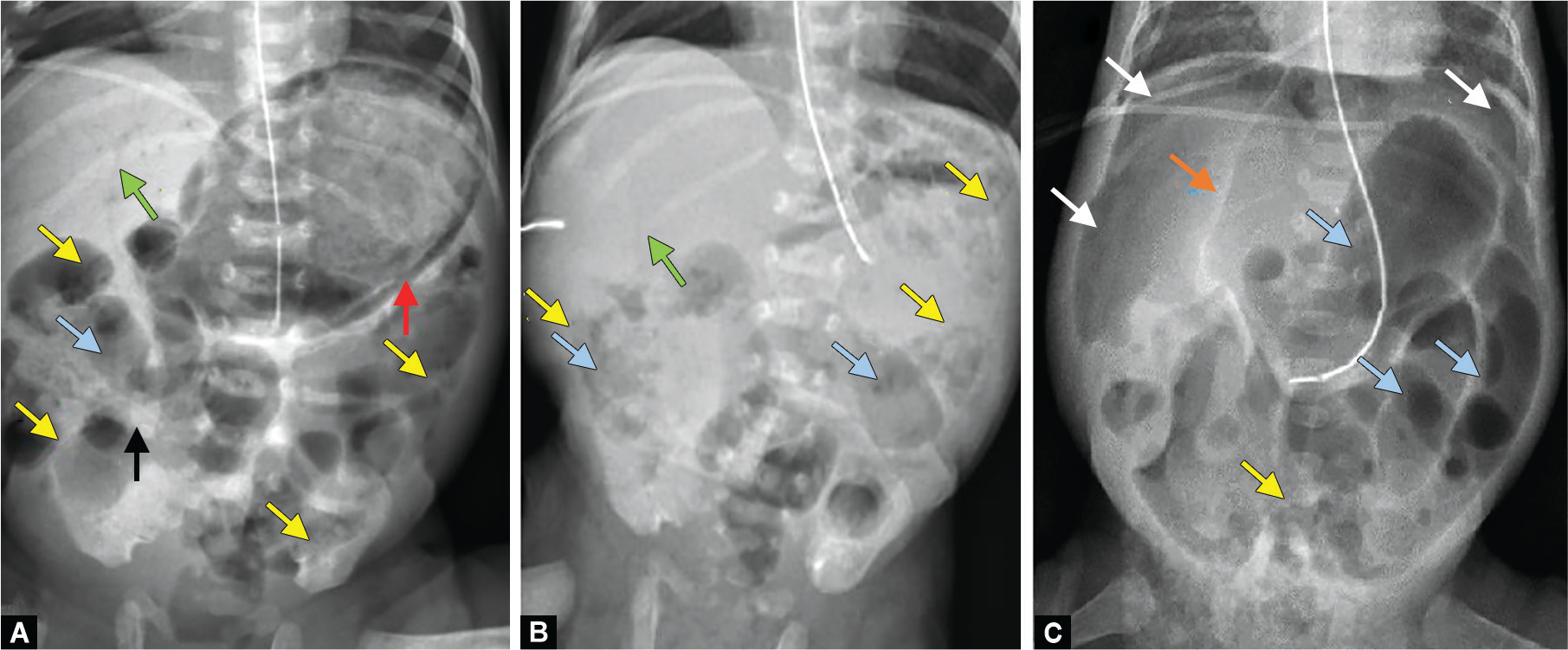
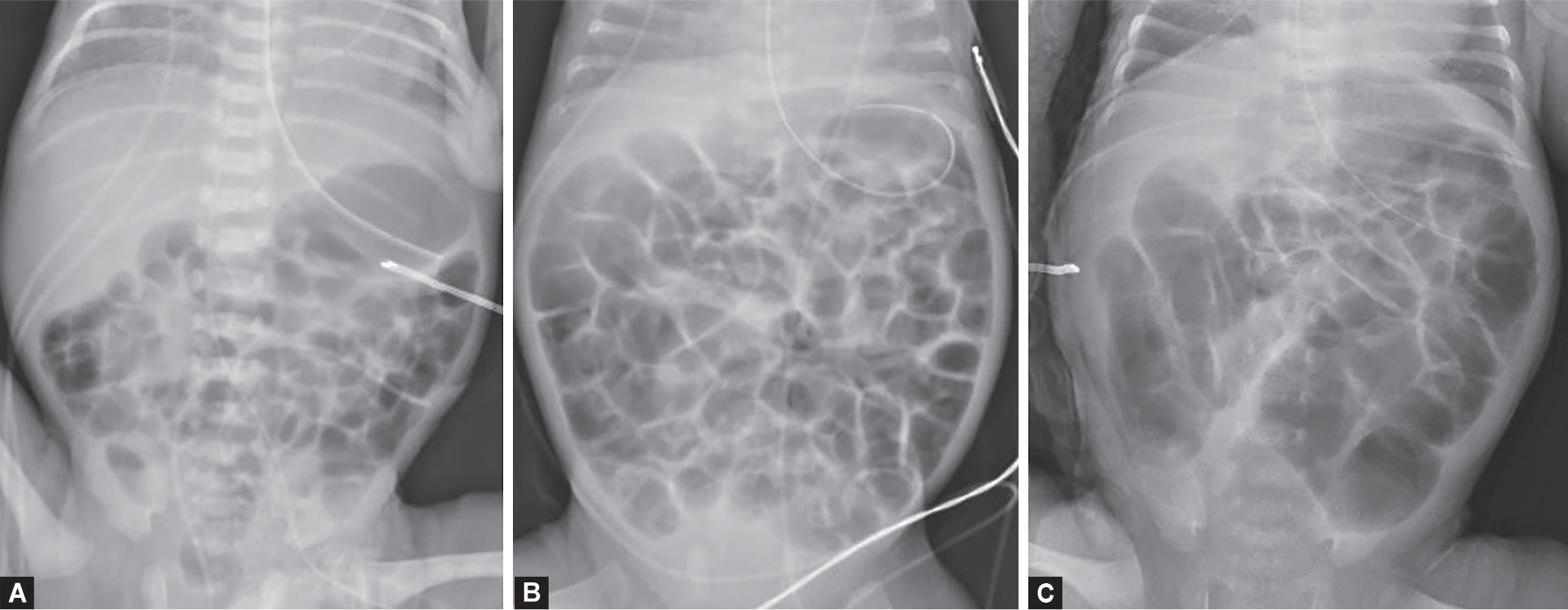
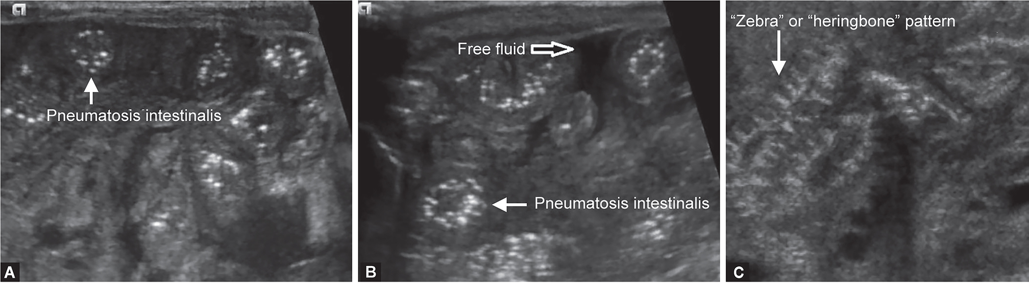
Necrotizing enterocolitis (NEC) is inflammatory bowel necrosis of preterm and critically ill infants. The disease is seen in 6-10% of preterm infants who weigh less than 1500 g at birth and carries considerable morbidity, mortality, and healthcare cost burden. Efforts focused on timely mitigation remain restricted due to challenges in early diagnosis as clinical features, and available laboratory tests remain nonspecific until late in the disease. There is renewed interest in the radiological and sonographic assessment of intestinal diseases due to technological advances making them safe, cost-efficient, and supporting Web-based transmission of images, thereby reducing time to diagnosis by disease experts. Most of our experience has been with plain abdominal radiography, which shows characteristic features such as pneumatosis intestinalis in up to 50-60% of patients. Many patients with advanced disease may also show features such as portal venous gas and pneumoperitoneum. Unfortunately, these features are not seen consistently in patients with early, treatable conditions, and hence, there has been an unfulfilled need for additional imaging modalities. In recent years, abdominal ultrasound (AUS) has emerged as a readily available, noninvasive imaging tool that may be a valuable adjunct to plain radiographs for evaluating NEC. AUS can allow real-time assessment of vascular perfusion, bowel wall thickness, with higher sensitivity in detecting pneumatosis, altered peristalsis, and characteristics of the peritoneal fluid. Several other modalities, such as contrast-enhanced ultrasound (CEUS), magnetic resonance imaging (MRI), and near-infrared spectroscopy (NIRS), are also emerging. In this article, we have reviewed the available imaging options for NEC evaluation.

 求助内容:
求助内容: 应助结果提醒方式:


