Evaluation of coronary arteries in congenital heart disease in children: diagnostic comparison of electrocardiogram-gated and non-electro-cardiogram-gated computed tomography cardiac angiography.
IF 0.9 Q4 RADIOLOGY, NUCLEAR MEDICINE & MEDICAL IMAGING
Kushaljit Singh Sodhi, Anmol Bhatia, Pratyaksha Rana, Shameema Farook, Akshay K Saxena, Harkant S Baryah, Anand K Mishra, Rohit Manoj
{"title":"Evaluation of coronary arteries in congenital heart disease in children: diagnostic comparison of electrocardiogram-gated and non-electro-cardiogram-gated computed tomography cardiac angiography.","authors":"Kushaljit Singh Sodhi, Anmol Bhatia, Pratyaksha Rana, Shameema Farook, Akshay K Saxena, Harkant S Baryah, Anand K Mishra, Rohit Manoj","doi":"10.5114/pjr.2022.123855","DOIUrl":null,"url":null,"abstract":"<p><strong>Purpose: </strong>To compare the visualization and anatomy of coronary arteries in children (≤ 2 years) with congenital heart disease (CHD) on non-electrocardiogram (ECG)-gated and ECG-gated computed tomography angiography (CTA).</p><p><strong>Material and methods: </strong>In this retrospective study, approved by the Ethics Committee of our institute, evaluation of coronary arteries in CHD was performed in 40 children on non-ECG-gated CTA and in 42 children on ECG-gated CTA. The origin and course of the right coronary artery (RCA), left main coronary artery (LMCA), left anterior descending (LAD) artery, and left circumflex (LCX) artery were evaluated by 2 paediatric radiologists independently.</p><p><strong>Results: </strong>ECG-gated CT scans yielded increased (additional) visualization of all the coronary arteries, when compared to non-ECG-gated CT scans. The RCA, LMCA, LAD artery, and LCX artery were visualized in 47.5%, 62.5%, 55%, and 32.5% of children, respectively, on non-ECG-gated studies, while they were visualized in 64.3%, 92.8%, 80.9%, and 62% children, respectively, on ECG-gated studies. The coronary artery anatomical variations were also supplementarily detected more in the ECG-gated group (23.8%) than in the non-ECG gated group (2.5%).</p><p><strong>Conclusions: </strong>ECG-gated CT cardiac angiography studies yield enhanced diagnostic outcomes for the evaluation of the coronary arteries in comparison to non-ECG-gated studies.</p>","PeriodicalId":47128,"journal":{"name":"Polish Journal of Radiology","volume":"87 ","pages":"e688-e693"},"PeriodicalIF":0.9000,"publicationDate":"2022-01-01","publicationTypes":"Journal Article","fieldsOfStudy":null,"isOpenAccess":false,"openAccessPdf":"https://ftp.ncbi.nlm.nih.gov/pub/pmc/oa_pdf/cb/9b/PJR-87-49806.PMC9834070.pdf","citationCount":"0","resultStr":null,"platform":"Semanticscholar","paperid":null,"PeriodicalName":"Polish Journal of Radiology","FirstCategoryId":"1085","ListUrlMain":"https://doi.org/10.5114/pjr.2022.123855","RegionNum":0,"RegionCategory":null,"ArticlePicture":[],"TitleCN":null,"AbstractTextCN":null,"PMCID":null,"EPubDate":"","PubModel":"","JCR":"Q4","JCRName":"RADIOLOGY, NUCLEAR MEDICINE & MEDICAL IMAGING","Score":null,"Total":0}
引用次数: 0
Abstract
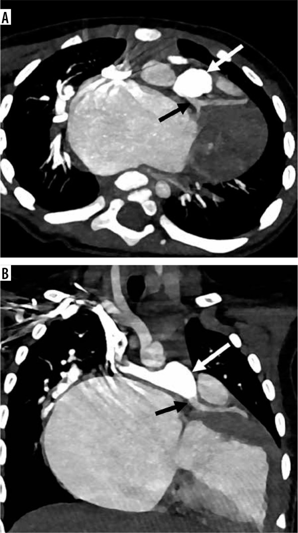
Purpose: To compare the visualization and anatomy of coronary arteries in children (≤ 2 years) with congenital heart disease (CHD) on non-electrocardiogram (ECG)-gated and ECG-gated computed tomography angiography (CTA).
Material and methods: In this retrospective study, approved by the Ethics Committee of our institute, evaluation of coronary arteries in CHD was performed in 40 children on non-ECG-gated CTA and in 42 children on ECG-gated CTA. The origin and course of the right coronary artery (RCA), left main coronary artery (LMCA), left anterior descending (LAD) artery, and left circumflex (LCX) artery were evaluated by 2 paediatric radiologists independently.
Results: ECG-gated CT scans yielded increased (additional) visualization of all the coronary arteries, when compared to non-ECG-gated CT scans. The RCA, LMCA, LAD artery, and LCX artery were visualized in 47.5%, 62.5%, 55%, and 32.5% of children, respectively, on non-ECG-gated studies, while they were visualized in 64.3%, 92.8%, 80.9%, and 62% children, respectively, on ECG-gated studies. The coronary artery anatomical variations were also supplementarily detected more in the ECG-gated group (23.8%) than in the non-ECG gated group (2.5%).
Conclusions: ECG-gated CT cardiac angiography studies yield enhanced diagnostic outcomes for the evaluation of the coronary arteries in comparison to non-ECG-gated studies.


 求助内容:
求助内容: 应助结果提醒方式:


