{"title":"Tumor–neuron interactions: A novel concept contributor to glioblastoma invasion","authors":"Dongling Pei, Zhenyu Zhang, Long Zhang","doi":"10.1002/mog2.17","DOIUrl":null,"url":null,"abstract":"<p>Recently, a study published in <i>Cell</i> by Venkataramani et al.<span><sup>1</sup></span> demonstrated that neuronal (NEU), neural progenitor (NPC)-like, and nonmesenchymal (non-MES)-like glioblastoma cells (GBCs) could drive brain tumor cell invasion. Those astrocyte (AS)-unconnected, invasive GBCs (“unconnected<sup>TUM/AC</sup>”) transitioned over time and infiltrated into other regions, largely consisting of tumor cell and AS-connected, stable GBCs (“connected<sup>TUM/AC</sup>”). This work elegantly indicated that GBCs are not only connected with each other via gap junctions but are also connected with neurons. In addition, NEU activity drives glioma progression via glutamatergic neuron-to-brain tumor synaptic communication and nonsynaptic paracrine stimulation.<span><sup>2, 3</sup></span> However, how these tumors integrate into complex NEU circuits remains unclear.</p><p>Glioblastomas are the most frequently occurring malignant type of primary brain tumor, comprising 12%–15% of all intracranial tumors. However, even with current standard therapies, the majority of patients succumb to the disease within 2 years of diagnosis.<span><sup>4</sup></span> Due to the high heterogeneity and invasion of glioblastoma, gross total resection, radiotherapy, and chemotherapy with DNA-alkylaing agent (temozolomide) are largely ineffective.<span><sup>5</sup></span> Suvà and co-workers<span><sup>6</sup></span> demonstrated that GBCs exist in four cellular states: NPC-like, oligodendrocyte progenitor (OPC)-like, AS-like, and MES-like states. Despite different subtypes, no specific treatment works more effectively. Thus, research into malignant gliomas is complex and challenging.</p><p>Increasing evidence has been found to illustrate the functional and synaptic integration of glioma into the brain network, facilitating brain tumor progression. The NEU activities function through gap junctions, neurotransmitters, ion channels, synapses, tumor microtubes (TMs), and NEU molecules to establish communication with glioma. As reported, there have been several recent discoveries that have described how neurons may form synaptic connections with brain tumor cells. Among cell–cell communication, gap junctions consisted of connexin proteins, which form conductive pores in the plasma membranes among adjacent NEU cells, increasing glioma cell invasiveness and migration.<span><sup>7, 8</sup></span> γ-Aminobutyric acid and glutamate are the predominant neurotransmitters that impair glioma cell growth.<span><sup>3, 9</sup></span> We aimed to provide a simplified explanation of crosstalk on neuron-glioma, found by Venkataramani.</p><p>To understand the rapid spread of malignant glioma, patient-derived human GBC cells were injected into mice via intracranial stereolocalization and the GBCs were separated into two subtypes: unconnected<sup>TUM/AC</sup> GBCs and connected<sup>TUM/AC</sup> GBCs. Connected<sup>TUM/AC</sup> GBCs were found to be anatomically connected with other tumor cells, AS, or both via ultralong and thin-membrane protrusions and TMs. Unexpectedly, the authors demonstrated that unconnected<sup>TUM/AC</sup> GBCs were more invasive than connected<sup>TUM/AC</sup> GBCs. This discovery supported the previous theory that communication between neurons and brain tumor cells drives brain tumor progression and metastasis in high-grade pediatric,<span><sup>2</sup></span> as well as adult oligodendroglioma.<span><sup>3</sup></span></p><p>Furthermore, intravital two-photon microscopy (IV2PM) and single-cell transcriptomics (scRNA-seq) identified that unconnected<sup>TUM/AC</sup> GBCs (dynamic and invasive GBCs) were predominately enriched for analogous neural cells (OPC-like, NEU-like, and NPC-like cells), while connected<sup>TUM/AC</sup> GBCs (stationary and gap-junction-coupled GBCs) mainly consisted of non-NEU cells (GPM/MTC/PPR), AS-like, and MES-like cells. Combining IV2PM and scRNA-seq, the authors aimed to provide a conceptual framework that described how unconnected<sup>TUM/AC</sup> GBMs infiltrate the surrounding healthy brain (Figure 1C). The NEU cell state of unconnected<sup>TUM/AC</sup> GBMs was mostly located in the tumor rim (Figure 1C), was easily transferred to distant regions in the brain environment, and normally formed synapses with other GBCs and AS by increasing the TMs. The activation of NEU signaling in stem-like neoplastic cells may result from tumor–neuron interactions.<span><sup>10</sup></span></p><p>To explain why NEU cell features coincide with brain tumor invasion, the authors utilized quantitative morphometric analysis and deep-learning-enabled intravital subcellular time-lapse imaging (DeepISTI) to show that TMs mainly improved the volume and surface area of GBMs, which increased the probability of interaction with neurons in the brain. In addition, not all TMs contributed to the invasive process; blind-ending TMs (lacking connection with other TMs) predominately improved the invasiveness of GBCs. The movement of blind-ending TMs mainly relies on three mechanisms: extension, retraction, and TM branching generation, which are similar to the migration of immature neurons. DeepISTI results again proved the neuron-like behavior of GBCs infiltrating via branching migration, locomotion, and translocation, similar to that of immature neurons. Even though the behavior was similar to axonal growth cone pathfinding, the unconnected<sup>TUM/AC</sup> GBM cells responded to NEU input for axonal migration by resembling an interneuron movement. Therefore, it was concluded that TM invasion follows a search-efficient mechanism and increases the invasion speed of GBMs via NEU activity (Figure 1A).</p><p>When invasive unconnected<sup>TUM/AC</sup> GBCs find a favorable location in normal brain tissue, the pioneering GBCs change their molecular properties and become a stationary cell type to form a gap-junction-coupled tumor–AS network. Dr. Venkataramani<span><sup>1</sup></span> describes this behavior as being similar to the colonization of the New World, where individual settlers traveled great distances, made contact with the locals, and finally settled down in their new location.</p><p>To explore the intracellular downstream mechanism, in vivo and in vitro experiments revealed that calcium transient caused by synaptic NEU activity is critical for GBC invasion. Moreover, blockage of calcium transients by calcium chelators (BAPTA-AM) or a CREB inhibitor (666-15) inhibited glioma growth, supporting the growth-promoting role of NEU membrane depolarization and calcium signaling in gliomas (Figure 1B). Thus, NEU activity is not only important for glioma growth but also plays a significant role in driving the migration of these deadly tumors. Furthermore, the unconnected<sup>TUM/AC</sup> GBM cells on the tumor edge exhibit an AMPA-receptor phenotype that is different from that expressed in the core of adult gliomas and is normally restricted to neural progenitor cells, making them particularly sensitive to the proliferative effects of glutamate, supporting a key role of neurogliomal synapses in tumor invasion. More importantly, Food and Drug Administration-approved AMPAR inhibition was proven effective in reducing TM length and branching points.<span><sup>1, 3</sup></span></p><p>In summary, this study leverages cutting-edge techniques, including single-cell sequencing, intravital time-lapse imaging, intravital calcium imaging, patch-clamp electrophysiology, correlative light and electron microscopy, spontaneous excitatory postsynaptic currents, and in vivo optogenetics to explore the hijacking mechanism of NEU interactions in GBM progression. The distant colonization of GBCs now has been illustrated in the context of NEU activity and how it drives glioma initiation and growth (Figure 1C).</p><p>As discussed earlier in this article, we argued that the glutamatergic effects could be reduced by interfering with the AMPAR inhibitor, which slows the tumor progression in the brain. Thus, identifying and having a better understanding of these mechanisms could help in the development of future novel therapeutic strategies for currently incurable glioblastoma.</p><p><b>Dongling Pei</b>: Visualization (lead); writing – original draft (lead). <b>Zhenyu Zhang</b>: Conceptualization (equal); writing – review and editing (supporting). <b>Long Zhang</b>: Conceptualization (lead); writing – review and editing (lead). All authors have read and approved the article.</p><p>Long Zhang is an editorial board member of MedComm – Oncology, but was not involved in the review of the journal, or decisions related to this manuscript. The remaining authors declare no conflict of interest.</p><p>Not applicable.</p>","PeriodicalId":100902,"journal":{"name":"MedComm – Oncology","volume":"1 2","pages":""},"PeriodicalIF":0.0000,"publicationDate":"2022-11-04","publicationTypes":"Journal Article","fieldsOfStudy":null,"isOpenAccess":false,"openAccessPdf":"https://onlinelibrary.wiley.com/doi/epdf/10.1002/mog2.17","citationCount":"0","resultStr":null,"platform":"Semanticscholar","paperid":null,"PeriodicalName":"MedComm – Oncology","FirstCategoryId":"1085","ListUrlMain":"https://onlinelibrary.wiley.com/doi/10.1002/mog2.17","RegionNum":0,"RegionCategory":null,"ArticlePicture":[],"TitleCN":null,"AbstractTextCN":null,"PMCID":null,"EPubDate":"","PubModel":"","JCR":"","JCRName":"","Score":null,"Total":0}
引用次数: 0
Abstract
Recently, a study published in Cell by Venkataramani et al.1 demonstrated that neuronal (NEU), neural progenitor (NPC)-like, and nonmesenchymal (non-MES)-like glioblastoma cells (GBCs) could drive brain tumor cell invasion. Those astrocyte (AS)-unconnected, invasive GBCs (“unconnectedTUM/AC”) transitioned over time and infiltrated into other regions, largely consisting of tumor cell and AS-connected, stable GBCs (“connectedTUM/AC”). This work elegantly indicated that GBCs are not only connected with each other via gap junctions but are also connected with neurons. In addition, NEU activity drives glioma progression via glutamatergic neuron-to-brain tumor synaptic communication and nonsynaptic paracrine stimulation.2, 3 However, how these tumors integrate into complex NEU circuits remains unclear.
Glioblastomas are the most frequently occurring malignant type of primary brain tumor, comprising 12%–15% of all intracranial tumors. However, even with current standard therapies, the majority of patients succumb to the disease within 2 years of diagnosis.4 Due to the high heterogeneity and invasion of glioblastoma, gross total resection, radiotherapy, and chemotherapy with DNA-alkylaing agent (temozolomide) are largely ineffective.5 Suvà and co-workers6 demonstrated that GBCs exist in four cellular states: NPC-like, oligodendrocyte progenitor (OPC)-like, AS-like, and MES-like states. Despite different subtypes, no specific treatment works more effectively. Thus, research into malignant gliomas is complex and challenging.
Increasing evidence has been found to illustrate the functional and synaptic integration of glioma into the brain network, facilitating brain tumor progression. The NEU activities function through gap junctions, neurotransmitters, ion channels, synapses, tumor microtubes (TMs), and NEU molecules to establish communication with glioma. As reported, there have been several recent discoveries that have described how neurons may form synaptic connections with brain tumor cells. Among cell–cell communication, gap junctions consisted of connexin proteins, which form conductive pores in the plasma membranes among adjacent NEU cells, increasing glioma cell invasiveness and migration.7, 8 γ-Aminobutyric acid and glutamate are the predominant neurotransmitters that impair glioma cell growth.3, 9 We aimed to provide a simplified explanation of crosstalk on neuron-glioma, found by Venkataramani.
To understand the rapid spread of malignant glioma, patient-derived human GBC cells were injected into mice via intracranial stereolocalization and the GBCs were separated into two subtypes: unconnectedTUM/AC GBCs and connectedTUM/AC GBCs. ConnectedTUM/AC GBCs were found to be anatomically connected with other tumor cells, AS, or both via ultralong and thin-membrane protrusions and TMs. Unexpectedly, the authors demonstrated that unconnectedTUM/AC GBCs were more invasive than connectedTUM/AC GBCs. This discovery supported the previous theory that communication between neurons and brain tumor cells drives brain tumor progression and metastasis in high-grade pediatric,2 as well as adult oligodendroglioma.3
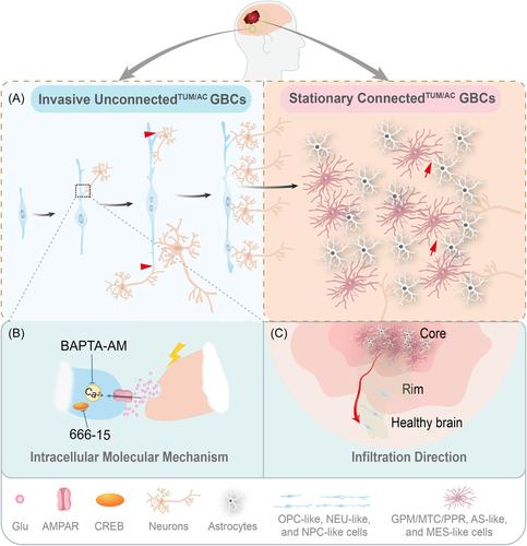
Furthermore, intravital two-photon microscopy (IV2PM) and single-cell transcriptomics (scRNA-seq) identified that unconnectedTUM/AC GBCs (dynamic and invasive GBCs) were predominately enriched for analogous neural cells (OPC-like, NEU-like, and NPC-like cells), while connectedTUM/AC GBCs (stationary and gap-junction-coupled GBCs) mainly consisted of non-NEU cells (GPM/MTC/PPR), AS-like, and MES-like cells. Combining IV2PM and scRNA-seq, the authors aimed to provide a conceptual framework that described how unconnectedTUM/AC GBMs infiltrate the surrounding healthy brain (Figure 1C). The NEU cell state of unconnectedTUM/AC GBMs was mostly located in the tumor rim (Figure 1C), was easily transferred to distant regions in the brain environment, and normally formed synapses with other GBCs and AS by increasing the TMs. The activation of NEU signaling in stem-like neoplastic cells may result from tumor–neuron interactions.10
To explain why NEU cell features coincide with brain tumor invasion, the authors utilized quantitative morphometric analysis and deep-learning-enabled intravital subcellular time-lapse imaging (DeepISTI) to show that TMs mainly improved the volume and surface area of GBMs, which increased the probability of interaction with neurons in the brain. In addition, not all TMs contributed to the invasive process; blind-ending TMs (lacking connection with other TMs) predominately improved the invasiveness of GBCs. The movement of blind-ending TMs mainly relies on three mechanisms: extension, retraction, and TM branching generation, which are similar to the migration of immature neurons. DeepISTI results again proved the neuron-like behavior of GBCs infiltrating via branching migration, locomotion, and translocation, similar to that of immature neurons. Even though the behavior was similar to axonal growth cone pathfinding, the unconnectedTUM/AC GBM cells responded to NEU input for axonal migration by resembling an interneuron movement. Therefore, it was concluded that TM invasion follows a search-efficient mechanism and increases the invasion speed of GBMs via NEU activity (Figure 1A).
When invasive unconnectedTUM/AC GBCs find a favorable location in normal brain tissue, the pioneering GBCs change their molecular properties and become a stationary cell type to form a gap-junction-coupled tumor–AS network. Dr. Venkataramani1 describes this behavior as being similar to the colonization of the New World, where individual settlers traveled great distances, made contact with the locals, and finally settled down in their new location.
To explore the intracellular downstream mechanism, in vivo and in vitro experiments revealed that calcium transient caused by synaptic NEU activity is critical for GBC invasion. Moreover, blockage of calcium transients by calcium chelators (BAPTA-AM) or a CREB inhibitor (666-15) inhibited glioma growth, supporting the growth-promoting role of NEU membrane depolarization and calcium signaling in gliomas (Figure 1B). Thus, NEU activity is not only important for glioma growth but also plays a significant role in driving the migration of these deadly tumors. Furthermore, the unconnectedTUM/AC GBM cells on the tumor edge exhibit an AMPA-receptor phenotype that is different from that expressed in the core of adult gliomas and is normally restricted to neural progenitor cells, making them particularly sensitive to the proliferative effects of glutamate, supporting a key role of neurogliomal synapses in tumor invasion. More importantly, Food and Drug Administration-approved AMPAR inhibition was proven effective in reducing TM length and branching points.1, 3
In summary, this study leverages cutting-edge techniques, including single-cell sequencing, intravital time-lapse imaging, intravital calcium imaging, patch-clamp electrophysiology, correlative light and electron microscopy, spontaneous excitatory postsynaptic currents, and in vivo optogenetics to explore the hijacking mechanism of NEU interactions in GBM progression. The distant colonization of GBCs now has been illustrated in the context of NEU activity and how it drives glioma initiation and growth (Figure 1C).
As discussed earlier in this article, we argued that the glutamatergic effects could be reduced by interfering with the AMPAR inhibitor, which slows the tumor progression in the brain. Thus, identifying and having a better understanding of these mechanisms could help in the development of future novel therapeutic strategies for currently incurable glioblastoma.
Dongling Pei: Visualization (lead); writing – original draft (lead). Zhenyu Zhang: Conceptualization (equal); writing – review and editing (supporting). Long Zhang: Conceptualization (lead); writing – review and editing (lead). All authors have read and approved the article.
Long Zhang is an editorial board member of MedComm – Oncology, but was not involved in the review of the journal, or decisions related to this manuscript. The remaining authors declare no conflict of interest.

 求助内容:
求助内容: 应助结果提醒方式:


