Elbrus Zarbaliyev, Mustafa Okumus, Payam Hacisalihoglu
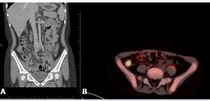
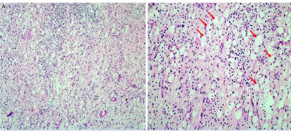
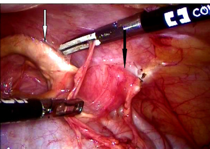
{"title":"Intraperitoneal \"golden yellow\" in a pediatric patient with Burkitt lymphoma: Xanthogranulomatous appendicitis.","authors":"Elbrus Zarbaliyev, Mustafa Okumus, Payam Hacisalihoglu","doi":"10.14744/nci.2022.78477","DOIUrl":null,"url":null,"abstract":"<p><p>Xanthogranulomatous inflammation is a rare chronic inflammatory reaction. Appendiceal involvement in the pediatric age group is extremely rare. We present a case of xanthogranulomatous appendicitis (XGA) that was detected incidentally during the excision of a residual intraabdominal mass in an 8-year-old male patient who was treated for Burkitt lymphoma. An 8-year-old male patient who had been diagnosed with Burkitt lymphoma underwent abdominal computerized tomography for evaluation after chemotherapy. An approximately 2.5 cm mass in the right lower quadrant of the abdomen was detected, and laparoscopic excision of the mass was planned. During the operation, it was noticed that the appendix (adjacent to the mass) was golden yellow in color and abnormal in appearance, so a synchronous appendectomy was performed. The pathology result of the mass was compatible with Burkitt lymphoma. Microscopic examination of the appendix revealed that the columnar surface epithelium had eroded and been replaced by fibrin and cell debris. Inflammatory cell infiltration rich in foamy histiocytes as well as lymphocytes and sparse neutrophils that form destructive aggregates was observed in all appendiceal layers. The final diagnosis of the appendectomy specimen was compatible with XGA. In very few XGA cases, the appendix is described as bright yellow or golden yellow. The diagnosis is usually made by the pathological examination after surgery. Though the diagnosis was made postoperatively in our case, there is now, for the first time in the literature, a view of the golden yellow color of XGA taken from an intraoperative video clip.</p>","PeriodicalId":19164,"journal":{"name":"Northern Clinics of Istanbul","volume":"5 1","pages":"579-582"},"PeriodicalIF":0.9000,"publicationDate":"2024-11-19","publicationTypes":"Journal Article","fieldsOfStudy":null,"isOpenAccess":false,"openAccessPdf":"https://www.ncbi.nlm.nih.gov/pmc/articles/PMC11622746/pdf/","citationCount":"0","resultStr":null,"platform":"Semanticscholar","paperid":null,"PeriodicalName":"Northern Clinics of Istanbul","FirstCategoryId":"1085","ListUrlMain":"https://doi.org/10.14744/nci.2022.78477","RegionNum":0,"RegionCategory":null,"ArticlePicture":[],"TitleCN":null,"AbstractTextCN":null,"PMCID":null,"EPubDate":"2024/1/1 0:00:00","PubModel":"eCollection","JCR":"Q3","JCRName":"MEDICINE, GENERAL & INTERNAL","Score":null,"Total":0}
引用次数: 0
Abstract
Xanthogranulomatous inflammation is a rare chronic inflammatory reaction. Appendiceal involvement in the pediatric age group is extremely rare. We present a case of xanthogranulomatous appendicitis (XGA) that was detected incidentally during the excision of a residual intraabdominal mass in an 8-year-old male patient who was treated for Burkitt lymphoma. An 8-year-old male patient who had been diagnosed with Burkitt lymphoma underwent abdominal computerized tomography for evaluation after chemotherapy. An approximately 2.5 cm mass in the right lower quadrant of the abdomen was detected, and laparoscopic excision of the mass was planned. During the operation, it was noticed that the appendix (adjacent to the mass) was golden yellow in color and abnormal in appearance, so a synchronous appendectomy was performed. The pathology result of the mass was compatible with Burkitt lymphoma. Microscopic examination of the appendix revealed that the columnar surface epithelium had eroded and been replaced by fibrin and cell debris. Inflammatory cell infiltration rich in foamy histiocytes as well as lymphocytes and sparse neutrophils that form destructive aggregates was observed in all appendiceal layers. The final diagnosis of the appendectomy specimen was compatible with XGA. In very few XGA cases, the appendix is described as bright yellow or golden yellow. The diagnosis is usually made by the pathological examination after surgery. Though the diagnosis was made postoperatively in our case, there is now, for the first time in the literature, a view of the golden yellow color of XGA taken from an intraoperative video clip.

 求助内容:
求助内容: 应助结果提醒方式:


