{"title":"Paracentral Acute Middle Maculopathy and Central Retinal Venous Occlusion following Electrical Injury.","authors":"Sahel Khazaei, Mehrdad Motamed Shariati, Naser Shoeibi, Mohammad Arjmand, Seyedeh Maryam Hosseini","doi":"10.1155/2022/3699667","DOIUrl":null,"url":null,"abstract":"<p><strong>Purpose: </strong>To report a case of central retinal vein occlusion (CRVO) and paracentral acute middle maculopathy (PAMM) following electric shock injury. <i>Case Description</i>. A 45-year-old male presented with a significant painless decreased vision in the right eye following an electrical injury of the right hand in his workplace. The best-corrected visual acuity (BCVA) of the right eye was 20/40. Funduscopic examination of the right eye revealed diffuse superficial and deep intraretinal hemorrhages, mild venous tortuosity, and an area of the pale retina. Optical coherence tomography (OCT) demonstrated hyperreflective band-like lesions in the middle retinal layers. Patchy areas of vascular flow void in deep capillary plexus seen in OCT angiography of the right eye were compatible with PAMM. Fluorescein angiography of the right eye was indicative of delayed venous filling suggestive of CRVO. The left eye was completely normal on exam and imaging.</p><p><strong>Conclusion: </strong>This report illustrates the occurrence of CRVO associated with PAMM following electric shock injury. Electrical injury leads to a wide range of retinal manifestations. Clinicians need to pay attention to any hyperreflectivity and thinning of middle retinal layers in OCT in cases with the presentation of sudden visual loss following electrical injuries.</p>","PeriodicalId":45023,"journal":{"name":"Public Archaeology","volume":"16 1","pages":"3699667"},"PeriodicalIF":0.9000,"publicationDate":"2022-04-16","publicationTypes":"Journal Article","fieldsOfStudy":null,"isOpenAccess":false,"openAccessPdf":"https://www.ncbi.nlm.nih.gov/pmc/articles/PMC9034943/pdf/","citationCount":"0","resultStr":null,"platform":"Semanticscholar","paperid":null,"PeriodicalName":"Public Archaeology","FirstCategoryId":"1085","ListUrlMain":"https://doi.org/10.1155/2022/3699667","RegionNum":4,"RegionCategory":"历史学","ArticlePicture":[],"TitleCN":null,"AbstractTextCN":null,"PMCID":null,"EPubDate":"2022/1/1 0:00:00","PubModel":"eCollection","JCR":"0","JCRName":"ARCHAEOLOGY","Score":null,"Total":0}
引用次数: 0
Abstract
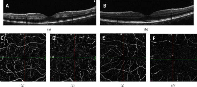
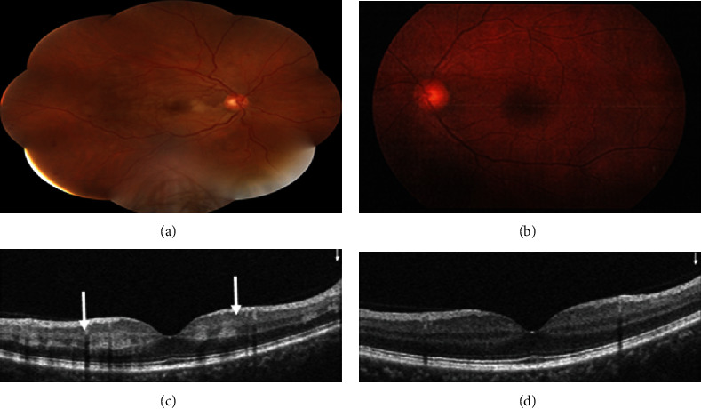
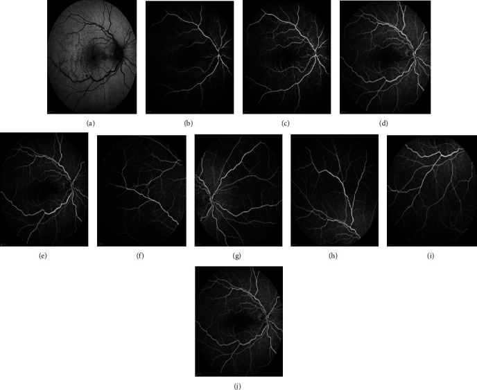
Purpose: To report a case of central retinal vein occlusion (CRVO) and paracentral acute middle maculopathy (PAMM) following electric shock injury. Case Description. A 45-year-old male presented with a significant painless decreased vision in the right eye following an electrical injury of the right hand in his workplace. The best-corrected visual acuity (BCVA) of the right eye was 20/40. Funduscopic examination of the right eye revealed diffuse superficial and deep intraretinal hemorrhages, mild venous tortuosity, and an area of the pale retina. Optical coherence tomography (OCT) demonstrated hyperreflective band-like lesions in the middle retinal layers. Patchy areas of vascular flow void in deep capillary plexus seen in OCT angiography of the right eye were compatible with PAMM. Fluorescein angiography of the right eye was indicative of delayed venous filling suggestive of CRVO. The left eye was completely normal on exam and imaging.
Conclusion: This report illustrates the occurrence of CRVO associated with PAMM following electric shock injury. Electrical injury leads to a wide range of retinal manifestations. Clinicians need to pay attention to any hyperreflectivity and thinning of middle retinal layers in OCT in cases with the presentation of sudden visual loss following electrical injuries.

 求助内容:
求助内容: 应助结果提醒方式:


