Anran Wang, Lin Yang, Weimin Wen, Song Zhang, Dongmei Hao, Syed G Khalid, Dingchang Zheng
{"title":"Quantification of radial arterial pulse characteristics change during exercise and recovery.","authors":"Anran Wang, Lin Yang, Weimin Wen, Song Zhang, Dongmei Hao, Syed G Khalid, Dingchang Zheng","doi":"10.1007/s12576-016-0515-7","DOIUrl":null,"url":null,"abstract":"<p><p>It is physiologically important to understand the arterial pulse waveform characteristics change during exercise and recovery. However, there is a lack of a comprehensive investigation. This study aimed to provide scientific evidence on the arterial pulse characteristics change during exercise and recovery. Sixty-five healthy subjects were studied. The exercise loads were gradually increased from 0 to 125 W for female subjects and to 150 W for male subjects. Radial pulses were digitally recorded during exercise and 4-min recovery. Four parameters were extracted from the raw arterial pulse waveform, including the pulse amplitude, width, pulse peak and dicrotic notch time. Five parameters were extracted from the normalized radial pulse waveform, including the pulse peak and dicrotic notch position, pulse Area, Area<sub>1</sub> and Area<sub>2</sub> separated by notch point. With increasing loads during exercise, the raw pulse amplitude increased significantly with decreased pulse period, reduced peak and notch time. From the normalized pulses, the pulse Area, pulse Area<sub>1</sub> and Area<sub>2</sub> decreased, respectively, from 38 ± 4, 61 ± 5 and 23 ± 5 at rest to 34 ± 4, 52 ± 6 and 13 ± 5 at 150-W exercise load. During recovery, an opposite trend was observed. This study quantitatively demonstrated significant changes of radial pulse characteristics during different exercise loads and recovery phases.</p>","PeriodicalId":22836,"journal":{"name":"The Journal of Physiological Sciences","volume":"7 1","pages":"113-120"},"PeriodicalIF":0.0000,"publicationDate":"2018-03-01","publicationTypes":"Journal Article","fieldsOfStudy":null,"isOpenAccess":false,"openAccessPdf":"https://www.ncbi.nlm.nih.gov/pmc/articles/PMC5799316/pdf/","citationCount":"0","resultStr":null,"platform":"Semanticscholar","paperid":null,"PeriodicalName":"The Journal of Physiological Sciences","FirstCategoryId":"1085","ListUrlMain":"https://doi.org/10.1007/s12576-016-0515-7","RegionNum":0,"RegionCategory":null,"ArticlePicture":[],"TitleCN":null,"AbstractTextCN":null,"PMCID":null,"EPubDate":"2016/12/27 0:00:00","PubModel":"Epub","JCR":"","JCRName":"","Score":null,"Total":0}
引用次数: 0
Abstract
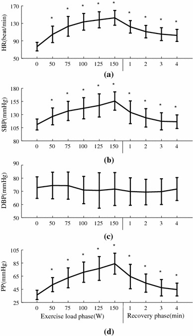
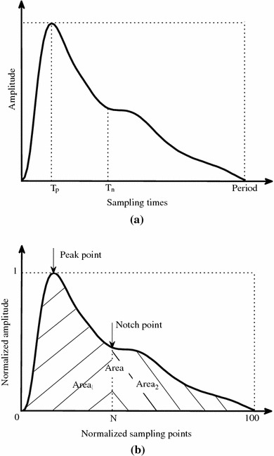
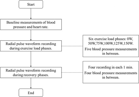
It is physiologically important to understand the arterial pulse waveform characteristics change during exercise and recovery. However, there is a lack of a comprehensive investigation. This study aimed to provide scientific evidence on the arterial pulse characteristics change during exercise and recovery. Sixty-five healthy subjects were studied. The exercise loads were gradually increased from 0 to 125 W for female subjects and to 150 W for male subjects. Radial pulses were digitally recorded during exercise and 4-min recovery. Four parameters were extracted from the raw arterial pulse waveform, including the pulse amplitude, width, pulse peak and dicrotic notch time. Five parameters were extracted from the normalized radial pulse waveform, including the pulse peak and dicrotic notch position, pulse Area, Area1 and Area2 separated by notch point. With increasing loads during exercise, the raw pulse amplitude increased significantly with decreased pulse period, reduced peak and notch time. From the normalized pulses, the pulse Area, pulse Area1 and Area2 decreased, respectively, from 38 ± 4, 61 ± 5 and 23 ± 5 at rest to 34 ± 4, 52 ± 6 and 13 ± 5 at 150-W exercise load. During recovery, an opposite trend was observed. This study quantitatively demonstrated significant changes of radial pulse characteristics during different exercise loads and recovery phases.

 求助内容:
求助内容: 应助结果提醒方式:


