Protodioscin protects PC12 cells against oxygen and glucose deprivation-induced injury through miR-124/AKT/Nrf2 pathway
IF 3.3
3区 生物学
Q3 CELL BIOLOGY
引用次数: 0
Abstract
The purpose of the current study was to demonstrate the neuroprotective effect of protodioscin (Prot) in an in vitro model of ischemia/reperfusion (I/R) and investigate the underlying molecular mechanism. After PC12 cells were exposed to oxygen and glucose deprivation (OGD) reperfusion, PI staining by flow cytometry was used to quantify the rate of apoptosis. The levels of hypoxia-inducible factor 1-alpha (HIF-1α), superoxide dismutase (SOD), glutathione peroxidase (GSH-Px), and malondialdehyde (MDA) were determined using commercially available kits. Intracellular reactive oxygen species (ROS) level was detected using the 20,70-dichlorodihy-drofluorescein diacetate (DCFH-DA) fluorescence assay. The expression levels of heat-shock proteins (HSP), PI3K, AKT, Nrf2, and miR-124 were tested by western blot or quantitative PCR. Prot significantly attenuated oxygen–glucose deprivation/reperfusion (OGD/R)-induced apoptotic death. Prot also reduced the oxidative stress as revealed by increasing the activities of SOD and GSH-Px, decreasing the levels of ROS and MDA. Moreover, mechanism investigations suggested that Prot prevented the decrease of HSP70, HSP32 (hemeoxygenase-1, HO-1), and PI3K protein expression, phosphorylation of AKT, and the accumulation of nuclear Nrf2. The level of miR-124 was decreased in PC12 cells, which was also effectively reversed by Prot treatment. Prot protected PC12 cells against OGD/R-induced injury through inhibiting oxidative stress and apoptosis, which could be associated with increasing HSP proteins expression via activating PI3K/AKT/Nrf2 pathway and miR-124 modulation.
原薯蓣皂苷通过 miR-124/AKT/Nrf2 通路保护 PC12 细胞免受氧和葡萄糖剥夺诱导的损伤。
本研究的目的是在体外缺血再灌注(I/R)模型中证明原薯蓣皂苷(Prot)的神经保护作用,并研究其潜在的分子机制。将 PC12 细胞暴露于缺氧和缺糖(OGD)再灌注后,用流式细胞仪进行 PI 染色以量化细胞凋亡率。使用市售试剂盒测定缺氧诱导因子 1-α(HIF-1α)、超氧化物歧化酶(SOD)、谷胱甘肽过氧化物酶(GSH-Px)和丙二醛(MDA)的水平。细胞内活性氧(ROS)水平用 20,70 二甲基二荧光素二乙酸酯(DCFH-DA)荧光测定法检测。热休克蛋白(HSP)、PI3K、AKT、Nrf2 和 miR-124 的表达水平通过 Western 印迹或定量 PCR 进行了检测。Prot明显减轻了氧-葡萄糖剥夺/再灌注(OGD/R)诱导的细胞凋亡。通过提高 SOD 和 GSH-Px 的活性、降低 ROS 和 MDA 的水平,Prot 还降低了氧化应激。此外,机理研究表明,Prot 能防止 HSP70、HSP32(血红素氧化酶-1,HO-1)和 PI3K 蛋白表达的减少、AKT 的磷酸化以及核 Nrf2 的积累。PC12细胞中miR-124的水平降低,Prot处理后也能有效逆转这一现象。Prot通过抑制氧化应激和细胞凋亡保护PC12细胞免受OGD/R诱导的损伤,这可能与通过激活PI3K/AKT/Nrf2通路和调控miR-124增加HSP蛋白表达有关。
本文章由计算机程序翻译,如有差异,请以英文原文为准。
求助全文
约1分钟内获得全文
求助全文
来源期刊

Cell Stress & Chaperones
生物-细胞生物学
CiteScore
7.60
自引率
2.60%
发文量
59
审稿时长
6-12 weeks
期刊介绍:
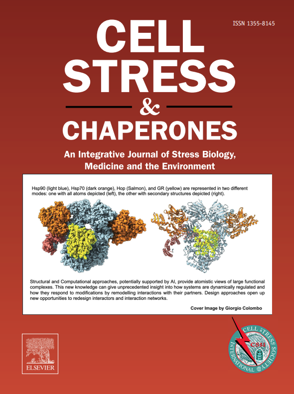
Cell Stress and Chaperones is an integrative journal that bridges the gap between laboratory model systems and natural populations. The journal captures the eclectic spirit of the cellular stress response field in a single, concentrated source of current information. Major emphasis is placed on the effects of climate change on individual species in the natural environment and their capacity to adapt. This emphasis expands our focus on stress biology and medicine by linking climate change effects to research on cellular stress responses of animals, micro-organisms and plants.
 求助内容:
求助内容: 应助结果提醒方式:


