{"title":"Myocardial perfusion SPECT and SPECT/CT in interventional cardiology","authors":"E. Trägårdh, P. Hasbak, B. Hesse","doi":"10.1002/cce2.76","DOIUrl":null,"url":null,"abstract":"<div>\n \n <p>Myocardial perfusion imaging with single-photon emission computed tomography (SPECT) is an important and widely used non-invasive imaging test for the diagnosis and semiquantification of myocardial ischemia. SPECT data, acquired after a stress test and at rest on a gamma camera, shows left ventricular tracer uptake during stress and at rest. The tracer distributions are proportionate to the relative, regional coronary-flow distributions, respectively. A stress-induced perfusion defect reflects myocardial ischemia, while a permanent defect, unchanged from stress to rest, indicates myocardial infarction. By ECG-gating global and regional left ventricular function can also be assessed. The three most important indications for SPECT in the setting of interventional cardiology include (1) the diagnosis of coronary artery disease in intermediate-risk patients, (2) the assessment of ischemia in patients with prior successful revascularization and recurrence of symptoms, and (3) the diagnosis of acute coronary syndrome in the emergency department. The amount of ischemia is related to the outcome of patients undergoing revascularization. It was observed that patients with an ischemic area of <10% of the left ventricle demonstrated by SPECT benefit from staying on optimal medical therapy, whereas patients with more ischemia on SPECT benefit from revascularization. Guidelines and appropriate use criteria for the indications of SPECT in relation to interventional cardiology have been published by the European Society and American College of Cardiology.</p>\n </div>","PeriodicalId":100331,"journal":{"name":"Continuing Cardiology Education","volume":"4 1","pages":"45-50"},"PeriodicalIF":0.0000,"publicationDate":"2018-07-27","publicationTypes":"Journal Article","fieldsOfStudy":null,"isOpenAccess":false,"openAccessPdf":"https://sci-hub-pdf.com/10.1002/cce2.76","citationCount":"2","resultStr":null,"platform":"Semanticscholar","paperid":null,"PeriodicalName":"Continuing Cardiology Education","FirstCategoryId":"1085","ListUrlMain":"https://onlinelibrary.wiley.com/doi/10.1002/cce2.76","RegionNum":0,"RegionCategory":null,"ArticlePicture":[],"TitleCN":null,"AbstractTextCN":null,"PMCID":null,"EPubDate":"","PubModel":"","JCR":"","JCRName":"","Score":null,"Total":0}
引用次数: 2
Abstract
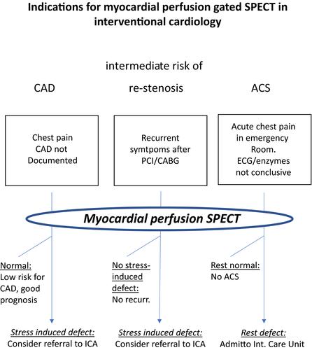
Myocardial perfusion imaging with single-photon emission computed tomography (SPECT) is an important and widely used non-invasive imaging test for the diagnosis and semiquantification of myocardial ischemia. SPECT data, acquired after a stress test and at rest on a gamma camera, shows left ventricular tracer uptake during stress and at rest. The tracer distributions are proportionate to the relative, regional coronary-flow distributions, respectively. A stress-induced perfusion defect reflects myocardial ischemia, while a permanent defect, unchanged from stress to rest, indicates myocardial infarction. By ECG-gating global and regional left ventricular function can also be assessed. The three most important indications for SPECT in the setting of interventional cardiology include (1) the diagnosis of coronary artery disease in intermediate-risk patients, (2) the assessment of ischemia in patients with prior successful revascularization and recurrence of symptoms, and (3) the diagnosis of acute coronary syndrome in the emergency department. The amount of ischemia is related to the outcome of patients undergoing revascularization. It was observed that patients with an ischemic area of <10% of the left ventricle demonstrated by SPECT benefit from staying on optimal medical therapy, whereas patients with more ischemia on SPECT benefit from revascularization. Guidelines and appropriate use criteria for the indications of SPECT in relation to interventional cardiology have been published by the European Society and American College of Cardiology.

 求助内容:
求助内容: 应助结果提醒方式:


