Johannes Hatzl, Daniel Henning, Niklas Hartmann, Dittmar Böckler, Christian Uhl
{"title":"A New Method for Common Femoral Arterial Access Using a Mixed Reality-Assisted Technique on a Phantom Model.","authors":"Johannes Hatzl, Daniel Henning, Niklas Hartmann, Dittmar Böckler, Christian Uhl","doi":"10.1177/15266028231208640","DOIUrl":null,"url":null,"abstract":"<p><strong>Purpose: </strong>The purpose of this study was to investigate the technical feasibility and usability of a mixed reality (MiR)-assisted common femoral arterial (CFA) access technique using a sonography-assisted registration method.</p><p><strong>Materials and methods: </strong>A total of 60 CFA punctures were performed on a phantom model by 2 observers. Thirty punctures were performed using MiR (MiR group) and 30 punctures were performed using a conventional sonography-guided access procedure (control group). In the MiR group, a virtual object was created based on a computed tomography (CT) angiography scan of the model and registered to the physical patient in an MiR environment utilizing a software prototype that allowed registration based on a sonography scan. Positional error assessment encompassed 4 measurements using cone beam CT scans: (1) distance of the needle tip to the centerline, (2) distance of the needle entry site from the mid-level of the ostium of the profound femoral artery, (3) angle of entry of the needle in coronal, and (4) sagittal planes. Technical success rates as well as positional errors were compared between both groups. In addition, the usability of the system was assessed according to the system usability scale (SUS).</p><p><strong>Results: </strong>Technical success was 96.7% and 100% in the MiR and control groups, respectively. The median distance between the needle tip and the centerline was 3.0 (interquartile range [IQR]: 2.0-4.6) in the MiR group and 3.2 mm (IQR: 2.3-3.9) (p=0.63) in the control group. Similarly, the median distance from the needle entry site to the mid-level of the ostium of the profound femoral artery was 3.0 mm (IQR: 2.0-5.0) in the MiR group and 4.5 mm (IQR: 2.0-7.8) (p=0.18) in the control group. The median coronal angles of needle entry were 7.5° (IQR: 6-11) and 6° (IQR: 2-12) (p=0.13), and the median sagittal angles were 50° (IQR: 47-51) and 51° (IQR: 50-55) (p<0.01) in the MiR and control groups, respectively. The mean SUS score provided by both observers was 51.3.</p><p><strong>Conclusion: </strong>The feasibility of an MiR-assisted CFA access technique could be demonstrated on a phantom model. Further studies are needed to investigate the technique beyond phantom model experiments and in different anatomical settings.Clinical ImpactThis study demonstrates the technical feasibility of a Mixed-Reality-assisted common femoral arterial access procedure on a phantom model. The positional accuracy was comparable to a conventional sonography-guided technique. However, there are several limitations that need to be resolved prior to potential implementation into clinical practice. Further studies are needed to investigate its performance beyond phantom model experiments and the prototypical application requires further technical refinement to increase its usability.</p>","PeriodicalId":50210,"journal":{"name":"Journal of Endovascular Therapy","volume":" ","pages":"1259-1266"},"PeriodicalIF":1.5000,"publicationDate":"2025-10-01","publicationTypes":"Journal Article","fieldsOfStudy":null,"isOpenAccess":false,"openAccessPdf":"https://www.ncbi.nlm.nih.gov/pmc/articles/PMC12433519/pdf/","citationCount":"0","resultStr":null,"platform":"Semanticscholar","paperid":null,"PeriodicalName":"Journal of Endovascular Therapy","FirstCategoryId":"3","ListUrlMain":"https://doi.org/10.1177/15266028231208640","RegionNum":2,"RegionCategory":"医学","ArticlePicture":[],"TitleCN":null,"AbstractTextCN":null,"PMCID":null,"EPubDate":"2023/11/2 0:00:00","PubModel":"Epub","JCR":"Q3","JCRName":"PERIPHERAL VASCULAR DISEASE","Score":null,"Total":0}
引用次数: 0
Abstract
Purpose: The purpose of this study was to investigate the technical feasibility and usability of a mixed reality (MiR)-assisted common femoral arterial (CFA) access technique using a sonography-assisted registration method.
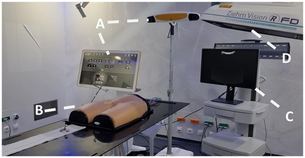
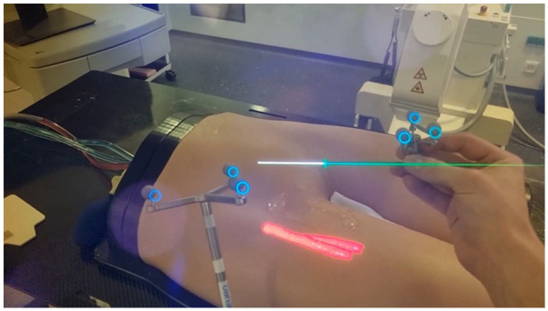
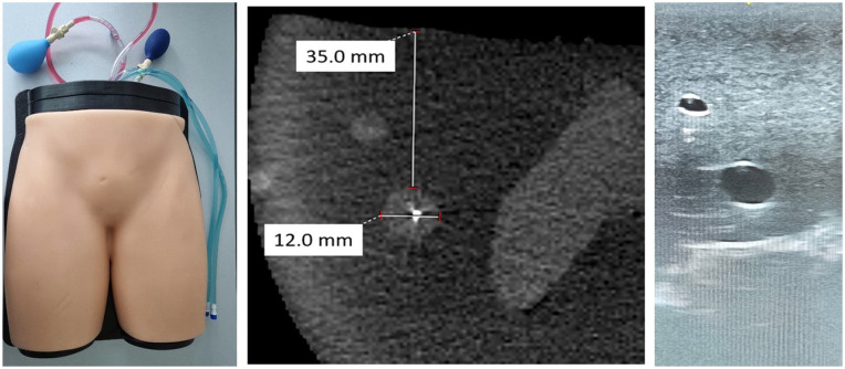
Materials and methods: A total of 60 CFA punctures were performed on a phantom model by 2 observers. Thirty punctures were performed using MiR (MiR group) and 30 punctures were performed using a conventional sonography-guided access procedure (control group). In the MiR group, a virtual object was created based on a computed tomography (CT) angiography scan of the model and registered to the physical patient in an MiR environment utilizing a software prototype that allowed registration based on a sonography scan. Positional error assessment encompassed 4 measurements using cone beam CT scans: (1) distance of the needle tip to the centerline, (2) distance of the needle entry site from the mid-level of the ostium of the profound femoral artery, (3) angle of entry of the needle in coronal, and (4) sagittal planes. Technical success rates as well as positional errors were compared between both groups. In addition, the usability of the system was assessed according to the system usability scale (SUS).
Results: Technical success was 96.7% and 100% in the MiR and control groups, respectively. The median distance between the needle tip and the centerline was 3.0 (interquartile range [IQR]: 2.0-4.6) in the MiR group and 3.2 mm (IQR: 2.3-3.9) (p=0.63) in the control group. Similarly, the median distance from the needle entry site to the mid-level of the ostium of the profound femoral artery was 3.0 mm (IQR: 2.0-5.0) in the MiR group and 4.5 mm (IQR: 2.0-7.8) (p=0.18) in the control group. The median coronal angles of needle entry were 7.5° (IQR: 6-11) and 6° (IQR: 2-12) (p=0.13), and the median sagittal angles were 50° (IQR: 47-51) and 51° (IQR: 50-55) (p<0.01) in the MiR and control groups, respectively. The mean SUS score provided by both observers was 51.3.
Conclusion: The feasibility of an MiR-assisted CFA access technique could be demonstrated on a phantom model. Further studies are needed to investigate the technique beyond phantom model experiments and in different anatomical settings.Clinical ImpactThis study demonstrates the technical feasibility of a Mixed-Reality-assisted common femoral arterial access procedure on a phantom model. The positional accuracy was comparable to a conventional sonography-guided technique. However, there are several limitations that need to be resolved prior to potential implementation into clinical practice. Further studies are needed to investigate its performance beyond phantom model experiments and the prototypical application requires further technical refinement to increase its usability.
期刊介绍:
The Journal of Endovascular Therapy (formerly the Journal of Endovascular Surgery) was established in 1994 as a forum for all physicians, scientists, and allied healthcare professionals who are engaged or interested in peripheral endovascular techniques and technology. An official publication of the International Society of Endovascular Specialists (ISEVS), the Journal of Endovascular Therapy publishes peer-reviewed articles of interest to clinicians and researchers in the field of peripheral endovascular interventions.
 求助内容:
求助内容: 应助结果提醒方式:


