Effect of Propofol on Expression of PKC mRNA in Pulmonary Injury Induced by Ischemia-Reperfusion in Rabbits
引用次数: 0
Abstract
Aim: This prospective study aimed at investigating the effect of propofol on the expression of protein kinase C (PKC) mRNA during pulmonary ischemia-reperfusion injury (PIRI) in the rabbits. Methods: Single lung ischemia-reperfusion animal model was administrated in vivo. The rabbits were randomly divided into three groups): sham-operated group (Sham); pulmonary I/R group (PIR) and PIR+propofol group (PIR+PPF). Changes of several parameters, including malondialdehyde (MDA) concentration, superoxide dismutase (SOD) activity, nitric oxide (NO) content, wet/dry ratio of lung tissue (W/D) and the index of quantitative assessment of histologic lung injury (IQA) were measured at 60 min after reperfusion. Meanwhile, the location and expression of PKC mRNA were observed, lung tissues were also harvested for histopathological evaluation. Results: As compared with PIR group, PKC mRNA is largely expressed in intima and extima of small pulmonary artery as well as thin-wall vessels (mostly small pulmonary veins). The average optical density values of PKC-α, δ and θ mRNA in small pulmonary veins in PIR+PPF group showed obviously higher than that in group PIR (all P<0.01); the activity of SOD increased, the concentration of MDA, W/D and IQA decreased at 60 min after reperfusion in lung tissue (P<0.01 and P<0.05). Abnormal morphological changes of the lung tissue were lessened markedly in PIR+PPF group. Conclusions: The results of this study suggested that propofol possesses and acts its notable protective effect on PIRI in rabbits by activating PKC-α, δ and θ mRNA expression in lung tissue, raising NO level, reducing OFR level and decreasing lipid peroxidation.异丙酚对兔缺血再灌注肺损伤中PKC mRNA表达的影响
目的:本前瞻性研究旨在探讨异丙酚对兔肺缺血再灌注损伤(PIRI)时蛋白激酶C (PKC) mRNA表达的影响。方法:活体建立单肺缺血再灌注动物模型。将家兔随机分为3组:假手术组(Sham);肺I/R组(PIR)和PIR+异丙酚组(PIR+PPF)。测定再灌注后60 min丙二醛(MDA)浓度、超氧化物歧化酶(SOD)活性、一氧化氮(NO)含量、肺组织干湿比(W/D)及组织学肺损伤定量评价指标(IQA)的变化。同时观察PKC mRNA的位置和表达,并采集肺组织进行组织病理学评价。结果:与PIR组相比,PKC mRNA在小肺动脉内膜、外膜及薄壁血管(以小肺静脉为主)中大量表达。PIR+PPF组小肺静脉PKC-α、δ、θ mRNA平均光密度值显著高于PIR组(均P<0.01);再灌注后60 min,肺组织SOD活性升高,MDA、W/D、IQA浓度降低(P<0.01和P<0.05)。PIR+PPF组肺组织异常形态学改变明显减轻。结论:本研究提示异丙酚通过激活兔肺组织PKC-α、δ和θ mRNA表达,提高NO水平,降低OFR水平,降低脂质过氧化,对兔PIRI具有显著的保护作用。
本文章由计算机程序翻译,如有差异,请以英文原文为准。
求助全文
约1分钟内获得全文
求助全文
来源期刊
CiteScore
0.80
自引率
0.00%
发文量
16309
期刊介绍:
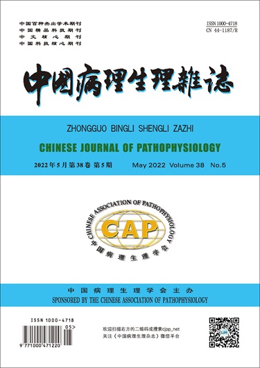
Chinese Journal of Pathophysiology is the only comprehensive advanced academic journal of pathophysiology in China, which is supervised by China Association for Science and Technology, sponsored by Chinese Pathophysiology Society and hosted by Jinan University. Journal of pathophysiology research (including experimental research and clinical research), thematic reviews, experimental technology innovation and improvement, focusing on the introduction of disease pathogenesis.
The current Chinese Journal of Science Pathophysiology has been included in the core Database of China Science Citation Database (CSCD), CNKI, Wanfang, Vipp, Superstar, Scopus, EBSCO, Chemical Abstracting (CA), JST, WHO Western Pacific Regional Medical Index (W PRIM) and other databases included.

 求助内容:
求助内容: 应助结果提醒方式:


