{"title":"Lactate: A critical regulator of cell proliferation via anaphase promoting complex remodeling","authors":"Qiqing Yang, Long Zhang, Jun Chen","doi":"10.1002/mog2.38","DOIUrl":null,"url":null,"abstract":"<p>In a study recently published in <i>Nature</i>, Liu et al. discovered that lactate directly inhibits SUMO-specific peptidase 1 (SENP1), resulting in the stabilization of anaphase promoting complex (APC) subunit 4 (APC4) SUMOylation, and transient binding of APC/cyclosome (APC/C) and ubiquitin conjugating enzyme E2 C (UBE2C), which promotes the ubiquitination and degradation of cyclin B1 and securin.<span><sup>1</sup></span> Furthermore, sustained accumulation of lactate was found to counteract the effects of anti-mitotic drugs by inducing mitotic slippage, which ultimately facilitates mitotic exit. This study shed light on a potential mechanism behind the observed high levels of lactate in rapidly dividing cells.</p><p>Cancer cells exhibit a unique metabolic phenotype characterized by increased glucose uptake and reliance on aerobic glycolysis to fuel their rapid proliferation. This metabolic shift contributes to lactate accumulation, which is closely associated with cell proliferation; however, the precise mechanism of the latter remains unclear. APC/C is a member of the ubiquitin ligase family that plays a crucial role in regulating the metaphase-to-anaphase transition and mitotic exit by assembling K11-linked ubiquitin chains on substrates such as cyclin B1 and securin.<span><sup>2</sup></span> A recently published work by Liu et al. uncovered a link between lactate and APC/C activity, and elucidated the significance of this connection in cell cycle and cell proliferation modulation.</p><p>To explore the direct effect of elevated lactate levels on the entire proteome, Liu et al. treated native human embryonic kidney cell lysates with 15 or 25 mM <span>l</span>-lactate before conducting thermal proteomic profiling. They observed a significant shift in the thermostability of UBE2C, an E2 enzyme recruited by APC/C upon structural reorganization of its subunits. However, it is unlikely that lactate binds directly to UBE2C because of its low affinity. Moreover, no change in the abundance or posttranslational modification of UBE2C was detected, suggesting that <span>l</span>-lactate might enhance the interaction between UBE2C and APC/C. To verify this hypothesis, cells were first synchronized to pro-metaphase, a period during which APC/C is inhibited due to its interaction with the mitotic checkpoint complex. Subsequently, Liu et al. incubated cell lysates with 15 mM <span>l</span>-lactate and performed immunoprecipitation of APC/C, revealing that <span>l</span>-lactate significantly enhanced the binding between UBE2C and APC/C. In addition, mass spectrometry analysis of APC/C showed a lactate-dependent elevation of SUMO2/3 conjunctions. Previous studies have shown that SUMOylation of APC4 on residues K772 and K798 results in a substantial rearrangement of the WHB domain in APC2, facilitating the binding of UBE2C to APC/C for an efficient APC/C activation.<span><sup>3, 4</sup></span> To further investigate the role of APC4 SUMOylation in lactate-mediated UBE2C–APC/C interactions, Liu et al. constructed APC4 K772/798R cells in which APC4 SUMOylation is abrogated. They found that the lactate-mediated interaction between UBE2C and APC/C was completely lost in these cells.</p><p>Liu et al. then investigated the mechanism by which <span>l</span>-lactate affects APC4 SUMOylation and proposed that lactate directly targets SENP1, a deSUMOylating enzyme critical for mitosis. Supporting this conjecture, results showed that deleting SENP1 recapitulated the effects of elevated lactate levels, including increased APC4 SUMOylation and strong binding of UBE2C and APC/C. Furthermore, increasing the lactate concentration in SENP1-deficient cells did not further enhance APC4 SUMOylation. Further analysis revealed that the active site of SENP1 is structurally similar to a zinc-binding pocket, and lactate displaced the His533 side chain of SENP1 to bind zinc. Moreover, Cys535 and Asn556 in SENP1 were essential for the effect of lactate on SENP1 activity. Lactate enhances SENP1 and zinc chelation by forming hydrogen bonds with Asn556 to inhibit SENP1 activity, whereas lactate-mediated APC4 SUMOylation is prevented in cells carrying the N556A mutation in SENP1 (Figure 1A).</p><p>To determine whether the newly discovered function of lactate plays a widespread role in the regulation of mitosis, Liu et al. tracked lactate levels in synchronized HeLa S3 and HCT116 cells, and found that they increased upon entry into mitosis. When endogenous lactate reached 15 mM, it triggered a timed remodeling of APC/C and degradation of cyclinB1 and securin, allowing for metaphase-to-anaphase transition and mitotic exit. These findings suggest that proliferating cells naturally accumulate lactate at the concentration required for APC/C remodeling upon entry into mitosis.</p><p>Antimitotic drugs can inhibit the rapid proliferation of cells and be used in cancer treatment. Nocodazole prevents the kinetochores of sister chromatids in metaphase from binding to mitotic spindle and activates the spindle assembly checkpoint (SAC) by interfering with microtubule polymerization. The activated SAC generates the mitotic checkpoint complex, which inhibits the activity of APC/C, leading to mitotic arrest.<span><sup>4, 5</sup></span> However, a robust increase in lactate levels can activate APC/C and significantly promote the degradation of cyclin B1, leading to mitotic slippage, a phenomenon that promotes the progression of mitosis even in the presence of an activated SAC (Figure 1B). Liu et al. also treated cells with <span>d</span>-lactate and pyruvate but did not reproduce the APC/C remodeling effect of <span>l</span>-lactate. To show that the function of <span>l</span>-lactate in the cell cycle relies on an appropriately oriented α-hydroxy group adjacent to the lactate carboxylate, cells were treated with <span>l</span>-2-hydroxyglutarate, which has a similar structure to that of <span>l</span>-lactate; Liu et al. observed that <span>l</span>-2-hydroxyglutarate enhanced the zinc-mediated inhibition of SENP1.</p><p>In summary, the study by Liu et al. presents a novel mechanism by which lactate regulates the cell cycle. Lactate accumulation indicates a nutrient-rich growth phase as the cell enters mitosis, in which lactate forms a complex with zinc at the SENP1 active site and stabilizes APC4 SUMOylation to stimulate the timed activation of APC/C. However, persistent lactate accumulation drives aberrant APC/C remodeling, leading to uncontrolled cell proliferation. Cancer cells accumulate large amounts of lactate to accelerate their proliferation, and some drugs have been found to reduce the accumulation of lactate in these cells. Classic drugs for treating diabetes, such as metformin and sodium-glucose cotransporter 2 inhibitors, have been shown to decrease lactate accumulation in cancer cells. Combining these drugs with microtubule polymerization inhibitors may yield better therapeutic effects in cancer treatment. Moreover, lactate accumulates not only in proliferating cells but also in adipocyte hypertrophy and exercising muscle. Therefore, future studies are needed to identify other SENP1 targets regulated by lactate and to better understand the significance of lactate signaling in various physiological processes.</p><p><b>Qiqing Yang</b>: Visualization (lead); writing—original draft (lead). <b>Long Zhang</b>: Conceptualization (lead); writing—review and editing (lead). <b>Jun Chen</b>: Conceptualization (equal); writing—review and editing (supporting). All authors have read and approved the final manuscript.</p><p>Author Long Zhang is an Editorial board member of MedComm – Oncology. Author Long Zhang was not involved in the journal's review of or decisions related to this manuscript. The remaining authors declare no conflict of interest.</p><p>Not applicable.</p>","PeriodicalId":100902,"journal":{"name":"MedComm – Oncology","volume":"2 2","pages":""},"PeriodicalIF":0.0000,"publicationDate":"2023-06-05","publicationTypes":"Journal Article","fieldsOfStudy":null,"isOpenAccess":false,"openAccessPdf":"https://onlinelibrary.wiley.com/doi/epdf/10.1002/mog2.38","citationCount":"0","resultStr":null,"platform":"Semanticscholar","paperid":null,"PeriodicalName":"MedComm – Oncology","FirstCategoryId":"1085","ListUrlMain":"https://onlinelibrary.wiley.com/doi/10.1002/mog2.38","RegionNum":0,"RegionCategory":null,"ArticlePicture":[],"TitleCN":null,"AbstractTextCN":null,"PMCID":null,"EPubDate":"","PubModel":"","JCR":"","JCRName":"","Score":null,"Total":0}
引用次数: 0
Abstract
In a study recently published in Nature, Liu et al. discovered that lactate directly inhibits SUMO-specific peptidase 1 (SENP1), resulting in the stabilization of anaphase promoting complex (APC) subunit 4 (APC4) SUMOylation, and transient binding of APC/cyclosome (APC/C) and ubiquitin conjugating enzyme E2 C (UBE2C), which promotes the ubiquitination and degradation of cyclin B1 and securin.1 Furthermore, sustained accumulation of lactate was found to counteract the effects of anti-mitotic drugs by inducing mitotic slippage, which ultimately facilitates mitotic exit. This study shed light on a potential mechanism behind the observed high levels of lactate in rapidly dividing cells.
Cancer cells exhibit a unique metabolic phenotype characterized by increased glucose uptake and reliance on aerobic glycolysis to fuel their rapid proliferation. This metabolic shift contributes to lactate accumulation, which is closely associated with cell proliferation; however, the precise mechanism of the latter remains unclear. APC/C is a member of the ubiquitin ligase family that plays a crucial role in regulating the metaphase-to-anaphase transition and mitotic exit by assembling K11-linked ubiquitin chains on substrates such as cyclin B1 and securin.2 A recently published work by Liu et al. uncovered a link between lactate and APC/C activity, and elucidated the significance of this connection in cell cycle and cell proliferation modulation.
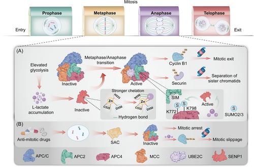
To explore the direct effect of elevated lactate levels on the entire proteome, Liu et al. treated native human embryonic kidney cell lysates with 15 or 25 mM l-lactate before conducting thermal proteomic profiling. They observed a significant shift in the thermostability of UBE2C, an E2 enzyme recruited by APC/C upon structural reorganization of its subunits. However, it is unlikely that lactate binds directly to UBE2C because of its low affinity. Moreover, no change in the abundance or posttranslational modification of UBE2C was detected, suggesting that l-lactate might enhance the interaction between UBE2C and APC/C. To verify this hypothesis, cells were first synchronized to pro-metaphase, a period during which APC/C is inhibited due to its interaction with the mitotic checkpoint complex. Subsequently, Liu et al. incubated cell lysates with 15 mM l-lactate and performed immunoprecipitation of APC/C, revealing that l-lactate significantly enhanced the binding between UBE2C and APC/C. In addition, mass spectrometry analysis of APC/C showed a lactate-dependent elevation of SUMO2/3 conjunctions. Previous studies have shown that SUMOylation of APC4 on residues K772 and K798 results in a substantial rearrangement of the WHB domain in APC2, facilitating the binding of UBE2C to APC/C for an efficient APC/C activation.3, 4 To further investigate the role of APC4 SUMOylation in lactate-mediated UBE2C–APC/C interactions, Liu et al. constructed APC4 K772/798R cells in which APC4 SUMOylation is abrogated. They found that the lactate-mediated interaction between UBE2C and APC/C was completely lost in these cells.
Liu et al. then investigated the mechanism by which l-lactate affects APC4 SUMOylation and proposed that lactate directly targets SENP1, a deSUMOylating enzyme critical for mitosis. Supporting this conjecture, results showed that deleting SENP1 recapitulated the effects of elevated lactate levels, including increased APC4 SUMOylation and strong binding of UBE2C and APC/C. Furthermore, increasing the lactate concentration in SENP1-deficient cells did not further enhance APC4 SUMOylation. Further analysis revealed that the active site of SENP1 is structurally similar to a zinc-binding pocket, and lactate displaced the His533 side chain of SENP1 to bind zinc. Moreover, Cys535 and Asn556 in SENP1 were essential for the effect of lactate on SENP1 activity. Lactate enhances SENP1 and zinc chelation by forming hydrogen bonds with Asn556 to inhibit SENP1 activity, whereas lactate-mediated APC4 SUMOylation is prevented in cells carrying the N556A mutation in SENP1 (Figure 1A).
To determine whether the newly discovered function of lactate plays a widespread role in the regulation of mitosis, Liu et al. tracked lactate levels in synchronized HeLa S3 and HCT116 cells, and found that they increased upon entry into mitosis. When endogenous lactate reached 15 mM, it triggered a timed remodeling of APC/C and degradation of cyclinB1 and securin, allowing for metaphase-to-anaphase transition and mitotic exit. These findings suggest that proliferating cells naturally accumulate lactate at the concentration required for APC/C remodeling upon entry into mitosis.
Antimitotic drugs can inhibit the rapid proliferation of cells and be used in cancer treatment. Nocodazole prevents the kinetochores of sister chromatids in metaphase from binding to mitotic spindle and activates the spindle assembly checkpoint (SAC) by interfering with microtubule polymerization. The activated SAC generates the mitotic checkpoint complex, which inhibits the activity of APC/C, leading to mitotic arrest.4, 5 However, a robust increase in lactate levels can activate APC/C and significantly promote the degradation of cyclin B1, leading to mitotic slippage, a phenomenon that promotes the progression of mitosis even in the presence of an activated SAC (Figure 1B). Liu et al. also treated cells with d-lactate and pyruvate but did not reproduce the APC/C remodeling effect of l-lactate. To show that the function of l-lactate in the cell cycle relies on an appropriately oriented α-hydroxy group adjacent to the lactate carboxylate, cells were treated with l-2-hydroxyglutarate, which has a similar structure to that of l-lactate; Liu et al. observed that l-2-hydroxyglutarate enhanced the zinc-mediated inhibition of SENP1.
In summary, the study by Liu et al. presents a novel mechanism by which lactate regulates the cell cycle. Lactate accumulation indicates a nutrient-rich growth phase as the cell enters mitosis, in which lactate forms a complex with zinc at the SENP1 active site and stabilizes APC4 SUMOylation to stimulate the timed activation of APC/C. However, persistent lactate accumulation drives aberrant APC/C remodeling, leading to uncontrolled cell proliferation. Cancer cells accumulate large amounts of lactate to accelerate their proliferation, and some drugs have been found to reduce the accumulation of lactate in these cells. Classic drugs for treating diabetes, such as metformin and sodium-glucose cotransporter 2 inhibitors, have been shown to decrease lactate accumulation in cancer cells. Combining these drugs with microtubule polymerization inhibitors may yield better therapeutic effects in cancer treatment. Moreover, lactate accumulates not only in proliferating cells but also in adipocyte hypertrophy and exercising muscle. Therefore, future studies are needed to identify other SENP1 targets regulated by lactate and to better understand the significance of lactate signaling in various physiological processes.
Qiqing Yang: Visualization (lead); writing—original draft (lead). Long Zhang: Conceptualization (lead); writing—review and editing (lead). Jun Chen: Conceptualization (equal); writing—review and editing (supporting). All authors have read and approved the final manuscript.
Author Long Zhang is an Editorial board member of MedComm – Oncology. Author Long Zhang was not involved in the journal's review of or decisions related to this manuscript. The remaining authors declare no conflict of interest.

 求助内容:
求助内容: 应助结果提醒方式:


