Calcium phosphate with submicron topography influences primary human macrophage response, enhancing downstream angiogenesis and osteogenesis in vitro
Abstract
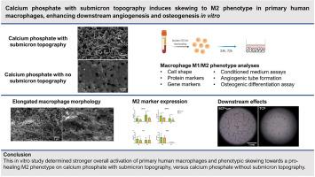
Calcium phosphates with submicron surface features have demonstrated superior performance to conventional calcium phosphates and equivalence to autologous bone in pre-clinical bone healing models. This is related to their ability to form bone in soft tissues, without the addition of cells and growth factors. It is hypothesized that a specific innate immune response to submicron topography contributes to the enhanced bone healing by these materials. Upregulation of pro-healing, anti-inflammatory ‘M2’ macrophages versus pro-inflammatory ‘M1’ macrophages on submicron-structured calcium phosphates may be involved. In this in vitro study, the response of primary human macrophages to different calcium phosphate bone graft substitutes was assessed. Primary CD14+ monocytes were isolated from human buffy coats and were seeded on two different calcium phosphate materials. The first material had a submicron topography of needle-shaped crystals (BCP<μm) while the second material had no submicron topography (TCP). Macrophage M1/M2 phenotype characterization by protein and gene expression markers at 24 h and 72 h indicated overall stronger macrophage activation and subtle phenotypic skewing towards the M2 phenotype on BCP<μm vs TCP. Moreover, macrophages exhibited an elongated morphology on BCP<μm, which is associated with the M2 phenotype, while macrophages on TCP primarily exhibited a spherical morphology. Conditioned medium of macrophages cultured on BCP<μm resulted in enhanced in vitro angiogenic tube formation and osteogenic differentiation of mesenchymal stromal cells, compared to conditioned medium from macrophages on TCP. Altogether, these findings suggest a potential role of M2 macrophage upregulation in the bone-induction mechanism of calcium phosphates with submicron surface topography.

 求助内容:
求助内容: 应助结果提醒方式:


