Emine Cansu Topçuoğlu, Tuğçe Çevik Sönmez, Tülay Koç, Ömer Fahrettin Göze
{"title":"Preserving periodontal tissue in the treatment of a large peripheral ossifying fibroma: a case study.","authors":"Emine Cansu Topçuoğlu, Tuğçe Çevik Sönmez, Tülay Koç, Ömer Fahrettin Göze","doi":"10.47162/RJME.64.3.14","DOIUrl":null,"url":null,"abstract":"<p><p>Peripheral ossifying fibroma (POF) is a reactive, benign gingival enlargement. Its etiology is not fully known. It can be seen in many different sizes in the mouth. The histopathological appearance of POF is mineralized tissue and fibrous proliferation. All relevant soft and hard tissues must be removed to prevent recurrence. Periodontal tissue remaining after excision is important for tooth preservation. With large lesions, the loss of healthy periodontal tissue is also large. Periodontal surgical approaches are important to preserve the remaining periodontal tissue. The positive effects of autogenously obtained titanium-prepared platelet-rich fibrin (T-PRF) and connective tissue graft (CTG) on soft tissue are well known. A 34-year-old woman presented with a fibrous and pedunculated gingival mass in the upper left canine premolar region. The operation was performed with complete excision of the lesion down to the bone along with the surrounding healthy tissue. Periodontal treatment of the large defect created after excision of a large POF lesion was performed with laterally positioned flap, CTG and T-PRF. The periodontal tissue and defect were noted to heal in a healthy manner at the 6-month follow-up. POF is a benign lesion; however, it has a high recurrence rate. Complete elimination of the lesion is crucial to prevent recurrence. Periodontal surgical methods and biomaterials applied after surgical excision are significant to maintain the periodontal health of the remaining teeth and tissues.</p>","PeriodicalId":54447,"journal":{"name":"Romanian Journal of Morphology and Embryology","volume":"64 3","pages":"427-430"},"PeriodicalIF":1.5000,"publicationDate":"2023-07-01","publicationTypes":"Journal Article","fieldsOfStudy":null,"isOpenAccess":false,"openAccessPdf":"https://www.ncbi.nlm.nih.gov/pmc/articles/PMC10720930/pdf/","citationCount":"0","resultStr":null,"platform":"Semanticscholar","paperid":null,"PeriodicalName":"Romanian Journal of Morphology and Embryology","FirstCategoryId":"3","ListUrlMain":"https://doi.org/10.47162/RJME.64.3.14","RegionNum":4,"RegionCategory":"医学","ArticlePicture":[],"TitleCN":null,"AbstractTextCN":null,"PMCID":null,"EPubDate":"","PubModel":"","JCR":"Q4","JCRName":"DEVELOPMENTAL BIOLOGY","Score":null,"Total":0}
引用次数: 0
Abstract
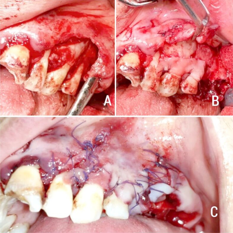
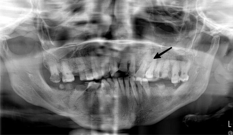
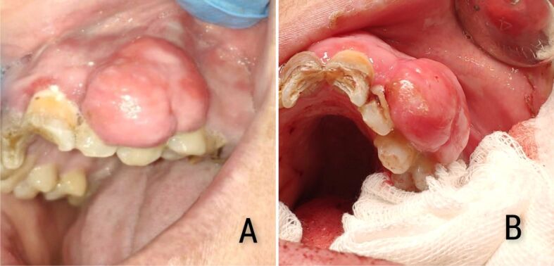
Peripheral ossifying fibroma (POF) is a reactive, benign gingival enlargement. Its etiology is not fully known. It can be seen in many different sizes in the mouth. The histopathological appearance of POF is mineralized tissue and fibrous proliferation. All relevant soft and hard tissues must be removed to prevent recurrence. Periodontal tissue remaining after excision is important for tooth preservation. With large lesions, the loss of healthy periodontal tissue is also large. Periodontal surgical approaches are important to preserve the remaining periodontal tissue. The positive effects of autogenously obtained titanium-prepared platelet-rich fibrin (T-PRF) and connective tissue graft (CTG) on soft tissue are well known. A 34-year-old woman presented with a fibrous and pedunculated gingival mass in the upper left canine premolar region. The operation was performed with complete excision of the lesion down to the bone along with the surrounding healthy tissue. Periodontal treatment of the large defect created after excision of a large POF lesion was performed with laterally positioned flap, CTG and T-PRF. The periodontal tissue and defect were noted to heal in a healthy manner at the 6-month follow-up. POF is a benign lesion; however, it has a high recurrence rate. Complete elimination of the lesion is crucial to prevent recurrence. Periodontal surgical methods and biomaterials applied after surgical excision are significant to maintain the periodontal health of the remaining teeth and tissues.
期刊介绍:
Romanian Journal of Morphology and Embryology (Rom J Morphol Embryol) publishes studies on all aspects of normal morphology and human comparative and experimental pathology. The Journal accepts only researches that utilize modern investigation methods (studies of anatomy, pathology, cytopathology, immunohistochemistry, histochemistry, immunology, morphometry, molecular and cellular biology, electronic microscopy, etc.).
 求助内容:
求助内容: 应助结果提醒方式:


