{"title":"Electroacupuncture may alleviate diabetic neuropathic pain by inhibiting the microglia P2X4R and neuroinflammation.","authors":"Si-Ying Qu, Han-Zhi Wang, Qun-Qi Hu, Yi-Qi Ma, Yu-Rong Kang, Li-Qian Ma, Xiang Li, Lu-Hang Chen, Bo-Yu Liu, Xiao-Mei Shao, Bo-Yi Liu, Jun-Ying Du, Yi Liang, Hong-Li Zhao, Yong-Liang Jiang, Jian-Qiao Fang, Xiao-Fen He","doi":"10.1007/s11302-023-09972-9","DOIUrl":null,"url":null,"abstract":"<p><p>Diabetic neuropathic pain (DNP) is a common and destructive complication of diabetes mellitus. The discovery of effective therapeutic methods for DNP is vitally imperative because of the lack of effective treatments. Although 2 Hz electroacupuncture (EA) was a successful approach for relieving DNP, the mechanism underlying the effect of EA on DNP is still poorly understood. Here, we established a rat model of DNP that was induced by streptozotocin (STZ) injection. P2X4R was upregulated in the spinal cord after STZ-injection. The upregulation of P2X4R was mainly expressed on activated microglia. Intrathecal injection of a P2X4R antagonist or microglia inhibitor attenuated STZ-induced nociceptive thermal hyperalgesia and reduced the overexpression of brain-derived neurotrophic factor (BDNF), interleukin-1β (IL-1β) and tumor necrosis factor-α (TNF-α) in the spinal cord. We also assessed the effects of EA treatment on the pain hypersensitivities of DNP rats, and further investigated the possible mechanism underlying the analgesic effect of EA. EA relieved the hyperalgesia of DNP. In terms of mechanism, EA reduced the upregulation of P2X4R on activated microglia and decreased BDNF, IL-1β and TNF-α in the spinal cord. Mechanistic research of EA's analgesic impact would be beneficial in ensuring its prospective therapeutic effect on DNP as well as in extending EA's applicability.</p>","PeriodicalId":20952,"journal":{"name":"Purinergic Signalling","volume":" ","pages":"533-547"},"PeriodicalIF":2.4000,"publicationDate":"2025-08-01","publicationTypes":"Journal Article","fieldsOfStudy":null,"isOpenAccess":false,"openAccessPdf":"https://www.ncbi.nlm.nih.gov/pmc/articles/PMC12454746/pdf/","citationCount":"0","resultStr":null,"platform":"Semanticscholar","paperid":null,"PeriodicalName":"Purinergic Signalling","FirstCategoryId":"3","ListUrlMain":"https://doi.org/10.1007/s11302-023-09972-9","RegionNum":4,"RegionCategory":"医学","ArticlePicture":[],"TitleCN":null,"AbstractTextCN":null,"PMCID":null,"EPubDate":"2023/10/23 0:00:00","PubModel":"Epub","JCR":"Q2","JCRName":"NEUROSCIENCES","Score":null,"Total":0}
引用次数: 0
Abstract
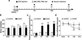
Diabetic neuropathic pain (DNP) is a common and destructive complication of diabetes mellitus. The discovery of effective therapeutic methods for DNP is vitally imperative because of the lack of effective treatments. Although 2 Hz electroacupuncture (EA) was a successful approach for relieving DNP, the mechanism underlying the effect of EA on DNP is still poorly understood. Here, we established a rat model of DNP that was induced by streptozotocin (STZ) injection. P2X4R was upregulated in the spinal cord after STZ-injection. The upregulation of P2X4R was mainly expressed on activated microglia. Intrathecal injection of a P2X4R antagonist or microglia inhibitor attenuated STZ-induced nociceptive thermal hyperalgesia and reduced the overexpression of brain-derived neurotrophic factor (BDNF), interleukin-1β (IL-1β) and tumor necrosis factor-α (TNF-α) in the spinal cord. We also assessed the effects of EA treatment on the pain hypersensitivities of DNP rats, and further investigated the possible mechanism underlying the analgesic effect of EA. EA relieved the hyperalgesia of DNP. In terms of mechanism, EA reduced the upregulation of P2X4R on activated microglia and decreased BDNF, IL-1β and TNF-α in the spinal cord. Mechanistic research of EA's analgesic impact would be beneficial in ensuring its prospective therapeutic effect on DNP as well as in extending EA's applicability.
期刊介绍:
Nucleotides and nucleosides are primitive biological molecules that were utilized early in evolution both as intracellular energy sources and as extracellular signalling molecules. ATP was first identified as a neurotransmitter and later as a co-transmitter with all the established neurotransmitters in both peripheral and central nervous systems. Four subtypes of P1 (adenosine) receptors, 7 subtypes of P2X ion channel receptors and 8 subtypes of P2Y G protein-coupled receptors have currently been identified. Since P2 receptors were first cloned in the early 1990’s, there is clear evidence for the widespread distribution of both P1 and P2 receptor subtypes in neuronal and non-neuronal cells, including glial, immune, bone, muscle, endothelial, epithelial and endocrine cells.
 求助内容:
求助内容: 应助结果提醒方式:


