Effects of Visual-Motor Illusion via Image Videos Showing Increased Exercise Intensity on the Tibial Anterior during Sit-to-Stand Movement: A Study of Healthy Participants.
{"title":"Effects of Visual-Motor Illusion via Image Videos Showing Increased Exercise Intensity on the Tibial Anterior during Sit-to-Stand Movement: A Study of Healthy Participants.","authors":"Junpei Tanabe, Kazu Amimoto, Katsuya Sakai, Motoyoshi Morishita","doi":"10.3390/neurolint15040081","DOIUrl":null,"url":null,"abstract":"<p><p>Visual-motor illusion (VMI) elicits kinesthetic sensation from visual stimulation. We have previously performed ankle motion VMI with resistance applied to the ankle joint on the paralyzed side (power-VMI (P-VMI)) and ankle motion VMI without resistance (standard-VMI (S-VMI)) to activate the tibialis anterior (TA) muscle in stroke-paralyzed patients and compared sit-to-stand (STS) durations, but these studies did not measure TA activity during the STS movement. The purpose of this study was to evaluate the effects of different intensities of visual stimuli presented during VMI on TA and STS movement. Healthy right-footed adults (<i>n</i> = 18) observed two different VMI videos of ankle dorsiflexion, including S-VMI and P-VMI, with an observation time of 2 min each. STS movement was evaluated before and after watching each video. Each participant performed both S-VMI and P-VMI interventions on the same day. Only P-VMI enhanced the integrated electromyogram of the TA, increased the angular velocities of the trunk forward inclination and the ankle dorsiflexion, and shortened the STS duration. Our results indicate that P-VMI facilitates the activation of TA during STS, and we believe that we have clarified the intervention mechanism of VMI.</p>","PeriodicalId":19130,"journal":{"name":"Neurology International","volume":"15 4","pages":"1290-1302"},"PeriodicalIF":3.2000,"publicationDate":"2023-10-20","publicationTypes":"Journal Article","fieldsOfStudy":null,"isOpenAccess":false,"openAccessPdf":"https://www.ncbi.nlm.nih.gov/pmc/articles/PMC10594414/pdf/","citationCount":"0","resultStr":null,"platform":"Semanticscholar","paperid":null,"PeriodicalName":"Neurology International","FirstCategoryId":"1085","ListUrlMain":"https://doi.org/10.3390/neurolint15040081","RegionNum":0,"RegionCategory":null,"ArticlePicture":[],"TitleCN":null,"AbstractTextCN":null,"PMCID":null,"EPubDate":"","PubModel":"","JCR":"Q2","JCRName":"CLINICAL NEUROLOGY","Score":null,"Total":0}
引用次数: 0
Abstract
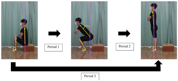
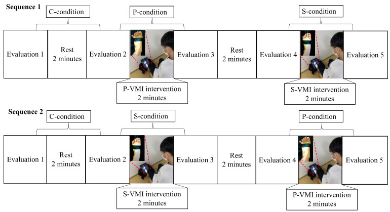
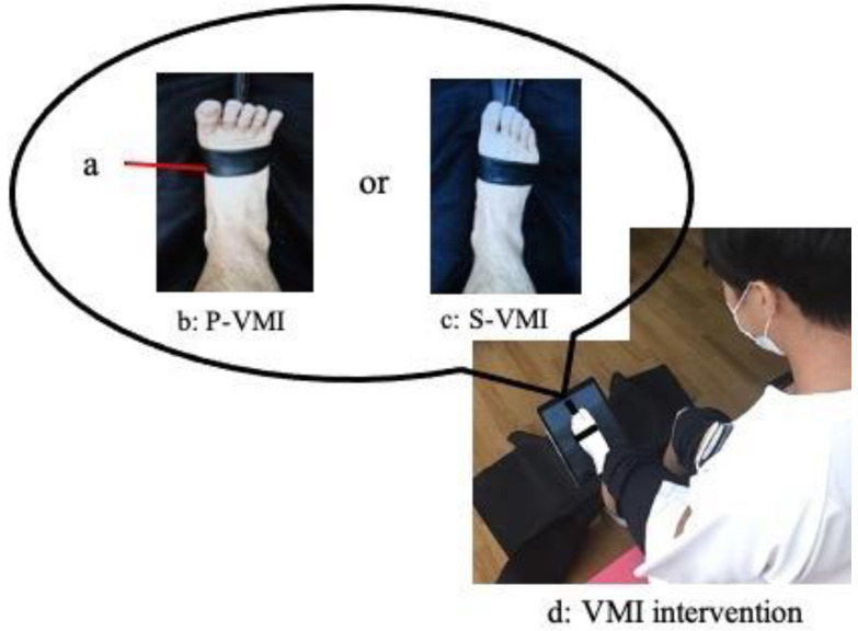
Visual-motor illusion (VMI) elicits kinesthetic sensation from visual stimulation. We have previously performed ankle motion VMI with resistance applied to the ankle joint on the paralyzed side (power-VMI (P-VMI)) and ankle motion VMI without resistance (standard-VMI (S-VMI)) to activate the tibialis anterior (TA) muscle in stroke-paralyzed patients and compared sit-to-stand (STS) durations, but these studies did not measure TA activity during the STS movement. The purpose of this study was to evaluate the effects of different intensities of visual stimuli presented during VMI on TA and STS movement. Healthy right-footed adults (n = 18) observed two different VMI videos of ankle dorsiflexion, including S-VMI and P-VMI, with an observation time of 2 min each. STS movement was evaluated before and after watching each video. Each participant performed both S-VMI and P-VMI interventions on the same day. Only P-VMI enhanced the integrated electromyogram of the TA, increased the angular velocities of the trunk forward inclination and the ankle dorsiflexion, and shortened the STS duration. Our results indicate that P-VMI facilitates the activation of TA during STS, and we believe that we have clarified the intervention mechanism of VMI.

 求助内容:
求助内容: 应助结果提醒方式:


