Vahid Ali, Hassan Kefayati, Mehdi Shafiee Ardestani, Afshin Pourahmad
{"title":"Synthesis and evaluation of new magneto-fluorescent carbon dot based on manganese citrate for MRI imaging.","authors":"Vahid Ali, Hassan Kefayati, Mehdi Shafiee Ardestani, Afshin Pourahmad","doi":"10.1007/s10334-023-01117-8","DOIUrl":null,"url":null,"abstract":"<p><strong>Objective: </strong>Medical imaging techniques have widely revolutionized the diagnosis and treatment of various health conditions. Among these techniques, magnetic resonance imaging (MRI) has stood out as a noninvasive and versatile tool. Now, a breakthrough innovation called \"manganese-carbon dots\" is poised to enhance MRI imaging and provide physicians with even greater insight into the human body.</p><p><strong>Materials and methods: </strong>In this study, one-pot hydrothermal method was used to fabricate magneto-fluorescent carbon quantum dots using manganese citrate, urea, and Mn2+. Manganese citrateAQ3 acted as a carbon source and contrast agent. TEM,XPS, FTIR, UV-Vis, fluorescent analysis confirmed the successful synthesis of magneto-fluorescent carbon quantum dots. The MTT assay was used to study its biocompatiblity, Finallay application of itscompound for mri imaging was investigated.</p><p><strong>Results: </strong>Characterization Techniques confirmed the succesful synthesis of product. MTT assay showed no toxicity of this product on HEK-293 cells. In addition, it exhibited high r1 relaxivity (7.4 mM-1 S-1) suggesting excellent potential of magneto-fluorescent carbon quantum dots as MRI T1 contrast agent and enabling specific imaging.</p><p><strong>Conclusion: </strong>Based on the results obtained, the synthesized carbon quantum dots could be used as fluorescence/MRI bimodal platform for in vivo imaging.</p>","PeriodicalId":18067,"journal":{"name":"Magnetic Resonance Materials in Physics, Biology and Medicine","volume":" ","pages":"139-148"},"PeriodicalIF":2.0000,"publicationDate":"2024-02-01","publicationTypes":"Journal Article","fieldsOfStudy":null,"isOpenAccess":false,"openAccessPdf":"","citationCount":"0","resultStr":null,"platform":"Semanticscholar","paperid":null,"PeriodicalName":"Magnetic Resonance Materials in Physics, Biology and Medicine","FirstCategoryId":"3","ListUrlMain":"https://doi.org/10.1007/s10334-023-01117-8","RegionNum":4,"RegionCategory":"医学","ArticlePicture":[],"TitleCN":null,"AbstractTextCN":null,"PMCID":null,"EPubDate":"2023/10/20 0:00:00","PubModel":"Epub","JCR":"Q3","JCRName":"RADIOLOGY, NUCLEAR MEDICINE & MEDICAL IMAGING","Score":null,"Total":0}
引用次数: 0
Abstract
Objective: Medical imaging techniques have widely revolutionized the diagnosis and treatment of various health conditions. Among these techniques, magnetic resonance imaging (MRI) has stood out as a noninvasive and versatile tool. Now, a breakthrough innovation called "manganese-carbon dots" is poised to enhance MRI imaging and provide physicians with even greater insight into the human body.
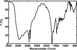
Materials and methods: In this study, one-pot hydrothermal method was used to fabricate magneto-fluorescent carbon quantum dots using manganese citrate, urea, and Mn2+. Manganese citrateAQ3 acted as a carbon source and contrast agent. TEM,XPS, FTIR, UV-Vis, fluorescent analysis confirmed the successful synthesis of magneto-fluorescent carbon quantum dots. The MTT assay was used to study its biocompatiblity, Finallay application of itscompound for mri imaging was investigated.
Results: Characterization Techniques confirmed the succesful synthesis of product. MTT assay showed no toxicity of this product on HEK-293 cells. In addition, it exhibited high r1 relaxivity (7.4 mM-1 S-1) suggesting excellent potential of magneto-fluorescent carbon quantum dots as MRI T1 contrast agent and enabling specific imaging.
Conclusion: Based on the results obtained, the synthesized carbon quantum dots could be used as fluorescence/MRI bimodal platform for in vivo imaging.
期刊介绍:
MAGMA is a multidisciplinary international journal devoted to the publication of articles on all aspects of magnetic resonance techniques and their applications in medicine and biology. MAGMA currently publishes research papers, reviews, letters to the editor, and commentaries, six times a year. The subject areas covered by MAGMA include:
advances in materials, hardware and software in magnetic resonance technology,
new developments and results in research and practical applications of magnetic resonance imaging and spectroscopy related to biology and medicine,
study of animal models and intact cells using magnetic resonance,
reports of clinical trials on humans and clinical validation of magnetic resonance protocols.

 求助内容:
求助内容: 应助结果提醒方式:


