Longitudinal imaging in Kleefstra syndrome—Brief report and literature review
Abstract
Background
Kleefstra syndrome (KS) is a rare genetic condition affecting the euchromatic histone methyltransferase 1 (EHMT1) gene, typically presenting with developmental delay, generalized hypotonia, distinctive facial dysmorphisms, and neuropsychiatric anomalies.
Methods
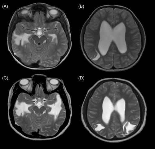
We collected longitudinal magnetic resonance imaging (MRI) data of a child with KS and cerebrospinal fluid (CSF) dissection over 16 years and reviewed the literature focusing on articles reporting MRI findings in KS based on a PubMed search.
Results
An 18-year-old female with KS presented with developmental delay at age 18 months and seizures at age 10 years. At age 23 months, MRI showed ventriculomegaly. Follow-up MRI nine years later showed CSF dissection from the right lateral ventricle, which progressed similarly in the contralateral side 3 years later and stabilized over time. To date, only 20 articles report MRI findings from 49 patients with KS. The brain imaging findings range from normal (about 25%) to various abnormalities, including ventriculomegaly, white matter signal abnormalities, and dysmorphic corpus callosum and brainstem structures. CSF dissection was not found in any cases with white matter or other abnormalities.
Conclusions
CSF dissection may be a rare neuroimaging finding of KS. Its etiology is unclear, but we speculate that epigenetic mutations could indirectly affect the development and integrity of ependymal cell lining along the ventricles. Alternatively, the long-standing hydrocephalus may have caused the development of pseudodiverticula and subsequent CSF dissection.

 求助内容:
求助内容: 应助结果提醒方式:


