{"title":"Issue Information TOC","authors":"","doi":"10.1002/cpph.50","DOIUrl":null,"url":null,"abstract":"<p><b>Cover</b>: In Kasprick et al. (https://doi.org/10.1002/cpph.55), the image shows analysis of skin sections. <b>(A)</b> Determination of titer from rabbits immunized with mCOL7<sup>vWFA2</sup>. Cryosections of normal mouse skin were incubated with serum diluted 1:4,000 1:8,000, 1:16,000, and 1:32,000 and then stained with FITC-AffiniPure donkey anti-rabbit IgG as described in Basic Protocol 3. As the serum specifically binds to the dermal-epidermal junction of the skin, a typical green line is visible (arrows). The line visible at the lowest tested concentration determines the titer (here, 1:8,000). <b>(B)</b> Analysis of H&E staining. Typical ear sections 12 days after first IgG injection in antibody transfer–induced EBA. Upper left picture shows a mild inflammation with less thickening and only a few inflammatory cells. Other pictures show typical thickening of the epidermal layers and massive inflammation. Arrows indicate split formation at dermal-epidermal junction; <sup>**</sup>indicates cartilage of the ear.\n\n <figure>\n <div><picture>\n <source></source></picture><p></p>\n </div>\n </figure></p>","PeriodicalId":10871,"journal":{"name":"Current Protocols in Pharmacology","volume":"84 1","pages":""},"PeriodicalIF":0.0000,"publicationDate":"2019-03-05","publicationTypes":"Journal Article","fieldsOfStudy":null,"isOpenAccess":false,"openAccessPdf":"https://sci-hub-pdf.com/10.1002/cpph.50","citationCount":"0","resultStr":null,"platform":"Semanticscholar","paperid":null,"PeriodicalName":"Current Protocols in Pharmacology","FirstCategoryId":"1085","ListUrlMain":"https://onlinelibrary.wiley.com/doi/10.1002/cpph.50","RegionNum":0,"RegionCategory":null,"ArticlePicture":[],"TitleCN":null,"AbstractTextCN":null,"PMCID":null,"EPubDate":"","PubModel":"","JCR":"Q2","JCRName":"Pharmacology, Toxicology and Pharmaceutics","Score":null,"Total":0}
引用次数: 0
Abstract
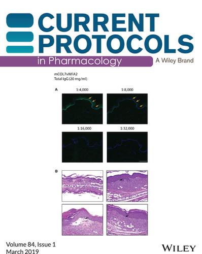
Cover: In Kasprick et al. (https://doi.org/10.1002/cpph.55), the image shows analysis of skin sections. (A) Determination of titer from rabbits immunized with mCOL7vWFA2. Cryosections of normal mouse skin were incubated with serum diluted 1:4,000 1:8,000, 1:16,000, and 1:32,000 and then stained with FITC-AffiniPure donkey anti-rabbit IgG as described in Basic Protocol 3. As the serum specifically binds to the dermal-epidermal junction of the skin, a typical green line is visible (arrows). The line visible at the lowest tested concentration determines the titer (here, 1:8,000). (B) Analysis of H&E staining. Typical ear sections 12 days after first IgG injection in antibody transfer–induced EBA. Upper left picture shows a mild inflammation with less thickening and only a few inflammatory cells. Other pictures show typical thickening of the epidermal layers and massive inflammation. Arrows indicate split formation at dermal-epidermal junction; **indicates cartilage of the ear.

 求助内容:
求助内容: 应助结果提醒方式:


