{"title":"Convoluted tumorous lesions at second portion of duodenum in a cirrhotic patient with massive upper gastrointestinal bleeding and shock","authors":"Wei-Chih Su, Chia-Chi Wang, Jiann-Hwa Chen","doi":"10.1002/aid2.13366","DOIUrl":null,"url":null,"abstract":"<p>This 49-year-old male, a victim of chronic hepatitis B-related liver cirrhosis, visited our emergency department due to hematemesis and tarry stool passage. Hypovolemic shock and severe anemia (Hb 4.8 mg/dL) were noted on arrival. After fluid resuscitation and blood transfusion, urgent esophagogastroduodenoscopy revealed no varices in the esophagus and cardiac portion of the stomach; however, some blood was retained at the proximal duodenum. After the scope was pushed down to the distal 2nd portion of the duodenum, convoluted tumorous lesions (Figure 1) with an erosion were noticed distal to ampulla vater. First, what is your diagnosis? Second, what will be your next step?</p><p>Ectopic duodenal varices at the second portion were diagnosed by endoscopy. It was confirmed by computed tomography (Figure 2), which revealed enhanced engorged vessels at the wall of second portion duodenum. The bleeding episode was successfully controlled by endoscopic injection sclerotherapy with histoacryl glue and somatostatin intravenous infusion.</p><p>Ectopic varices are defined as dilated portosystemic collateral veins located in unusual sites other than the gastroesophageal region and constitute 1% to 5% of all variceal bleeds.<span><sup>1</sup></span> These lesions could locate in different sites, including the duodenum, small bowel, rectum, anastomotic site, and stoma with high interobserver variability in their distribution.<span><sup>2</sup></span> In a large study of 173 patients from Japan, Watanabe et al.<span><sup>3</sup></span> mentioned that the duodenum (32.9%) is the second most common site, and 82.5% of them are located in descending part. Currently, there are no clear guidelines on the management of ectopic varices. Endoscopic treatment, including endoscopic injection sclerotherapy and endoscopic variceal ligation, was the most frequent modality for acute duodenal variceal bleeding, and interventional radiology therapy such as transjugular intrahepatic portosystemic shunt or surgery could be used as rescue therapy. The successful rate of endoscopic treatment alone for acute duodenal variceal bleeding is 73.3%. However, 53.3% patients experience rebleeding within 1 year.<span><sup>4</sup></span></p><p>Each author contributed to the manuscript. <b>Wei-Chih Su</b>: Conceptualization, Writing—original draft. <b>Chia-Chi Wang</b>: Conceptualization, Writing—review & editing, <b>Jiann-Hwa Chen</b>: Supervision, Writing—review & editing.</p><p>The authors declare no conflicts of interest.</p><p>The case report was approved by the Institutional Review Board (11-CR-105) of Taipei Tzu Chi Hospital, Buddhist Tzu Chi Medical Foundation.</p>","PeriodicalId":7278,"journal":{"name":"Advances in Digestive Medicine","volume":"11 1","pages":"49-50"},"PeriodicalIF":0.4000,"publicationDate":"2023-07-17","publicationTypes":"Journal Article","fieldsOfStudy":null,"isOpenAccess":false,"openAccessPdf":"https://onlinelibrary.wiley.com/doi/epdf/10.1002/aid2.13366","citationCount":"0","resultStr":null,"platform":"Semanticscholar","paperid":null,"PeriodicalName":"Advances in Digestive Medicine","FirstCategoryId":"1085","ListUrlMain":"https://onlinelibrary.wiley.com/doi/10.1002/aid2.13366","RegionNum":0,"RegionCategory":null,"ArticlePicture":[],"TitleCN":null,"AbstractTextCN":null,"PMCID":null,"EPubDate":"","PubModel":"","JCR":"Q4","JCRName":"GASTROENTEROLOGY & HEPATOLOGY","Score":null,"Total":0}
引用次数: 0
Abstract
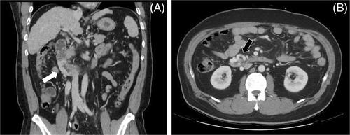
This 49-year-old male, a victim of chronic hepatitis B-related liver cirrhosis, visited our emergency department due to hematemesis and tarry stool passage. Hypovolemic shock and severe anemia (Hb 4.8 mg/dL) were noted on arrival. After fluid resuscitation and blood transfusion, urgent esophagogastroduodenoscopy revealed no varices in the esophagus and cardiac portion of the stomach; however, some blood was retained at the proximal duodenum. After the scope was pushed down to the distal 2nd portion of the duodenum, convoluted tumorous lesions (Figure 1) with an erosion were noticed distal to ampulla vater. First, what is your diagnosis? Second, what will be your next step?
Ectopic duodenal varices at the second portion were diagnosed by endoscopy. It was confirmed by computed tomography (Figure 2), which revealed enhanced engorged vessels at the wall of second portion duodenum. The bleeding episode was successfully controlled by endoscopic injection sclerotherapy with histoacryl glue and somatostatin intravenous infusion.
Ectopic varices are defined as dilated portosystemic collateral veins located in unusual sites other than the gastroesophageal region and constitute 1% to 5% of all variceal bleeds.1 These lesions could locate in different sites, including the duodenum, small bowel, rectum, anastomotic site, and stoma with high interobserver variability in their distribution.2 In a large study of 173 patients from Japan, Watanabe et al.3 mentioned that the duodenum (32.9%) is the second most common site, and 82.5% of them are located in descending part. Currently, there are no clear guidelines on the management of ectopic varices. Endoscopic treatment, including endoscopic injection sclerotherapy and endoscopic variceal ligation, was the most frequent modality for acute duodenal variceal bleeding, and interventional radiology therapy such as transjugular intrahepatic portosystemic shunt or surgery could be used as rescue therapy. The successful rate of endoscopic treatment alone for acute duodenal variceal bleeding is 73.3%. However, 53.3% patients experience rebleeding within 1 year.4
Each author contributed to the manuscript. Wei-Chih Su: Conceptualization, Writing—original draft. Chia-Chi Wang: Conceptualization, Writing—review & editing, Jiann-Hwa Chen: Supervision, Writing—review & editing.
The authors declare no conflicts of interest.
The case report was approved by the Institutional Review Board (11-CR-105) of Taipei Tzu Chi Hospital, Buddhist Tzu Chi Medical Foundation.
期刊介绍:
Advances in Digestive Medicine is the official peer-reviewed journal of GEST, DEST and TASL. Missions of AIDM are to enhance the quality of patient care, to promote researches in gastroenterology, endoscopy and hepatology related fields, and to develop platforms for digestive science. Specific areas of interest are included, but not limited to: • Acid-related disease • Small intestinal disease • Digestive cancer • Diagnostic & therapeutic endoscopy • Enteral nutrition • Innovation in endoscopic technology • Functional GI • Hepatitis • GI images • Liver cirrhosis • Gut hormone • NASH • Helicobacter pylori • Cancer screening • IBD • Laparoscopic surgery • Infectious disease of digestive tract • Genetics and metabolic disorder • Microbiota • Regenerative medicine • Pancreaticobiliary disease • Guideline & consensus.

 求助内容:
求助内容: 应助结果提醒方式:


