{"title":"Abstract","authors":"","doi":"10.1002/jin2.24","DOIUrl":null,"url":null,"abstract":"<p><b>S001</b></p><p><b>S001 The British Society for Nanomedicine</b></p><p>Speaker: Professor R. Steven Conlan</p><p><i>Swansea University Medical School, Singleton Park, Swansea, SA2 8PP, UK</i></p><p><b>Abstract</b>: With the global benefits of the new science of nanomedicine growing each year, the British Society for Nanomedicine enables open access for industry, academia, clinicians and the public to news and details of ongoing research throughout the UK. Our mission includes the direct explanation of the ongoing science and commercial developments to allow the public to understand and stay in touch with this exciting area as it impacts future global healthcare. The British Society for Nanomedicine runs the Journal of Interdisciplinary Nanomedicine (JOIN), an international peer-reviewed academic journal that aims to report truly interdisciplinary nanomedicine research. The British Society for Nanomedicine represents UK nanomedicine interests at a UK, EU and International level including the ETP nanomedicine, and European national nanomedicines platform. The British Society for Nanomedicine is proud to welcome you to ENM17 in London.</p><p><b>S002</b></p><p><b>S002 The French Society for Nanomedicine (SFNano)</b></p><p>Speaker: Dr Nathalie Mignet</p><p><i>University Paris Descartes, CNRS, Faculty of Pharmacy, Paris, France</i></p><p><b>Abstract</b>: The French Society for Nanomedicine (SFNano) is a non-lucrative association whose objectives are to favour progress and knowledge diffusion in the Nanomedicine domain. For these purposes, SFNano organises seminars, workshops and discussions in order to favour a french network, and broader, in collaboration with European nanomedicine societies. SFNano has more than 300 members.</p><p><b>S003</b></p><p><b>S003 Spanish Platform for Nanomedicine (NanoMed Spain)</b></p><p>Speaker: Dr. Teresa Sanchis</p><p><i>Spanish Platform for Nanomedicine (NanoMed Spain) – Institute for Bioengineering of Catalonia, Baldiri Reixach 1008028, Barcelona, (Spain)</i></p><p><b>Abstract</b>: The Spanish Platform for Nanomedicine (NanoMed Spain - http://nanomedspain.net/) is a forum that brings together 150 public research centres, hospitals, companies and government representatives active in nanomedicine. Nanomed Spain is an instrument to coordinate entities involved in R&D+i, fundamental to the transfer of results to industry and the health system in this highly multidisciplinary field. It is also a means of connection to facilitate the internationalization of initiatives and projects, with the aim of improving the competitiveness of Spanish companies in this emerging field.</p><p>Industry in the biomedical and biotechnology sector plays a leading role in the Platform, very actively supported by technology centers, research organizations, universities and hospitals, as well as by national public administration.</p><p>The mission of Nanomed Spain is to promote and facilitate public-private partnerships in research and innovation in nanomedicine in Spain, with the aim of accelerating the development of innovative therapeutics and diagnostics based on the capabilities offered by nanotechnology applied to health care.</p><p><b>S004</b></p><p><b>S004 German Platform NanoBioMedicine</b></p><p>Speaker: Klaus-Michael Weltring</p><p><i>Gesellschaft für Bioanalytik Münster e. V, Mendelstr. 17D-48149, Münster</i></p><p><b>S005</b></p><p><b>S005 NanoMed North - a nanomedicine consortium</b></p><p>Speaker: Ulf G Andersson</p><p><i>NanoMed North – Medeon Science Park & Incubator, Medeon Science Park – 205 12, Malmo, Sweden</i></p><p><b>Abstract</b>: The presentation will include a short presentation of the NanoMed North consortium including purpose, type of members and activities. Furthermore, some of the ongoing nanomedicine activities among the members of NanoMed North will be briefly presented.</p><p><b>S006</b></p><p><b>S006 Overview of the European Nanomedicine Characterisation Laboratory (EU-NCL)</b></p><p>Speaker: Simon Baconnier</p><p><i>CEA, 17 rue des Martyrs38000, Grenoble, France</i></p><p><b>Abstract</b>: The use of nanotechnologies in healthcare promises to tackle major medical challenges. However, the manufacturing process of nanomedicines is potentially complex and inconsistencies must be carefully assessed before clinical applications can be considered. Furthermore, safety concerns related to the use of nanomaterials must be addressed as early as possible during product development.</p><p>The European Nanomedicine Characterization laboratory (EU-NCL) will address these issues by providing the critical infrastructure and characterization services required to analyze physical and chemical attributes, in vitro biological properties, and in vivo characteristics of nanomedicines under development.</p><p>With this objective, EU-NCL will integrate a high diversity of materials to be analyzed. Actually, the current nanomedicines can be organic, inorganic, metal based, combined or loaded with active compound. Thus, EU-NCL will adapt and optimize its characterization capacities and strategies to the complexity of the product to be characterized as well as their different uses (Diagnosis, Treatment, targeted or not). On top of that, there is a strong need for high quality data, produced with standardized methods in line with regulatory requirements and standardization needs supported by robust and established standards and controls. This rigorous frame is the only way toward quality level that may serve the users of EU-NCL in their future clinical development.</p><p><b>S007</b></p><p><b>S007 The Science of Chemical Characterisation</b></p><p>Speaker: Sven Even Borgos</p><p><i>Sem Sælands v 2A, N-7034, Trondheim, Norway</i></p><p><b>Abstract</b>: Nanomedicines owe much of their tremendous potential to their nanometre size range, surface structure and – importantly – their <i>spatially structured chemical composition</i>. API encapsulation inside nanoparticles shields the APIs from premature metabolism in the body, and conversely, protects the body against off-target toxicities. The carrier composition determines release rates of the APIs, either intrinsic or triggered by external factors like heating. Surface chemical functionalisation, e.g. by antibody attachment, can form the basis of active and highly specific targeting in the body, whereas e.g. PEGylation of the surface will generally have a major effect on the nanomedicine circulation half-life.</p><p>The unique properties of nanomedicines do also, however, causes some unique challenges in their characterisation. For composition analyses, the full solubilisation of all components can be demanding, e.g. the release of intact, complex moieties like surface ligands that are covalently attached to polymer networks. Furthermore, release of API in complex systems like blood is subject to multiple simultaneous equilibria, necessitating advanced mass spectrometry based strategies in quantification of the various API forms.</p><p>This presentation aims to describe some nanomedicine-specific challenges – and solutions – in chemical analysis, with emphasis on the chemical characterisation performed within the European Nanomedicine Characterisation Laboratory (EU-NCL).</p><p><b>S008</b></p><p><b>S008 The Science of Biocompatibility Testing</b></p><p>Speaker: Neill Liptrott</p><p><i>The University of Liverpool, 70 Pembroke Place, Block H (first floor), Liverpool, L69 3GF, UK</i></p><p><b>Abstract</b>: Nanomedicines promise to revolutionise therapy across a number of diseases either through improved bioavailability or systemic distribution. However, there is a paucity of information regarding putative adverse interactions in biological systems, which is important for the development and regulation of safe and effective nanomedicines. The interaction of nanoparticles with various components of the immune system has been well documented and therefore assessment of these interactions is vital for successful translation to the clinic. Prediction of such reactions is important for development of robust preclinical systems for selection of viable leads, but is hampered by a limited understanding of the mechanisms and signalling pathways involved in the recognition of engineered nanoparticles by the host immune system. The mechanisms involved in this recognition are complex and varied including, but not limited to, altered cytokine expression, generation of reactive oxygen species, absorption of blood proteins (e.g. complement) which induce phagocyte activation and triggering of various signalling complexes. In this presentation, we will discuss the work on biocompatibility being conducted at the University of Liverpool and how it relates to the European Nanomedicine Characterisation Laboratory (EU-NCL).</p><p><b>S009</b></p><p><b>S009 Regulatory Challenges of Nanomedicines</b></p><p>Speaker: Ka-Wai Wan</p><p><i>MHRA, MHRA, 151 Buckingham Palace Road, London, SW1W 9SZ, UK</i></p><p><b>Abstract</b>: Nanotechnology has greatly advanced in the past decade and provides immense potential for the development of improved therapeutic and diagnostic tools for the treatment, prevention and diagnosis of various diseases of the central nervous system, cardiovascular system and cancer. Due to their small size in dimension, it is believed that nanoparticles are able to interact with cells at the molecular level more efficiently and provide better targeting ability towards desired cells and tissues with greater precision and efficacy. Nano-based delivery systems using polymers (e.g. polymeric micelles and polymer-drug conjugates) and lipids (e.g. liposomes, and solid lipid nanoparticles can help: i) increase solubility; ii) improve tumour targeting; (iii) decrease toxicity; (iv) overcome drug resistance, and v) prolong circulation half-life by modifying the surfaces of these nanoparticulate drug delivery systems and increasing drug payload to the target cells. The complexity of nanomedicines requires a holistic assessment of the quality, safety and efficacy of the product. This talk will focus on the challenges of developing these advanced drug delivery systems to match with the current regulatory expectation.</p><p><b>S010</b></p><p><b>S010 Polymeric nanomedicines</b></p><p>Speaker: Cameron Alexander</p><p><i>Nottingham University, School of Pharmacy</i></p><p><b>Abstract</b>: Polymers are of interest in the field of nanomedicine, either as inert carriers for drugs, genes or cells, or intrinsically as therapeutics. The advantages of polymers include the ability to contain multiple chemical functionalities, to assemble into controllable structures and to distribute in the body more selectively than conventional small molecule drugs. However, there are also disadvantages to polymer nanomedicines, mainly relating to difficulties in their transport across biological barriers, but also in their reproducible synthesis and scale-up at appropriate cost. This talk will focus on some examples of polymer nanomedicines in the laboratory, and also on our continuing efforts to introduce materials chemistries to nanomedicines which are effective, sustainable and affordable.</p><p><b>S011</b></p><p><b>S011 Multi-modal nanotools for early cancer diagnostics and treatment</b></p><p>Speaker: Yuri Volkov</p><p><i>Trinity College Dublin, Trinity Centre for Health Sciences, James's Street, Dublin 8</i></p><p><b>Abstract</b>: Current clinical cancer diagnostic systems commonly suffer from insufficient specificity and sensitivity. Several original nanotechnological platforms have been developed recently, enabling to perform highly sensitive tests for cancer markers and cells detection exploiting magnetic, optical, plasmonic and non-linear optical advanced nanoscale material properties. The devices based on these principles can operate in minimally invasive and miniaturised volume formats offering cost-effective technological solutions enabling to reach a qualitatively new level of diagnostic accuracy and imaging quality.</p><p>Optimistic expectations are also associated with the applications of nanoparticles as a new class of multifunctional drug delivery systems, arising both from the opportunities of precision targeting and from the fact that the finite, but tunable size of the engineered nanostructures used as drug delivery vehicles can impose discrete nanoscale drug distribution barriers at the level of cells, tissues and entire organism, thereby eliminating undesirable side effects pertinent to most contemporary chemotherapeutic drugs.</p><p>On the other hand, a steady worldwide rise in manufacturing and medical use of nanomaterials emphasizes the requirements for thorough assessment of health outcomes associated with novel nanoparticle applications and the necessity of a robust safety-ensuring approach in nanomedicines design and development. Here we will provide an overview of several nanoparticle application scenarios for improved diagnostics and advanced therapeutic use, along with the contemporary approaches to safety screening of nanomaterials with promising biomedical application potential.</p><p>This work has been supported, in parts, by the European Commission under “NAMDIATREAM” Project (<i>Nanotechnological toolkits for multi-modal disease diagnostics and treatment monitoring</i>, GA#246479), “MULTIFUN” Project (<i>Multifunctional nanoparticles for cancer treatment</i>, GA#262943), and “NOCANTHER” Project <i>(Nanomedicine Upscaling for Early Clinical Phases of Multimodal Cancer Therapy</i>, GA#685795).</p><p><b>S012</b></p><p><b>S012 Plasmonic and magnetic NPS for biomedical applications</b></p><p>Speaker: Nguyen T. K. Thanh</p><p><i>Biophyics group, Department of Physics and Astronomy and UCL Healthcare Biomagnetic and Nanomaterials Laboratory, University College London, UK</i></p><p>References</p><p>1. \n <span>Pallares, R. M.</span>, <span>Bosman, M.</span>, <span>Thanh, N.T.K.*</span>, and <span>Su, X.</span> (<span>2016</span>) <span>Plasmonic multi-logic gate platform based on sequence-specific binding of estrogen receptors and gold nanorods</span>. <i>Nanoscale.</i> <span>8</span>: <span>19919</span>–<span>20126</span>.</p><p>2. \n <span>Thanh, N. T. K.</span> (<span>2016</span>) <span>Preface of Theme issue ”Multifunctional nanostructures for diagnosis and therapy of diseases’</span>. <i>Interface Focus</i>, <span>6</span>: <span>20160077</span>.</p><p>3. \n <span>Baber, R.</span>, <span>Mazzei, L.</span>, <span>Thanh, N. T. K.</span>, <span>Gavriilidis, A.</span> (<span>2016</span>) <span>Synthesis of silver nanoparticles using microfluidic impinging jet reactors</span>. <i>Journal of Flow Chemistry.</i> <span>6</span>: <span>268</span>-<span>278</span>.</p><p>4. \n <span>Pallares, R. M.</span>, <span>Lim, S. H.</span>, <span>Thanh, N.T.K.*</span>, and <span>Su, X.</span> (<span>2016</span>) <span>Growth of Anisotropic Gold Nanoparticles in Photoresponsive Fluid and Application to UV Exposure Sensing and Erythema Prediction</span>. <i>Nanomedicine.</i> <span>11</span>: <span>2845</span>-<span>2860</span></p><p>5. \n <span>Mameli, V.</span>, <span>Musinu, A.</span>, <span>Ardu, A.</span>, <span>Ennas, G.</span>, <span>Peddis, D.</span>, <span>Niznansky, D.</span>, <span>Sangregorio, C.</span>, <span>Innocenti, C.</span>, <span>Thanh, N. T. K.*</span> and <span>Cannas, C.</span> (<span>2016</span>) <span>Studying the exclusive effect of Zn-substitution on the magnetic and hyperthermic properties of cobalt ferrite nanoparticles</span>. <i>Nanoscale.</i> <span>8</span>, <span>10124</span>-<span>10137</span>.</p><p>6. \n <span>Monteforte, M.</span>, <span>Kobayashi, S.</span>, <span>Tung, L. D.</span>, <span>Higashimine, K.</span>, <span>Mott, D. M.</span>, <span>Maenosono, S.</span>, <span>Thanh, N. T. K.</span>, and <span>Robinson, I. K.</span> (<span>2016</span>) <span>Quantitative Two Dimensional Strain Mapping of Small Core-Shell FePt@Fe<sub>3</sub>O<sub>4</sub> Nanoparticles</span>. <i>New Journal of Physics.</i> <span>18</span>: <span>033016</span></p><p>7. \n <span>Hervault, A.</span>, <span>Lim, M.</span>, <span>Boyer, C.</span>, <span>Dunn, A.</span>, <span>Mott, D.</span>, <span>Maenosono, S.</span> and <span>Thanh, N. T. K.*</span> (<span>2016</span>) <span>Doxorubicin loaded dual pH- and thermo-responsive magnetic nanocarrier for combined magnetic hyperthermia and targeted controlled drug delivery applications</span>. <i>Nanoscale.</i> <span>8</span>: <span>12152</span>-<span>12161</span></p><p>8. \n <span>Hachani, R.</span>, <span>Lowdell, M.</span>, <span>Birchall, M.</span>, <span>Hervault, A.</span>, <span>Merts, D.</span>, <span>Begin-Colin, S.</span>, <span>Thanh, N. T. K.*</span> <span>Polyol synthesis, functionalisation, and biocompatibility studies of superparamagnetic iron oxide nanoparticles for potential MRI contrast agents</span>. (<span>2016</span>) <i>Nanoscale</i>. <span>8</span>, <span>3278</span>-<span>3287</span>.</p><p>9. \n <span>Pallares, R. M.</span>, <span>Su, X.</span>, <span>Lim, S. H.</span>, <span>Thanh, N. T. K.*</span> (<span>2016</span>) <span>Fine-Tuning Gold Nanorods Dimensions and Plasmonic Properties Using the Hofmeister Salt Effects</span>. <i>Journal of Material Chemistry C.</i> <span>4</span>: <span>53</span>-<span>61</span></p><p>10. \n <span>Blanco-Andujar, C.</span>, <span>Southern, P.</span>, <span>Ortega, D.</span>, <span>Nesbitt, S.A.</span>, <span>Pankhurst, Q.A.</span>, and <span>Thanh, N.T.K.</span> (<span>2016</span>) <span>Real-time tracking of delayed-onset cellular apoptosis induced by intracellular magnetic hyperthermia</span>. <i>Nanomedicine.</i> <span>11</span>: <span>121</span>-<span>136</span>.</p><p><b>S013</b></p><p><b>S013 Preclinical models to investigate magnetic drug targeting in atherothrombotic disease</b></p><p>Speaker: Iwona Cicha</p><p><i>University Hospital Erlangen, Glueckstr. 10a91054, Erlangen, Germany</i></p><p><b>Abstract</b>: Cardiovascular diseases (CVD) are responsible for the majority of deaths worldwide. Recent years brought about a widespread interest in the potential applications of nanotechnology for the treatment of CVD. However, despite intensive research efforts in the field of cardiovascular nanomedicine, no specific nanoparticle-based system has yet been approved for diagnosis or therapy of CVD.</p><p>Among the wide variety of nanosystems which are being studied for the purpose of medical applications, magnetic nanoparticles represent a versatile platform that can be potentially utilised both as a diagnostic (contrast) agent and as a drug delivery system. For magnetic nanoparticles, a promising strategy of drug delivery, which results in increased drug payloads in the target tissue, at the same time reducing their systemic dose and toxicity, is based on so-called magnetic drug targeting (MDT). In this approach, conjugation of superparamagnetic iron oxide nanoparticles (SPIONs) with drugs in combination with an external magnetic field is used to target the particles to the diseased tissues or vasculature regions. The efficacy of this approach has been demonstrated in cancer and inflammatory diseases, characterised by an enhanced permeability of the microvessel endothelium, which facilitates the extravasation of nano-sized particles. However, in CVD settings, magnetic capture under flow conditions characteristic for larger vessels may vary greatly depending on the nanoparticle characteristics, the magnetic field gradients and the flow dynamics. It is therefore important to investigate the possibility of accumulation of magnetic particles under physiologic-like flow conditions in the experimental models prior to the <i>in vivo</i> MDT application. In this talk, the <i>in vitro</i> and <i>ex vivo</i> flow models to investigate the efficacy of magnetic targeting utilising different types of SPIONs will be highlighted. Furthermore, some animal models of atherosclerosis that are suitable to assess the capture of magnetic particles within large arteries will be discussed. One of the important therapeutic aims of cardiovascular nanomedicine is to move from systemic thrombolytic drug medication towards the targeted therapeutics that minimise side effects and improve treatment efficacy in stroke. MDT finds the application also in this field. The <i>in vitro</i> and <i>in vivo</i> preclinical models suitable for investigation of the magnetic targeting of tissue plasminogen activator (tPA)-conjugated SPIONs will be presented.</p><p>The reliable preclinical model systems for MDT investigations in atherothrombotic disease can provide critical information to predict SPION behaviour and the targeting efficacy <i>in vivo</i>, thus facilitating the selection of the suitable candidate nanoparticles for the future bedside applications.</p><p><b>S014</b></p><p><b>S014 Nanotechnological surface modification for medical devices</b></p><p>Speaker: Tomasz Ciach</p><p><i>Warsaw University of Technology, Faculty of Chemical and Process Engineering, Warynskiego 100-645, Warsaw, Poland</i></p><p><b>Abstract</b>: Contact area of a medical device or an implant with human tissue is like a front line between two different domains, between perfectly operating living system and lifeless product of our crude technology, always trying to mimic the first one in some limited extend. The processes taking place on this front line frequently decide on the war escalation followed by inflammatory response and implant rejection or on temporary ceasefire, but piece is always fragile there. These processes determine success or complete failure of complex medical procedure, and may even bring lethal consequences for a patient. That's why proper rational design of medical device's surface is so important. According to Vroman, first water molecules and small proteins interact with the surface being slowly replaced by bigger objects. If we could influence this process, or build artificial structure mimicking desired stadium of this process, we can save the piece on the front. Most of these surface driven processes originates at nanometric scale range, so we should employ nanotechnology.</p><p>Obtained materials were tested to determine stability, water contact angle and the friction type and coefficient with mammalian tissue (urinary mucosa). Coated samples were exposed to E. coli and P. mirabilis to determine bacteria adhesion and ability of bacteria to travel along coated surface, what happens in bacterial infection of catheterized urinary tracks. Coated surfaces were partially resistant to bacteria adhesion and biofilm formation. P. Mirabilis shows much smaller ability to travel across the coated polymer surface. Coatings were also tested in static and dynamic contact with human serum and with full blood. Protein adhesion and various signs of platelet activation were determined. Hydrogel coatings effectively prevents protein adhesion, platelet activation and platelet clusters formation. Some samples were implanted to rabbits to check toxicity and long term interaction with animal tissue. Further investigation reviled the lack of inflammation and foreign body response in the surrounding tissue as well as lack of toxic effects on remote organs. Finally, human endothelial cell adhesion to the modified surface was investigated showing that some amino acid sequences can promote endothelium adhesion and anchoring to the surface. This technology may result in the production of hybrid implants for permanent blood contact, lifeless mechanically robust systems covered by self-healing active coating of the patients own cells.</p><p>Obtained results reviles that nanoscale surface modification can help in rational design and manufacturing of highly biocompatible and biologically active implants.</p><p><b>S015</b></p><p><b>S015 Targeted nanomedicines for cancer therapy</b></p><p>Speaker: Christine Dufès</p><p><i>Strathclyde Institute of Pharmacy and Biomedical Sciences, University of Strathclyde, 161 Cathedral Street, Glasgow, G4 0RE, UK</i></p><p><b>Abstract</b>: The possibility of using genes as medicines to treat cancer is limited by the lack of safe and efficacious delivery systems able to deliver therapeutic genes selectively to tumours by intravenous administration, without secondary effects to healthy tissues. In order to remediate to this problem, we investigated if the conjugation of the generation 3 diaminobutyric polypropylenimine dendrimer to transferrin and lactoferrin, whose receptors are overexpressed on numerous cancers, could result in a selective gene delivery to tumours after intravenous administration, leading to an increased therapeutic efficacy. The intravenous administration of transferrin-bearing and lactoferrin-bearing polypropylenimine dendriplexes resulted in gene expression mainly in the tumours. Consequently, the intravenous administration of the transferrin-bearing delivery system complexed to a therapeutic DNA encoding tumour necrosis factor (TNF)α led to 90% tumour suppression over one month on A431 epidermoid tumours. It also resulted in tumour suppression for 60% of PC-3 and 50% of DU145 prostate tumours. Furthermore, the intravenous administration of the lactoferrin-bearing targeted dendriplexes encoding TNFα led to the complete suppression of 60% of A431 tumours and up to 50% of B16-F10 skin tumours over one month. Transferrin- and lactoferrin-bearing polypropylenimine dendrimers are therefore highly promising delivery systems for cancer therapy.</p><p><b>S016</b></p><p><b>S016 Imaging of nanoparticles in rat lung tissue by means of LA-ICP-MS</b></p><p>Speaker: Dr. Michael Sperling</p><p><i>University of Münster, Institute for Inorganic and Analytical Chemistry, Corrensstr. 3048149, Münster, Germany</i></p><p><b>Abstract</b>: Laser-ablation coupled with inductively plasma mass spectrometry is a very sensitive technique for the spatial resolved determination of elements. While originally developed for the determination of elements in solid samples, the technique has found application for imaging mass spectrometry and is today widely used for bioimaging. While the spatial resolution is typical in the range of a few µm, it can be shown that the technique is able to detect single nanoparticles in tissue sections, and can be used to study the distribution of nanoparticles in tissues. By using special elemental tags as markers, also some cell types can be visualized. Using these capabilities, the deposition, fate and transport of nanoparticles in the lungs of rats and their interaction will the tissue can be studied. We used this technique to gain information about the toxicity of different nanomaterials reaching the organism via the lungs. The capabilities of the techniques and the type of information accessible will be discussed by using some example nanomaterials.</p><p><b>S017</b></p><p><b>S017 Thinking about target product attributes in basic nanomedicine research can smooth the road to translation</b></p><p>Speaker: Lea Ann Dailey</p><p><i>Martin Luther University Halle-Wittenberg, Wolfgang-Langenbeck-Str. 406120, Halle (Saale), Germany</i></p><p><b>Abstract</b>: The creation of drug product attributes, sometimes known as target product specifications, is a common practice in the early research and development phase of conventional medicines and biopharmaceuticals, but not often applied to basic nanomedicine research. Encompassing a detailed concept of the dosage form, route of administration safety, efficacy, identity, strength, performance, quality of the drug product at release and during storage through the end of its shelf-life, target product attributes guide the development process, ensuring a high quality and consistency of the medicine. Relevant examples of target product profiles are provided for comparison and nanomaterial-specific considerations will be reviewed.</p><p><b>S018</b></p><p><b>S018 Future Direction of Nanomedicine Therapeutics</b></p><p>Speaker: Chris Scott</p><p><i>Queens University Belfast, CCRCB 97 Lisburn Road</i></p><p><b>Abstract</b>: In this presentation, we will examine the current position of nanomedicine and discuss some of the challenges we face in attempting to translate these therapeutic approaches. We will present the various research that our laboratory has been involved in, and in particular focus on the strategies that we have taken to exploit the natural properties of nanoparticles, representing new paradigms in how nanomedicine can achieve therapeutic effectiveness.</p><p><b>S019</b></p><p><b>S019 The ETPN Nanomedicine Translation Hub</b></p><p>Speaker: Boisseau Patrick</p><p><i>ETPN, 10 rue Vauquelin75005, Paris, France</i></p><p><b>Abstract</b>: Innovation in nanomedicine is mostly taking place in start-ups, SMEs and spin-off from academia. Entrepreneurs usually have an outstanding knowledge about their product, its characteristics, its properties, its efficacy in vitro or sometimes in vivo. This is related to the fact that they are either at the origin of the technology or have joined the team who was at the origin. However, these entrepreneurs start facing some major difficulties when translating their proof of concept towards the market becasuye translation is another world than R&D. These difficulties are for instance in the ultra-fine characterisation of their product and its scale up manufacturing in GMP grade. Moreover, young entrepreneurs are sometimes missing coaching and mentoring on industrializing their product, on developing their start-up, on making the right decision at the right time.</p><p>Oral Abstracts</p><p>Fundamental Nanomedicine</p><p><b>O001</b></p><p><b>O001 Lanthanide-based nanosensors reveal ROS concentration kinetics: from single cell signaling to tissue inflammation.</b></p><p>Nicolas Pétri*<sup>1</sup>, Mouna Abdesselem<sup>1</sup>, Valérie Rouffiac<sup>2</sup>, Rivo Ramodiharilafy<sup>1</sup>, Thierry Gacoin<sup>3</sup>, Corinne Laplace<sup>2</sup>, Cedric Bouzigues<sup>1</sup> and Antigoni Alexandrou<sup>1</sup></p><p><sup>1</sup><i>Laboratoire d'Optique et Biosciences, Ecole polytechnique, CNRS, INSERM, Université Paris-Saclay, Palaiseau;</i> <sup>2</sup><i>Plateforme d'Imagerie et de Cytométrie, UMS AMMICA, Gustave Roussy, Villejuif;</i> <sup>3</sup><i>Laboratoire de Physique de la Matière Condensée, Ecole polytechnique, CNRS, Université Paris-Saclay, Palaiseau</i></p><p>Reactive oxygen species (ROS), especially hydrogen peroxide (H2O2) are essential in cell signaling. At micromolar level, they are implied in normal processes in different pathways, but their overproduction is associated with pathological phenotypes, such inflammation or some cancers. Understanding this phenomenon requires a quantitative, space- and time-resolved description of their production in vivo, which was however inaccessible due to the lack of adequate sensors.</p><p>We thus devise ROS-sensitive lanthanide-based nanoparticles (YaG:Ce, Gd0.6Eu0.4VO4) to achieve this purpose. Simultaneous monitoring of nanoparticles luminescence1 allow us to measure quantitatively H2O2 concentration (0.5 μM accuracy in the 1-10 μM range) with a 500 ms time resolution in living cells, which revealed the kinetics of NADPH oxidase activation. We then used Gd0.6Eu0.4VO4 nanoprobes in order to characterize skin inflammation in a mouse ear swelling irritation model. The molecular imaging of ROS was combined with optical angiography (with TRITC-Dextran) of the vascularisation modifications during the evolution of the inflammation process.</p><p>We first show that we can detect ROS inproduction in response to inflammation. This experiment revealed a two-step ROS production kinetics with a concentration elevation immediately following inflammation induction and a second occurring after ~5 min. Our further comparison between mouse strains furthermore highlighted, that this response is highly dependent of the genetic background. Our approaches illustrate how lanthanide nanoparticle-based sensors are a powerful tool to dynamically probe molecular mechanisms shaping the oxidative response of cells and tissues. This work paves the way to determine absolute in vivo ROS concentration, which will lead to a better understanding of inflammation dynamics at the molecular scale.</p><p>References</p><p>[<b>1</b>] \n <span>M. Abdesselem</span>, <span>R. Ramodiharilafy</span>, <span>L. Devys</span>, <span>T. Gacoin</span>, <span>A. Alexandrou</span>, <span>C. Bouzigues</span>, <span>2016</span>, <span>Fast quantitative ROS detection based on dual-color single rare-earth nanoparticle imaging reveals signaling pathway kinetics in living cells</span>, <i>Nanoscale</i></p><p><b>O002</b></p><p><b>O002 Multi-Pores. Controlling and measuring the flow of charged species through tunable nanopores producing a rapid, multiplex assay.</b></p><p>Mark Platt</p><p><i>Chemistry Dept, Loughborough University</i></p><p><b>Background</b>: Point-of-need analytical devices have important applications in environmental, food security, forensic, biological warfare and the outbreak of contagious disease. Such sensors save time, overheads and lives, and to meet this demand a variety of technology platforms have emerged. Nanopore technologies offer single particle analysis, being used to sequence DNA, detect proteins, cells or nanomaterials. They even offer controlled and preferred ion flow enabling current rectifiers and ion sensors. Changing the size, length and shape of the pores has enabled a range of analytes to be quantified and characterised.</p><p><b>Methods</b>: The principle is remarkably simple; two reservoirs are filled with conductive solutions, each containing an electrode, which are then separated by an aperture “the pore”. The sample is added to one of the reservoirs and an ionic current is passed between the electrodes and through the pore. If an analyte passes through the pore it occludes the ionic current causing a transient current decrease known as a “blockade event”. The magnitude of the blockade event provides the information needed to determine the size of the analyte, and the number of blockades per unit time provides information on the analyte concentration. The technique being used is currently referred to as Tunable Resistive Pulse Sensing, TRPS. TRPS uses a polymer pore, and the dimensions of the pore can be changed in real time to suite the sample. TRPS is much more versatile than solid state pore equivalents, but there are limitations to how much each pore can be stretched, thus users typically match a membrane with a pore size to the sample of interest.</p><p><b>Results</b>: Here we present some of our recent work developing multiplexed assays using aptamer modified nanomaterials and pores to compare the use resistive pulses or rectification ratios on a tunable pore platform. We compare their ability quantify the cancer biomarker Vascular Endothelial Growth Factor (VEGF). Secondly by tuning the ligands and the setup we then show how the translocation speed, conductive and resistive pulse magnitude, can be used to infer the surface charge of a nanoparticle, and act as a specific transduction signal for the binding of metal ions to ligands on the particles surface, used to extract and detect copper (II) ions (Cu2+) from solution. Other data will include the measurements of the protein corona around nanomaterials in serum, plasma and whole blood. Finally, we show data from samples that contain bacteria and bacteriophage and strategies to quickly quantify them.</p><p><b>Conclusions</b>: The development of new quantitative methods for the determination of biomarker concentrations is of significant interest to research, clinical and commercial sectors. The work described within is truly interdisciplinary covering physical/analytical chemistry, and biological sciences.</p><p><b>O003</b></p><p><b>O003 Micropatterned surfaces for advancement of diagnostics: Antigen Interface Generated by Dip-Pen Nanolithography and Polymer Pen Lithography</b></p><p>Sylwia Sekula-Neuner<sup>1</sup>, Ravi Kumar<sup>1</sup>, Emmanuel Oppong<sup>2</sup>, Alice Bonicelli<sup>2</sup>, Per Nik Hedde<sup>3</sup>, Gerd U. Nienhaus<sup>3</sup>, Michael Hirtz<sup>1</sup>, Andrew C. B. Cato<sup>2</sup> and Harald Fuchs<sup>1,4</sup></p><p><sup>1</sup><i>Karlsruhe Institute of Technology (KIT), Institute of Nanotechnology (INT) and Karlsruhe Nano Micro Facility (KNMF), Herman-von-Helmholtz-Platz 1, 76344 Eggenstein-Leopoldshafen, Germany, d Center for Nanotechnology (CeNTech), Wilhelm-Klemm Straße 1048149, Münster, Germany;</i> <sup>2</sup><i>Karlsruhe Institute of Technology (KIT), Institute of Toxicology and Genetics (ITG), Herman-von-Helmholtz-Platz 176344, Eggenstein-Leopoldshafen, Germany;</i> <sup>3</sup><i>Karlsruhe Institute of Technology (KIT), Institute of Applied Physics and Center for Functional Nanostructures (CFN), Wolfgang-Gaede-Str.1a76131, Karlsruhe, Germany;</i> <sup>4</sup><i>University of Münster, Institute of Physics</i></p><p><b>Background</b>: Bio-functional nano- and microarrays play an ever more increasing role in the fields of medicine and biology. However, in basic research the complexity of the experiments is often limited by constraints in microarray fabrication regarding feature size, pattern complexity, possible choices of deposited material, and throughput.</p><p><b>Methods</b>: We are presenting two printing techniques for the flexible fabrication of micropatterns: dip-pen nanolithography (DPN) and polymer pen lithography (PPL). DPN makes use of atomic force microscope (AFM) tips, which are coated with different inks and transfer the material onto the substrate via capillary forces. To enhance throughput and to pattern large areas 1D arrays, with typically 12 to 50 cantilevers, can be substituted by 2D arrays, for massive parallelization (55 000 cantilevers). PPL is a complementary approach combining features of DPN and microcontact printing (μCP). It makes use of 2-dimensionally aligned elastomeric PDMS pens, shaped as pyramids, which are moulded from a silicon master. Appropriate levelling of pens relative to a sample surface allows for generation of homogenous or gradient nano- and micropatterns under well-controlled lateral movements with a piezoelectric system. Both techniques allow for a generation of sophisticated pattern designs and are especially versatile in choice of bioactive functionalization.</p><p><b>Conclusions</b>: The profiling of allergic responses is a powerful tool in biomedical research and in judging therapeutic outcome in patients suffering from allergy. Specific and sensitive recognition of the various doses of allergen by dedicated IgE molecules, as well as the quick response of living mast cells to allergen arrays show the potential of bio-functional micropatterns in diagnostic research. Click-chemistry bound allergens produced by PPL technique can be easily integrated in microfluidic chips and utilized as a platform for mast cell activation studies and cell sorting. Additionally, such setup allows for easy handling of other analytes like serum or drug solutions and incorporation into an automated read out system.</p><p>References</p><p>1. \n <span>R. Kumar</span>, et al., <span>Click-Chemistry Based Allergen Arrays Generated by Polymer Pen Lithography for Mast Cell Activation Studies</span>, <i>Small</i>, <span>2016</span>, <span>12</span>, <span>5330</span>.</p><p>2. \n <span>R. Kumar</span>, et al., <span>Multi-Color Polymer Pen Lithography for Oligonucleotide Arrays</span>, <i>Chem. Comm.</i>, <span>2016</span>, <span>52</span>, <span>12310</span>.</p><p>3. \n <span>F. Brinkmann</span>, et al., <span>A Versatile Microarray Platform for Capturing Rare Cells</span>, <i>Sci. Rep.</i> <span>5</span>, <span>2015</span>, <span>15342</span>.</p><p>4. \n <span>E. Oppong</span>, et al., <span>Localization and Dynamics of Glucocorticoid Receptor at the Plasma Membrane of Activated Mast Cells</span>, <i>Small</i>, <span>2014</span>, <span>10</span>, <span>1991</span>.</p><p>5. \n <span>F. Brinkmann</span>, et al., <span>Interdigitated Multicolored Bioink Micropatterns by Multiplexed Polymer Pen Lithography</span>, <i>Small</i>, <span>2013</span>, <span>9</span>, <span>3266</span>.</p><p>6. \n <span>S Sekula-Neuner</span>, et al., <span>Allergen arrays for antibody screening and immune cell activation profiling generated by parallel lipid dip-pen nanolithography</span>, <i>Small</i>, <span>2012</span>, <span>8</span>, <span>585</span>.</p><p>Oral Abstracts</p><p>Preclinical Nanomedicine</p><p><b>O004</b></p><p><b>O004 An Efficient Method for Radiolabelling and <i>in vivo</i> PET Imaging of Preformed Liposomal Nanomedicines</b></p><p>M S. Edmonds<sup>1</sup>, A. Volpe<sup>1</sup>, H. Shmeeda<sup>2</sup>, A.C. Parente-Pereira<sup>3</sup>, L.K. Meszaros<sup>1</sup>, J. Bagunya-Torres<sup>1</sup>, I. Szanda<sup>1</sup>, G. W. Severin<sup>4</sup>, P.J. Blower<sup>1</sup>, J. Maher<sup>3</sup>, G. Fruhwirth<sup>1</sup>, A. Gabizon<sup>1,2</sup> and R. T. M. de Rosales<sup>1</sup></p><p><sup>1</sup><i>King's College London, Division of Imaging Sciences & Biomedical Engineering, London, United Kingdom;</i> <sup>2</sup><i>Shaare Zedek Medical Center and Hebrew University, Oncology Institute, Jerusalem, Israel;</i> <sup>3</sup><i>King's College London, Department of Research Oncology, London, United Kingdom;</i> <sup>4</sup><i>The Hevesy Lab, Technical University of Denmark, 4000, Roskilde, Denmark</i></p><p><b>Background</b>. The clinical value of nanomedicines can be improved by introducing patient selection strategies based on positron emission tomography (PET) [1]. Thus, a broad method to radiolabel and track nanomedicines with PET radionuclides could have a wide impact in nanomedicine. We hypothesised that preformed liposomal nanomedicines could be efficiently radiolabelled if the encapsulated drug has metal-chelating properties by using hydroxyquinoline ionophores [2].</p><p><b>Methods</b>. Preformed Liposomes of known clinical/preclinical therapeutic activity (liposomal alendronate (PLA), liposomal alendronate/doxorubicin (PLAD) and liposomal doxorubicin (DOXIL/CAELYX)) were labelled with <sup>89</sup>Zr (t<sub>1/2</sub> = 3.2 d), <sup>64</sup>Cu (t<sub>1/2</sub> = 13 h) and <sup>52</sup>Mn (t<sub>1/2</sub> = 5.6 d) using hydroxyquinoline ionophores. PET-CT imaging was performed in two tumour models of breast cancer/ovarian cancer: i) a metastatic breast cancer model (MTLn3E-hNIS-GFP) that stably expresses the hNIS reporter gene traceable using [<sup>99m</sup>TcO<sub>4</sub>]<sup>-</sup> and GFP/RFP fluorescence, and (ii) ovarian cancer (SKOV3). Ex vivo biodistribution studies and histology/autoradiography were performed at the end of the imaging studies.</p><p><b>Results</b>. High radiolabelling yields (>98%) and specific activities were achieved. In vitro stabilities in human serum were >85-95% after 48h/37°C. <sup>89</sup>Zr/<sup>64</sup>Cu-liposomes were imaged in the two tumour models of breast cancer/ovarian cancer. Imaging data over 7 days shows the expected biodistribution for long-circulating stealth nanoparticles: uptake was found in the spleen, liver, primary tumours (5-10%ID/g) and blood (8-10%ID/g). Uptake in metastatic lymph nodes was significantly higher (16%ID/g) than in non-metastatic LNs (6% ID/g). The ex vivo biodistribution and histology experiments confirmed the results from the in vivo imaging.</p><p><b>Conclusions</b>. A new and efficient method to radiolabel preformed liposomes with PET radiometals has been developed [<b>2</b>]. Liposomes radiolabelled using this method can be tracked in vivo for several days allowing accurate quantification/biodistribution measurements and has the potential to be used as a theranostic tool in clinical nanomedicine.</p><p>References</p><p>[<b>1</b>] \n <span>Kunjachan, S.</span>; <span>Ehling, J.</span>; <span>Storm, G.</span>; <span>Kiessling, F.</span>; <span>Lammers, T.</span> <span>Noninvasive Imaging of Nanomedicines and Nanotheranostics: Principles, Progress, and Prospects</span>. <i>Chemical Reviews</i> <span>2015</span>, <span>115</span> (<span>19</span>), <span>10907</span>.</p><p>[<b>2</b>] \n <span>Edmonds, S.</span>; <span>Volpe, A.</span>; <span>Shmeeda, H.</span>; <span>Parente-Pereira, A. C.</span>; <span>Radia, R.</span>; <span>Baguña-Torres, J.</span>; <span>Szanda, I.</span>; <span>Severin, G. W.</span>; <span>Livieratos, L.</span>; <span>Blower, P. J.</span>; <span>Maher, J.</span>; <span>Fruhwirth, G. O.</span>; <span>Gabizon, A.</span> and <span>R. T. M. Rosales</span>. <span>Exploiting the Metal-Chelating Properties of the Drug Cargo for In Vivo Positron Emission Tomography Imaging of Liposomal Nanomedicines</span>. <i>ACS Nano</i> <span>2016</span>, <span>10</span> (<span>11</span>), <span>10294</span>.</p><p><b>O006</b></p><p><b>Protamine nanocapsules for microRNA replacement therapy in colorectal cancer cells</b></p><p>Sonia Reimondez-Troitiño<sup>1,2</sup>, José Vicente Gonzalez-Aramundiz<sup>2</sup>, Juan Ruíz-Bañobre<sup>1</sup>, Carmen Abuín-Redondo<sup>1</sup>, M.J. Alonso<sup>2</sup>, R. Lopez-Lopez<sup>1</sup>, Noemi Csaba<sup>2</sup> and María de la Fuente<sup>1</sup></p><p><sup>1</sup><i>Nano-oncology Unit, Translational Medical Oncology Group, Health Research Institute of Santiago de Compostela (IDIS), SERGAS, Santiago de Compostela, Spain;</i> <sup>2</sup><i>Center for Research in Molecular Medicine and Chronic Diseases (CIMUS), Health Research Institute of Santiago de Compostela (IDIS), Department of Pharmacy and Pharmaceutical Technology, School of Pharmacy, Univ. of Santiago de Compostela, Campus Vida, Santiago de Compostela, Spain</i></p><p><b>Background:</b> MicroRNAs (miRNAs) are noncoding RNAs that regulate different target genes and are key players in tumorigenesis, either playing a role as oncogenes or as tumor suppressors [1]. Gene replacement therapies by delivering oncosuppressor miRNAs, as well as therapies targeting oncogenic miRNAs, have been proposed to develop novel therapeutic interventions to overcome cancer resistance and tumour dissemination [2,3]. Successful delivery of gene therapies is still a challenging task. We propose here the use of protamine nanocapsules, for a combined delivery of miRNAs and curcumine, for restitution of tumour suppressor miR145.</p><p><b>Methods:</b> Protamine nanocapsules, previously developed by our group [4,5], were optimized to allow an efficient association of mature miR145 and delivery to colorectal cancer cells. Curcumine was encapsulated into the same formulation. The uptake of protamine nanocapsules and associated drugs by colorectal cancer cells was determined by confocal microscopy. The effect of these formulations to mediate an increase in the intracellular levels of miRNA145 was determined by qRT-PCR. The expression of proteins involved in miRNA1445 regulation, and therapeutic effects in terms of cell proliferation and migration, were determined by WB, cell counting, and wound-healing assays, respectively.</p><p><b>Results:</b> Selection of the surfactants that are included in the formulation resulted to be critical to achieve a suitable efficiency of binding of nucleic acids (pDNA, siRNA and miRNA) to protamine nanocapsules. In general, PEGylated surfactants hamper the association of nucleic acids, with the exception of Tween®80. Protamine nanocapsules present a good interaction with colorectal cancer cells and promote the internalization of both associated miRNA and encapsulated curcumine. Overall, the levels of intracellular miR145 were upregulated after transfection with miR145-loaded and curcumine-loaded protamine nanocapsules. An increase in the transfection efficiency of miR145 was observed when miR145-loaded protamine nanocapsules were coated with a second layer of protamine. Increasing intracellular levels of miR145 mediated the downregulation of pAKT, pERK, and IGFR, and interfere with cell proliferation and wound closure. Combining both drugs mature miRNA145 and curcumine into a single formulation, show a slight improvement in the intracellular levels of miRNA145 and a stronger decrease of cell proliferation.</p><p><b>Conclusions:</b> We have successfully optimized the properties of protamine nanocapsules for an efficient association of miRNA and curcumine, to promote their delivery to colorectal cancer cells. Restored miRNA145 levels after treatment with miRNA145- and curcumine-loaded protamine nanocapsules, due to the intracellular accumulation of both drugs, lead to the inhibition of cell proliferation and migration ability of transfected cells. We can conclude that protamine nanocapsules have the potential for the development of combinatory therapies aimed to restore the levels of oncosuppresor miRNAs by complementary routes.</p><p>References</p><p>[<b>1</b>] \n <span>Lotterman, C. D.</span> et al. <span>2008</span>. <i>Cell Cycle</i> <span>7</span>: <span>2493</span>–<span>2499</span>.</p><p>[<b>2</b>] \n <span>Gambari, R.</span> et al. <span>2016</span>. <i>Int J Oncol</i> <span>49</span>: <span>1</span>–<span>28</span>.</p><p>[<b>3</b>] \n <span>Jia, L.</span>, and <span>Yang, A.</span> <span>2016</span>. <i>Adv Exp Med Biol</i> <span>927</span>: <span>265</span>–<span>295</span>.</p><p>[<b>4</b>] \n <span>González-Aramundiz, J. V.</span> et al. <span>2017</span>. <i>J Control Release</i> <span>245</span>: <span>62</span>–<span>69</span>.</p><p>[<b>5</b>] \n <span>Beloqui, A.</span> et al. <span>2016</span>. <i>Colloids Surf B Biointerfaces</i> <span>143</span>: <span>327</span>–<span>335</span>.</p><p><b>O005</b></p><p><b>O005 Targeting Hypoxia in 3D Tumour Spheroids Using Novel Copper-Tirapazamine Liposomes</b></p><p>Vera Silva</p><p><i>University of East Anglia, Norwich Research Park</i></p><p><b>Background</b>: Hypoxia plays a key role in promoting angiogenesis, metastasis, and drug resistance. Tirapazamine (TPZ) is the most advanced hypoxia-activated prodrug and has shown great specificity and potency in inhibiting tumour growth. It is currently in phase III clinical trials to treat non-small cell lung cancer and cervical cancer, and its efficacy in vivo has been limited due to its rapid metabolism, and inadequate diffusion in the tumour mass. This project offers a new strategy to enhance the therapeutic efficacy of TPZ by developing a novel liposome-based delivery system, that efficiently encapsulates TPZ as a cupric-complex [Cu(TPZ)2]. The system developed herein could offer an enhanced penetration in tumour tissues, leading to a higher therapeutic efficacy in cancer patients.</p><p><b>Methods</b>: Cu(TPZ)2 complexes were prepared to improve the encapsulation of TPZ in liposomes. Next, a remote loading method was developed to stably encapsulate Cu(TPZ)2 in different liposomal formulations. Liposome physicochemical properties (size, surface charge, and stability) and morphology were determined using dynamic light scattering (DLS), and transmission electron microscopy (TEM), respectively. The cytotoxicity of TPZ, Cu(TPZ)2 and Cu(TPZ)2–loaded liposomes were assessed in vitro using 2D and 3D prostate tumour models. The development of hypoxia was validated in both models, using the CYTO-ID® Hypoxia/Oxidative Stress Detection kit. Cytotoxicity was assessed using resazurin cell viability assay, and spheroid growth delay assay.</p><p><b>Results</b>: In this work, we systematically evaluated the effect of buffer pH, incubation temperature, time, and lipid content on Cu(TPZ)2 complexes loading into liposomes. Our results showed high encapsulation (>70%, determined by HPLC) for all lipid formulations prepared. Temperature, buffers and the incubation time dramatically affected the final encapsulation efficiency. We also observed that drug loading increased at higher drug-to-lipid ratios, and good drug retention was observed over-time (4°C up to 1 month). Cu(TPZ)2 complexes and liposomal formulations maintained their selectivity under hypoxia. More interestingly, higher toxicity was observed with our liposomal formulations, compared to the free drug. Furthermore, their toxicity was dependent on the cell line, drug concentration, and the incubation time used.</p><p><b>Conclusion</b>: This is the first study showing Cu(TPZ)2 loading into a wide range of liposomal formulations, with enhanced toxicity in 3D tumour spheroids. These results are encouraging to assess the therapeutic efficacy of these novel liposomal formulations in vivo models, which could offer a promising approach to target hypoxia in advanced prostate cancer patients.</p><p><b>Acknowledgements</b>: This work was supported by Prostate Cancer UK (Grant CDF-12-002), the Engineering and Physical Sciences Research Council (EPSRC) (EP/M008657/1), and University of East Anglia.</p><p>Oral Abstracts</p><p>Translational Nanomedicine</p><p><b>O007</b></p><p><b>O006 Magnetic Blood Purification: From Concept to Clinic</b></p><p>M. Lattuada<sup>1</sup>, Q. Ren<sup>2</sup>, M. Galli<sup>3</sup>, G.B. Pier<sup>4</sup> and I.K. Herrmann*<sup>2</sup></p><p><sup>1</sup><i>Adolphe Merkele Institute, University of Fribourg, Fribourg, Switzerland;</i> <sup>2</sup><i>Department Materials Meet Life, Swiss Federal Laboratories for Materials Science and Technology (Empa), Lerchenfeldstrasse 59014, St. Gallen, Switzerland;</i> <sup>3</sup><i>Dipartimento di Chimica, Università degli Studi di Milano, Via Golgi 1920133, Milano, Italy;</i> <sup>4</sup><i>Brigham and Women's Hospital, Harvard Medical School, 181 Longwood Avenue, Boston, MA, 02115, USA</i></p><p><b>Background</b>: Sepsis is a potentially life-threatening condition that requires immediate medical attention. Early administration of antibiotics has direct impact on patient outcome.1 However, sepsis is difficult to differentiate from non-infectious systemic inflammation (SIRS), a condition that is very common in intensive care unit patients. Treating all patients who show SIRS symptoms without proper diagnosis not only increases costs, but leads to other complications and increased microbial resistance, which is equally undesirable. There is a major clinical need to better tailor antibiotic therapy. Here, we report on the design of functional magnetic capturing agents for theranostic magnetic separation-based blood purification.</p><p><b>Methods</b>: First, we optimized magnetic capturing agent-pathogen interactions using a mathematical model. Based on these results, we assembled iron oxide/polymer hybrid nanoclusters functionalized with a newly developed human IgG1 monoclonal antibody against poly-N-acetylglucosamine (PNAG), which binds to all but one of the pathogens responsible for the 10 most frequent nosocomial infections.2 We then employed these beads to capture and remove pathogens from body fluids, without the need of prior pathogen identification.</p><p><b>Results</b>: Bacteria quantification in the supernatant and on the particle surface revealed that bacteria were efficiently captured by PNAG-antibody-functionalized beads with capturing efficacies > 98%. Subsequently, we demonstrate rapid and sensitive identification of bacteria on magnetic beads recovered from the magnetic separator.3 We show quantitative capturing efficacies and degradation of the beads under physiological conditions, both being critical parameters for effective translation of the approach into clinical settings. Additionally, we will discuss potential hurdles encountered when translating magnetic blood purification into clinics. We will present data on an in vivo magnetic blood purification rodent model and on short (1 week) and long term (1 year) in vivo studies.</p><p><b>Conclusions</b>: The present theranostic approach could significantly help to reduce the overuse of antibiotics by allowing speedy detection and identification of the causing pathogens and at the same time providing an effective treatment modality by decreasing the bacterial load to bridge the time before appropriate antibiotics can be administered. By rationally designing the magnetic particles (based on modelling of binding times and safety considerations), we address safety risks at an early stage.</p><p>References</p><p>1. \n <span>D. C. Angus</span> and <span>T. Poll</span>, <i>New England Journal of Medicine</i>, <span>2013</span>, <span>369</span>, <span>840</span>-<span>851</span>.</p><p>2. \n <span>C. Cywes-Bentley</span>,.., <span>G. B. Pier</span>, <i>Proceedings of the National Academy of Sciences</i>, <span>2013</span>, <span>110</span>, <span>E2209</span>-<span>E2218</span>.</p><p>3. \n <span>M. Lattuada</span>, …, <span>I.K. Herrmann</span>, <i>Journal of Materials Chemistry B</i>, <span>2016</span>, <span>4</span>, <span>7080</span>-<span>7086</span>.</p><p><b>O008</b></p><p><b>O007 The Development of Bioinspired Star-Shaped Polypeptide Nanoparticles for Sustained Delivery of an Angiogenic Growth Factor to the Ischaemic Myocardium.</b></p><p>J O'Dwyer<sup>1,2,4</sup>, R Murphy<sup>3</sup>, A Heise<sup>3,5</sup>, GP Duffy<sup>2,5,6,7</sup> and SA Cryan<sup>1,2,4,5</sup></p><p><sup>1</sup><i>School of Pharmacy;</i> <sup>2</sup><i>Tissue Engineering Research Group;</i> <sup>3</sup><i>Department of Pharmaceutical and Medicinal Chemistry, Royal College of Surgeons in Ireland (RCSI), Dublin 2, Ireland;</i> <sup>4</sup><i>Trinity Centre for Bioengineering, Trinity College Dublin, Ireland;</i> <sup>5</sup><i>Centre for Research in Medical Devices (CURAM), NUIG & RCSI, Ireland;</i> <sup>6</sup><i>Advanced Materials and Bioengineering Research Centre (AMBER), Trinity College Dublin and RCSI;</i> <sup>7</sup><i>Anatomy, School of Medicine, College of Medicine, Nursing and Health Sciences, National University of Ireland Galway</i></p><p><b>Background</b>: The absence of a pharmacological agent to cure heart failure combined with a five year mortality rate of 50% means that new treatment modalities are urgently required. One area of particular interest is the use of protein growth factors and stem cells to regenerate or repair damaged heart tissue<sup>1</sup>. Success of growth factor based therapeutics has thus far been limited by their rapid degradation <i>in vivo</i>. Star-shaped polypeptides have recently been identified as potentially useful nanocarriers for such growth factors due to the flexibility of their structure and ability to facilitate sustained release of cargo<sup>2</sup>. The aim of this project is to incorporate the angiogenic growth factor Vascular Endothelial Growth Factor (VEGF) in a star-shaped polypeptide to create a sustained release nanosystem. This nanosystem and stem cells can then be incorporated into a hydrogel formulation for minimally invasive delivery to the ischaemic myocardium.</p><p><b>Methods</b>: A star-shaped polyglutamic acid (PGA) polypeptide capable of binding electrostatically to VEGF was synthesised. Optimal conditions for growth factor loading to prepare PGA:VEGF nanoparticles were determined by assessing particle size using Dynamic Light Scattering, Nanotracking analysis and Atomic Force Microscopy. Release of VEGF from the PGA:VEGF particles was investigated using a Float-A-Lyzer system. Toxicity of the nanoparticles was assessed on both human Mesenchymal Stem Cells (hMSC's) and human Umbilical Vein Endothelial Cells (hUVEC's) using Live/Dead and MTS assays. The angiogenic potential of the nanoparticles was determined by assessing their ability to promote microvessel formation and migration of hUVEC's on Matrigel and Scratch assays <i>in vitro</i>. The translational potential and suitability for <i>in vivo</i> nanoparticle delivery of a series of hydrogels formulated using either poly-L-lysine based star-shaped polypeptides or crosslinked hyaluronic acid was assessed using rheology, injectability testing and Live/Dead staining of hMSC's incorporated in the gels.</p><p><b>Results</b>: Particles formed at PGA:VEGF ratios between 30:1 and 100:1 were approximately 200nm in size and VEGF release from the nanoparticles was detected for up to 28 days. Particles were not toxic to either hMSC's or hUVEC's. The PGA:VEGF nanoparticle system was capable of inducing on average 33% more microvessel formation than cells alone on a Matrigel assay and significantly improved hUVEC migration on a Scratch assay (p<0.01). Storage moduli of the hydrogels tested varied from 1-5kPa depending on the formulation. All hydrogels could be injected at forces below the average human pinch strength. Addition of nanoparticles to the hydrogel formulations reduced the storage modulus of the gels however this effect was more significant for the star polypeptide based hydrogels.</p><p><b>Conclusions</b>: A biocompatible, bioactive PGA:VEGF nanoparticle system has been fabricated which is capable of releasing encapsulated VEGF for up to 28 days. Hydrogels characterised showed promising translational potential as vehicles for the pro-angiogenic nanomedicine. Further work will focus on the ability of this prototype system to aid controlled release of other angiogenic growth factors and the integration of the nanoparticle loaded hydrogel with a suitable device for <i>in vivo</i> delivery.</p><p>References</p><p>\n 1: <span>O'Neill, H.S.</span>, et al., <i>Adv Mater.</i> <span>2016 Jul</span>; <span>28</span>(<span>27</span>): <span>5648</span>-<span>61</span>.</p><p>\n 2: <span>Yan, Y</span>, et al., <i>Acta Biomat.</i> <span>2012</span> (<span>8</span>): <span>2113</span>-<span>2120</span>.</p><p>Poster Abstracts</p><p>Fundamental Nanomedicine</p><p><b>P001</b></p><p><b>P001 Direct Detection of Human Cytomegalovirus using Low-cost Zinc Oxide Nanobiosensors</b></p><p>Abdulaziz K. Assaifan<sup>1</sup>, Jonathan S. Lloyd<sup>1</sup>, Siamak Samavat<sup>1</sup>, Davide Deganello<sup>2</sup>, Richard J. Stanton<sup>3</sup> and Kar Seng Teng<sup>1</sup></p><p><sup>1</sup><i>College of Engineering, Swansea University, Bay Campus, Fabian Way, Crymlyn Burrows, Swansea, SA1 8EN, United Kingdom;</i> <sup>2</sup><i>Welsh Centre for Printing and Coating, College of Engineering, Swansea University, Bay Campus, Fabian Way, Crymlyn Burrows, Swansea, SA1 8EN, United Kingdom;</i> <sup>3</sup><i>Institute of Infection & Immunity, School of Medicine, Cardiff University, Henry Wellcome Building, Heath Park, Cardiff, CF14 4XN, United Kingdom</i></p><p><b>Background</b>: Human cytomegalovirus (HCMV) is one of the common viruses that belong to the herpes family of viruses. There are 50 to 80% of adults in the UK infected with HCMV. It can stay in the body for the rest of an adult life and does not have any detrimental effect. However, immunocompromised patients, such as patients with cancer or HIV can develop diseases such as liver failure, pneumonitis, esophagitis and CMV retinitis due to HCMV infection. Every year in the UK, 5000 newborns are infected with HCMV and many resulted in deafness, blindness and learning difficulties at later stages. Current detection techniques of HCMV, such as ELISA, PCR and virus isolation culture, are expensive, time consuming and require specialized lab technicians and they are unsuitable for screening of all newborns to allow early intervention. Therefore, rapid, low-cost and easy-to-use point of care diagnostic device for HCMV detection is highly desirable to enable screening of newborns for HCMV. Cost of the technology is an important factor for commercial viability when comes to large-scale screening. During this study, a facile method for the fabrication of nanotextured surface on a flexographic printed zinc oxide thin film for non-invasive, low cost, rapid detection of HCMV is demonstrated. Zinc oxide thin films were formed by printing zinc acetate ink on a flexible substrate and subsequently annealed at 300ᴼC. During the printing and drying processes, nanotextured surface with features of 100 to 700nm in length, 58 ± 18nm in width and 20 to 60nm height was formed. The devices were then functionalized with pp65-antibody to detect pp65-protein which is the most abundant tegument proteins within extracellular virus particles of HCMV. Non-faradaic electrochemical impedance spectroscopy (EIS) was used to detect HCMV. This platform offers the opportunity to detect different proteins associated with different viruses.</p><p><b>Methodology</b>: Silver ink was first printed onto the polyimide substrate as interdigitated electrodes using flexographic printing technique. After curing the printed sliver ink, zinc acetate precursor ink was then printed onto the electrodes. Zinc acetate was printed six times and annealed at 150ᴼC for 30s after each print. Pyrolytic decomposition of zinc acetate to zinc oxide thin film occurred by annealing the printed precursor ink at 300ᴼC for 30 minutes. Zinc oxide surface was subsequently treated with(3-Aminopropyl)triethoxysilane (APTES). Glutaraldehyde was then used to bind to APTES and thus offering aldehyde groups for the pp65-antibody to bind to. The surface of the biosensor was blocked to enhance selectivity and avoid non-specific binding using bovine serum albumin (BSA) and ethanolamine. Six different concentrations of pp65-protein in PBS were incubated on the biosensor and changes in phase and capacitance were extracted from the EIS study. Collagen, glycoprotein B and BSA were used to test the biosensor selectivity.</p><p><b>Results</b>: The nanobiosensor showed a change in phase of -3.5ᴼ and a change in capacitance of up to -1uF when incubated with 6 different concentrations of pp65-protein diluted in PBS. Furthermore, the nanobiosensor showed good stability when incubated six times successively with only PBS. Selectivity tests showed that the biosensor is highly selective when tested in two solutions, one consists of pp65-protein, gB, BSA and collagen and the other having all proteins except pp65-protein. The results also demonstrated a low detection limit of 5pg/ml and this is due to the high surface area to volume ratio of the nanotextured surface formed at the ZnO thin film.</p><p><b>Conclusions</b>: The novel use of flexographic printing technique in developing the nanotextured surface for highly sensitive and selective biosensing is commercially attractive as it provides low-cost, easy to use and mass producible point of care diagnostic devices in the detection of pathogens. The use of non-Faradaic EIS offers rapid detection of HCMV at early stages of the disease without the use of redox probes.</p><p><b>P002</b></p><p><b>P002 Dual-stimuli responsive injectable nanogel/solid drug nanoparticle nanocomposites for release of poorly soluble drugs</b></p><p>Adam R. Town<sup>1</sup>, Marco Giardiello<sup>1</sup>, Rohan Gurjar<sup>2</sup>, Marco Siccardi<sup>2</sup>, Michael. E. Briggs<sup>1</sup> and Tom O. McDonald<sup>1</sup></p><p><sup>1</sup><i>Department of Chemistry, University of Liverpool;</i> <sup>2</sup><i>Department of Molecular and Clinical Pharmacology, University of Liverpool</i></p><p><b>Background</b>: Prolonged administration of drugs for long term conditions faces the issue of poor patient compliance when frequent repeated administration of drug is required, e.g. a daily oral dose. Pre-formed sustained release implants overcome this issue but require invasive surgery. In situ-forming implants (ISFI) are easier to administer, allow site specific delivery and offer ease of manufacture, however, ISFIs suffer from issues such as high burst release of drug, toxicity and stability of the system. Hence there is a need for an ISFI capable of sustained drug release whilst avoiding the current drawbacks of ISFI systems [1]. We have developed a composite nanogel/solid drug nanoparticle [2] system capable of forming an aggregate drug delivery depot in response to the dual triggers of salt and temperature, Figure 1.</p><p><b>Methods</b>: Poly(N-isopropylacrylamide) (PNIPAM) nanogels were synthesised via precipitation polymerisation. These were characterised using techniques including DLS, laser Doppler electrophoresis and SEM. Their responsive aggregation was observed via DLS and visually on the macroscopic scale. Injection behaviour of the nanogels in vitro was also performed. Nanogels aggregates were imaged using SEM and atomic force microscopy (AFM). Nanogel/solid drug nanoparticle nanocomposite were then tested for their ability to provide sustained drug release, via a release experiment using HPLC to quantify drug release over 120 days.</p><p><b>Results</b>: The two nanogel species produced were both monodisperse species of 550 nm. The nanogels aggregated in phosphate buffered saline when heated above their volume phase transition temperature of 32 °C, giving a dual trigger response. This was demonstrated via DLS and on a macroscopic scale. They were also injectable from a syringe through a standard 20G hypodermic needle into 37 °C agarose gel, upon which aggregation of the nanogels occurs rapidly. The composite material formed from nanogels and solid drug nanoparticles (SDN), upon triggered aggregation was able to efficiently entrap the SDN payload so that substantial burst release of drug is avoided. The composites also provide long term sustained release of drug, with a tuneable release rate depending on the formulation of drug and the polymer composition of the two nanogels species used in the composite.</p><p>References</p><p>[1] \n <span>S. Kempe</span>, <span>K. Mäder</span>, <i>Journal of Controlled Release</i>, <span>2012</span>, <span>161</span>, <span>668</span>-<span>679</span>.</p><p>[2] \n <span>M. Giardiello</span>, <span>N. J. Liptrott</span>, <span>T. O. McDonald</span>, <span>D. Moss</span>, <span>M. Siccardi</span>, <span>P. Martin</span>, <span>D. Smith</span>, <span>R. Gurjar</span>, <span>S. P. Rannard</span> & <span>A. Owen</span>, <i>Nature Communications</i>, <span>2016</span>, <span>7</span>.</p><p><b>P003</b></p><p><b>P003 Nanotechnology-based approaches and treatment of paediatric diseases</b></p><p>Alessandro Paolini</p><p><i>Bambino Gesù Children's Hospital-IRCCS, Gene Expression – Microarrays Laboratory, Viale di San Paolo 1500146, Rome, Italy</i></p><p><b>Background</b>: The advent of nanotechnology (development of materials with at least one dimension of <100 nm) has the potential to revolutionize many forms of industry, and offers novel possibilities for biomedical applications and drug delivery. A variety of nanoparticles made by lipids, polymers and metals have already been evaluated as delivery systems for siRNAs, miRNA mimics/antagonists and other molecules. We have recently published our nanotechnology approach to the delivery of microRNAs to modulate angiogenesis. The final aim of our project is to treat children affected by pulmonary hypertension. However, this is not the only application that we have identified. We are discovering that some of the compounds we used to deliver microRNAs are also able to deliver peptides (maybe also small proteins) into cells, opening new therapeutic perspectives. The advent of 3D printing technology coupled to the biomedical field is also opening other unexpected possibilities.</p><p><b>Methods</b>: Polyamidoamine dendrimers (PAMAM) alone or conjugated with specific fluorochromes (i.e., FITC, Rhodamine or Near-InfraRed dyes) are able to deliver DNA, RNA and peptides into cells, Moreover, they have the intrinsic property to be seen by confocal microscopy in vitro or by optical imaging techniques in vivo. 3D printing technology has been also employed to prepare scaffolds and devices for in vitro culturing experiments and we assessed their biocompatibility.</p><p><b>Results</b>: We have found that these compounds are promising nanodelivery vectors for several applications and we think that many other applications can be envisaged in a near future. Moreover, we coupled 3D printing technology to drug delivery vectors and we designed novel systems with combined properties.</p><p><b>Conclusion</b>: Unexpectedly, our preliminary findings have shown that many other so-far-unexplored applications can be investigated deeply to exploit and potentially apply them to the treatment of many different paediatric diseases.</p><p><b>P004</b></p><p><b>P004 Insights in the development of polymer contrast agents for MRI</b></p><p>Alfonso Maria Ponsiglione*<sup>1,2</sup>, Paolo Antonio Netti<sup>1,2,3</sup> and Enza Torino<sup>2,3</sup></p><p><sup>1</sup><i>Dept. of Chemical, Materials and Production Engineering, University “Federico II”, Naples, Italy;</i> <sup>2</sup><i>Istituto Italiano di Tecnologia, - Center for Advanced Biomaterials for Healthcare, Naples, Italy;</i> <sup>3</sup><i>Interdisciplinary Research Center on Biomaterials, University “Federico II”, Naples, Italy</i></p><p><b>Background</b>: Macromolecular Gadolinium Contrast Agents (GdCAs) offer high relaxivities and are thus promising GdCAs for Magnetic Resonance Imaging (MRI). In the last decades, indeed, several strategies have been developed to improve the relaxivity of GdCAs by using polymers and nanostructured materials [1-2] and, more recently, the crucial role of polymer matrices in enhancing the relaxometric properties of GdCAs has been widely recognised [3-5]. Unfortunately, despite these promising properties of macromolecular GdCAs, a comprehensive knowledge of the complex phenomena, which rule the interaction between polymers and GdCAs and are responsible for the relaxation enhancement, is still lacking. The fundamental understanding of those energetic and thermodynamic contributions, capable to boost the efficacy of commercial GdCAs by exploiting the properties of FDA approved biopolymers, is of crucial importance also because the relaxivity of clinically-used GdCAs is far below its theoretical limit and could be largely increased. The in-depth knowledge and the proper control of the behaviour of a <i>library</i> of different biopolymers combined with GdCAs could significantly contribute to the development of new advanced nanostructures for MRI.</p><p><b>Methods</b>: Through an NMR relaxometer, we investigate changes in the relaxivity of Gd-DTPA in different biopolymer solutions by varying both the type of polymer used and the structure of the polymer matrix (crosslinked and non-crosslinked). We perform isothermal calorimetry measurements to characterize the thermodynamic properties of the investigated polymer/Gd-DTPA solutions.</p><p><b>Results</b>: Our results demonstrate that Gd-DTPA relaxivity is dependent on the structure of the polymer matrix as well as on its composition. The relaxivity can be moderately tuned by increasing the concentration of the polymer or by crosslinking the matrix. Therefore, these polymer architectures can perform differently by tailoring their structural parameters.</p><p><b>Conclusions</b>: Capitalizing on recent advances in nanotechnology together with a more fundamental insight in the behavior of these biopolymer systems could pave the way to the engineering of better and safer MRI CAs. Our findings about the interaction between biopolymer matrices and CAs could provide a flexible platform affording ample control over the physical attributes of the CAs and allow developing novel polymer-based CAs with increased relaxivity without inducing any chemical modification to the CA's structure and biocompatibility.</p><p>References</p><p>[1] \n <span>Courant, T.</span>, <span>Roullin, G.V.</span>, <span>Cadiou, C.</span>, <span>Callewaert, M.</span>, <span>Andry, M.C.</span>, et al. (<span>2013</span>). <span>Biocompatible nanoparticles and gadolinium complexes for MRI applications</span>. <i>Comptes Rendus Chimie</i> <span>16</span>, <span>531</span>-<span>539</span>.</p><p>[2] \n <span>Russo, M.</span>, <span>Bevilacqua, P.</span>, <span>Netti, P.A.</span> & <span>Torino, E.</span> (<span>2016</span>). <span>A Microfluidic Platform to design crosslinked Hyaluronic Acid Nanoparticles (cHANPs) for enhanced MRI</span>. <i>Sci. Rep.</i> <span>6</span>.</p><p>[3] \n <span>Bagher-Ebadian, H.</span>, <span>Paudyal, R.</span>, <span>Nagaraja, T. N.</span>, <span>Croxen, R. L.</span>, <span>Fenstermacher, J. D.</span>, et al. (<span>2011</span>). <span>MRI estimation of gadolinium and albumin effects on water proton</span>. <i>Neuroimage</i>, <span>54</span>, <span>S176</span>-<span>S179</span>.</p><p>[4] \n <span>Soleimani, A.</span>, <span>Martínez, F.</span>, <span>Economopoulos, V.</span>, <span>Foster, P. J.</span>, <span>Scholl, T. J.</span>, et al. (<span>2013</span>). <span>Polymer cross-linking: a nanogel approach to enhancing the relaxivity of MRI contrast agents</span>. <i>Journal of Materials Chemistry B</i>, <span>1</span>(<span>7</span>), <span>1027</span>-<span>1034</span>.</p><p>[5] \n <span>Ponsiglione, A.M.</span>, <span>Russo, M.</span>, <span>Netti, P.A.</span> & <span>Torino, E.</span> (<span>2016</span>). <span>Impact of biopolymer matrices on relaxometric properties of contrast agents</span>. <i>Interface Focus</i> <span>6</span>, <span>20160061</span>.</p><p><b>P005</b></p><p><b>P005 Thermoresponsive copolymer: (HPMA-CO-(APMA-R))-co-PEG polymer synthesis and physiochemical characterization</b></p><p>Ali Alsuraifi</p><p><i>Keele University, School of Pharmacy, Hornbeam Building, Keele University</i></p><p><b>Introduction</b>: A limitation associated with cancer treatment arises from the problems in directing highly cytotoxic agents to the diseased tissues, low solubility in aqueous media and poor bioavailability. Many drug delivery systems have been devised to address this problem, including thermoresponsive polymers. Thermoresponsive polymers are a class of smart polymers that respond to change in temperature. This property makes this type of polymers are useful materials in a wide range of applications specially, in the field of drug delivery. In this study, a novel HPMA-CO- AMPA-R thermoresponsive copolymer has been prepared, which has the potential to act both drug delivery system and enhance the solubility of some poor water-soluble drugs.</p><p><b>Methods</b>: Hydrophobic groups were grafted onto the primary amine group of APMA monomers using palmitoyl, dansyl, cholesteryl and oxadiazole to incorporate into the HPMA copolymer at varied molar ratio. Block copolymer was preparing from these derivatives via copolymerize it's with PEG to improve the thermo-responsibility behaviour of the polymer. The polymers were characterized by analytical methods, including FTIR, NMR and zeta sizer. Propofol, griseofulvin and prednisolone were loaded into these derivatives. Solubilising capacity and in vitro drug release were analysed by HPLC.</p><p><b>Results</b>: The result illustrate that all the HPMA derivatives were able to improve the solubility of these hydrophobic drugs. The PEG part addition shows a significant effect on the in vitro release behaviour in different temperatures.</p><p><b>Conclusion</b>: These preliminary findings indicate that this polymer may have potential as a stimuli responsive polymer in heat initiated drug delivery. A second generation polymer is now being developed with metallic hybrid iron oxide-gold nanoparticles incorporated into the intrinsic structure to act as a seed for heat initiated drug release as well as conferring imaging capability.</p><p><b>P006</b></p><p><b>P006 Functionalized Gold-based Nanostructures for Breast Cancer and Uveal Melanoma Treatment</b></p><p>Beatriz Álvarez Rodríguez<sup>1</sup>, Paula Milán<sup>1</sup>, Ana Belén Latorre<sup>1</sup>, Jose Lombardia<sup>1</sup>, Eduardo García Garrido<sup>1</sup>, Alfonso Latorre<sup>1</sup>, Ana Lázaro-Carrillo<sup>2</sup>, Macarena Calero<sup>2</sup>, Angeles Villanueva<sup>2</sup> and Álvaro Somoza<sup>1</sup></p><p><sup>1</sup><i>Instituto Madrileño de Estudios Avanzados en Nanociencia (IMDEA Nanociencia), & Nanobiotecnología (IMDEA-Nanociencia), Unidad Asociada al Centro Nacional de Biotecnología (CSIC), 28049, Madrid, Spain;</i> <sup>2</sup><i>Departamento de Biología. Facultad de Ciencias, Universidad Autónoma de Madrid, Darwin n°228049, Madrid, Spain</i></p><p><b>Background</b>: Gold-based nanostructures have reported excellent properties as nanocarriers due to the ease of preparation, functionalization and their low toxicity. However, the functionalization with multiple bioactive elements is not very well explored although this approach is the one applied in the clinic to prevent resistance and relapses. Particularly, we are applying these approaches to develop therapies for the treatment of Uveal Melanoma and Breast Cancer. Uveal melanoma is a rare disease accounting for 5% of all melanomas and 0.1% of all cancer deaths. It is the most common primary intraocular malignant tumor in adults resulting in liver metastasis in 85% of the cases, half of which end up in death. This overwhelming scene has raised up a considerable interest in the development of novel approaches for the treatment of such disease. On the other hand, a recent approach for breast cancer involves the use of two different drugs conjugated to a polymer. Based on our experience we envisioned that gold-based nanostructures are excellent candidates to prepare multifunctionalised derivatives for the treatment of diseases, such as breast cancer.</p><p><b>Methods</b>: We have explored the functionalization of gold nanoparticles and gold nanoclusters with different bioactive structures, such as drugs and nucleic acids. These processes involve the conjugation of these elements directly to the gold or selected stabilizing agents using sulfur-based derivatives. The structures were studied by DLS and SEM techniques. The release of these bioactive substances was studied under different conditions mimicking the cell environment (e.g. pH and redox). In these preparations, we have used four different drugs and nucleic acids (microRNA mimics and siRNAs). The biological activity was evaluated by standard methods, such as Alamarblue, microscopy, and immunofluorescence.</p><p><b>Conclusions</b>: Our results suggest that the versatility of gold based nanostructures provides an excellent platform for the preparation of multifunctional structures, which are more active in the treatment of uveal melanoma and breast cancer than the individual bioactive elements.</p><p><b>Acknowledgments</b>: This work was supported by the Spanish Ministry of Economy and Competitiveness (SAF2014-56763-R) and Asociación Española Contra el Cáncer (Proyectos Singulares 2014).</p><p><b>P007</b></p><p><b>P007 Antiretroviral Solid Drug Nanoparticles: Potential Long Acting Formulations Obtained Through Co-formulation with Inactive Hydrophobic Compounds</b></p><p>Andrew B. Dwyer<sup>1</sup>, Alison C. Savage<sup>1</sup>, Lee M. Tatham<sup>2</sup>, Andrew Owen<sup>2</sup> and Steven P. Rannard<sup>1</sup></p><p><sup>1</sup><i>Materials Innovation Factory, University of Liverpool, Crown StreetL69 7ZD, United Kingdom;</i> <sup>2</sup><i>Department of Molecular and Clinical Pharmacology, University of Liverpool, 70 Pembroke Place, Block H, Liverpool, L69 3GF, United Kingdom</i></p><p><b>Background</b>: At present, there is no cure for HIV/AIDS, but antiretroviral therapy (ART) has been hugely successful in suppressing viral replication by significantly reducing the plasma viral load and increasing the CD4+ T cell count. As a result, this has changed the prognosis from that of high mortality to a chronic, yet manageable, disease. However, ART remains suboptimal; patients are orally administered a combination of three or more drugs on a daily basis, which can potentially lead to poor patient-compliance as a result of undesired side effects and consequent pill-fatigue. Numerous nanoformulation platforms have been investigated to enhance the oral bioavailability of poorly water soluble antiretroviral drugs and thus improve their pharmacokinetic profile,<sup>1,2</sup> with long acting formulations posing as one particularly interesting option; maintaining therapeutic concentrations within systemic circulation though either weekly, monthly or quarterly dosing regimens could potentially help improve adherence to ART and ultimately prevent viral rebound and drug-resistance.</p><p><b>Methods and Results</b>: With this in mind, our research has involved the formulation of antiretroviral solid drug nanoparticles (SDNs) <i>via</i> an emulsion templated freeze-drying technique using clinically acceptable polymers and surfactants;<sup>3,4</sup> however, a key feature of our formulations that differs from those previously reported within our research group involves the coformulation of inactive hydrophobic compounds in order to slow the rate of drug-release. Through a rapid screening process of various polymer-surfactant excipient sets, antiretroviral SDN's consisting of both the active and inactive components were obtained with drug loadings as high as 70 wt%. These amorphous materials were readily dispersed in water to give aqueous nanodispersions with hydrodynamic diameters ranging between 85-190 nm, and negative zeta potentials ranging from -6 to -35 mV.</p><p><b>Conclusions</b>: Antiretroviral SDNs have been coformulated with inactive hydrophobic compounds <i>via</i> an emulsion templated freeze-drying technique, with the aim of slowing the rate of drug-release; should these materials prove to be viable as long-acting formulations, it is believed such nanomaterials would greatly improve patient adherence to ART.</p><p>References</p><p>1. \n <span>M. Siccardi</span>, <span>P. Martin</span>, <span>T. O. McDonald</span>, <span>N. J. Liptrott</span>, <span>M. Giardiello</span>, <span>S. Rannard</span> and <span>A. Owen</span>, <i>Ther. Deliv.</i>, <span>2013</span>, <span>4</span>, <span>153</span>–<span>156</span>.</p><p>2. \n <span>L. M. Tatham</span>, <span>S. P. Rannard</span> and <span>A. Owen</span>, <i>Ther. Deliv.</i>, <span>2015</span>, <span>6</span>, <span>469</span>–<span>490</span>.</p><p>3. \n <span>T. O. McDonald</span>, <span>M. Giardiello</span>, <span>P. Martin</span>, <span>M. Siccardi</span>, <span>N. J. Liptrott</span>, <span>D. Smith</span>, <span>P. Roberts</span>, <span>P. Curley</span>, <span>A. Schipani</span>, <span>S. H. Khoo</span>, <span>J. Long</span>, <span>A. J. Foster</span>, <span>S. P. Rannard</span> and <span>A. Owen</span>, <i>Adv. Healthc. Mater.</i>, <span>2014</span>, <span>3</span>, <span>400</span>–<span>411</span>.</p><p>4. \n <span>P. Martin</span>, <span>M. Giardiello</span>, <span>T. O. McDonald</span>, <span>D. Smith</span>, <span>M. Siccardi</span>, <span>S. P. Rannard</span> and <span>A. Owen</span>, <i>Mol. Pharm.</i>, <span>2015</span>, <span>12</span>, <span>3556</span>–<span>3568</span>.</p><p><b>P008</b></p><p><b>P008 Use of Anionic Calix[4]resorcinarenes for Drug Solubilisation</b></p><p>Clare Hoskins<sup>1</sup>, May Abdulrahman<sup>1</sup>, Ali Alsuraifi<sup>1</sup>, Afroditi Papachristou<sup>1</sup>, Man Ho<sup>1</sup>, Jon Preece<sup>2</sup> and Anthony Curtis<sup>1</sup></p><p><sup>1</sup><i>Institute for Science and Technology in Medicine, Keele University, Keele, ST5 5BG, UK;</i> <sup>2</sup><i>School of Chemistry, University of Birmingham, Edgbaston, Birmingham, B15 2TT, UK</i></p><p><b>Background</b>: Calix[4]resorcinarenes are cyclic tetramers prepared readily from the reaction of resorcinol with an aldehyde. The <i>rccc</i> conformational isomer is typically represented as a cone. Unfunctionalised calix[4]resorcinarenes are hydrophobic in nature. However, they may be functionalised readily to confer stability in aqueous media. Calix[4]resorcinarenes possess a hydrophobic pocket for host-guest interaction with organic molecules. These hydrophobic-hydrophobic interactions can be exploited in the solubilisation of hydrophobic drugs. In this study two tetrasulfonated- and two octacarboxylated calix[4]resorcinarenes have been evaluated for their potential as a solubilising agents for model hydrophobic drugs.</p><p>Structural characterisation was carried out using standard spectroscopic methods. Nanoparticulate characterisation was carried out using photon correlation spectroscopy, surface tension measurement and UV spectroscopy. Propofol and griseofulvin were incorporated into the calix[4]resorcinarenes using probe sonication. Excess drug was removed using a 0.45 µm syringe filter. Drug complexation was analysed using UV spectroscopy. Initial drug feed and excipient concentration were both varied in order to determine optimal formulation parameters.</p><p><b>Results</b>: Spectra of compounds <b>1 - 6</b> all show evidence of a highly symmetrical structure typical of calix[4]resorcinarenes in the <i>rccc</i> conformation and confirm the presence of the functional groups. When compared to published data the octacarboxylated calix[4]resorcinarenes <b>5</b> and <b>6</b> exhibit different properties to the sulfonated calix[4]resorcinarenes <b>3</b> and <b>4</b>. In particular, particle size measurements show that sulfonated compound <b>4</b> forms particles of 92 nm diameter at a concentration of 0.01 mg/mL in water, 240 nm diameter at 1 mg/mL and 363 nm diameter at 5 mg/mL, indicating that supramolecular structures are formed at higher concentrations. Octacarboxylated calix[4]resorcinarene <b>6</b> forms particles of a constant size over this range of concentrations. At a concentration of 5 mg/mL in water calix[4]resorcinarene <b>6</b> forms particles with a diameter of 125 nm. Critical aggregation concentration determination shows that calix[4]resorcinarene <b>4</b> forms aggregates at concentrations of 0.0195 mg/mL and 0.625 mg/mL. However, both calix[4]resorcinarenes <b>5</b> and <b>6</b> form aggregates at a concentration of 0.5 mg/mL. This suggests that aggregation of the highly anionic calix[4]resorcinarenes only occurs at a relatively high concentration. Sulfonated calix[4]resorcinarenes have been shown to increase the solubility of propofol in aqueous media. Drug loading studies show there is an increase in loading at higher concentrations of calix[4]resorcinarene <b>4</b> which, when combined with the particle size measurements reinforces the hypothesis that supramolecular species are formed in which drug molecules are surrounded by the calix[4]resorcinarene <b>4</b> rather than an individual drug molecule being hosted within the hydrophobic cavity. Studies are ongoing to fully characterise the drug loading capacity of calix[4]resorcinarenes <b>5</b> and <b>6</b>.</p><p><b>Conclusions</b>: This study highlights the potential of sulfonated- and polycarboxylated calix[4]resorcinarenes as drug solubilising agents. This study highlights the as drug solubilising agents. Further work is ongoing to further elucidate the clinical relevance of these systems.</p><p><b>P009</b></p><p><b>P009 Xanthate functionalised hyperbranched polydendrons: a versatile platform in potential drug delivery applications</b></p><p>C.I.M Armstrong, A. B. Dwyer, P. Chambon and S. P. Rannard*</p><p><i>University of Liverpool, Crown St, Liverpool, L69 7ZD</i></p><p>References</p><p>1. \n <span>F. L. Hatton</span>, <span>P. Chambon</span>, <span>T. O. McDonald</span>, <span>A. Owen</span> and <span>S. P. Rannard</span>, <i>Chemical Science</i>, <span>2014</span>, <span>5</span>, <span>1844</span>-<span>1853</span>.</p><p>2. \n <span>F. L. Hatton</span>, <span>L. M. Tatham</span>, <span>L. R. Tidbury</span>, <span>P. Chambon</span>, <span>T. He</span>, <span>A. Owen</span> and <span>S. P. Rannard</span>, <i>Chemical Science</i>, <span>2015</span>, <span>6</span>, <span>326</span>-<span>334</span>.</p><p>3. \n <span>H. E. Rogers</span>, <span>P. Chambon</span>, <span>S. E. R. Auty</span>, <span>F. Y. Hern</span>, <span>A. Owen</span> and <span>S. P. Rannard</span>, <i>Soft Matter</i>, <span>2015</span>, <span>11</span>, <span>7005</span>-<span>7015</span>.</p><p>4. \n <span>S. E. Auty</span>, <span>O. C. Andrén</span>, <span>F. Y. Hern</span>, <span>M. Malkoch</span> and <span>S. P. Rannard</span>, <i>Polymer Chemistry</i>, <span>2015</span>, <span>6</span>, <span>573</span>-<span>582</span>.</p><p>5. \n <span>F. Y. Hern</span>, <span>S. Auty</span>, <span>O. Andren</span>, <span>M. Malkoch</span> and <span>S. Rannard</span>, <span>Polymer Chemistry</span>, <span>2017</span>.</p><p>6. \n <span>H. E. Rogers</span>, <span>P. Chambon</span>, <span>S. E. Auty</span>, <span>F. Y. Hern</span>, <span>A. Owen</span> and <span>S. P. Rannard</span>, <i>Soft Matter</i>, <span>2015</span>, <span>11</span>, <span>7005</span>-<span>7015</span>.</p><p>7. \n <span>S. García-Gallego</span>, <span>D. Hult</span>, <span>J. V. Olsson</span> and <span>M. Malkoch</span>, <i>Angewandte Chemie</i>, <span>2015</span>, <span>127</span>, <span>2446</span>-<span>2449</span>.</p><p>8. \n <span>P. Stenström</span>, <span>O. C. Andrén</span> and <span>M. Malkoch</span>, <i>Molecules</i>, <span>2016</span>, <span>21</span>, <span>366</span>.</p><p><b>P010</b></p><p><b>P010 Non-spherical nanoparticles as potential drug delivery systems</b></p><p>Edyta Lewandowska and Tom McDonald</p><p><i>University of Liverpool, Department of Chemistry, Liverpool, United Kingdom</i></p><p><b>Background</b>: Every four minutes someone in the UK dies from cancer. In 2014, there were around 980 cases diagnosed every day.<sup>1</sup> The use of nanoscale carriers for drug delivery has been shown to improve the treatment of a range of diseases.<sup>2</sup> Such nanocarriers can be obtained with different sizes, geometries and surface properties, and these factors are important in controlling the interaction between nanomaterials and living cells. One promising type of material is carbon nanotubes (CNTs). CNTs have potential in biological applications due to their interesting properties like size, structure, surface area, high mechanical strength and high thermal conductivity.<sup>4</sup> As interesting application of CNTs in the literature is for use in cancer therapy, for example, complexes have been synthesised between carbon nanotubes and anticancer drug (doxorubicin). The results showed increased cell death for human cancer breast cells treated with the complexes compared to application of doxorubicin alone.<sup>5</sup></p><p><b>Methods</b>: Non-spherical nanocomposities (PCL/CNTs) consisting of multiwalled carbon nanotubes (CNTs) and polycaprolactone (PCL) were synthesised by a nanoprecipitation method. Particles were characterised by dynamic light scattering (DLS), scanning electron microscope (SEM) and asymmetric flow field flow fractionation (AF4).</p><p>References</p><p>1 \nhttp://www.cancerresearchuk.org/</p><p>2 \n <span>S. Slomkowski</span>, M.Gosecki, <i>Curr. Pharm. Biotechnol.</i>, <span>2011</span>, <span>11</span>, <span>1823</span>–<span>1839</span>.</p><p>3 \n <span>N. Lewinski</span>, <span>V. Colvin</span> and <span>R. Drezek</span>, <i>Small</i>, <span>2008</span>, <span>4</span>, <span>26</span>–<span>49</span>.</p><p>4 \n <span>W. Cheung</span>, <span>F. Pontoriero</span>, <span>O. Taratula</span>, <span>A. M. Chen</span>, <span>H. He</span>, <span>L. Lacerda</span>, <span>A. Bianco</span>, <span>M. Prato</span> and <span>K. Kostarelos</span>, <i>Adv. Drug Deliv. Rev.</i>, <span>2006</span>, <span>58</span>, <span>633</span>–<span>649</span>.</p><p>5 \n <span>H. Ali-Boucetta</span>, <span>K. T. Al-Jamal</span>, <span>D. McCarthy</span>, <span>M. Prato</span>, <span>A. Bianco</span> and <span>K. Kostarelos</span>, <i>Chem. Commun.</i>, <span>2008</span>, <span>459</span>–<span>461</span>.</p><p><b>P011</b></p><p><b>P011 Nanostructured lipid nanocarriers functionalized with WGA protein: development and characterization</b></p><p>Gabriela Hädrich, Juliana Bidone, Raphael Boschero, Helder Ferreira Teixeira, Alexandre Dal Bó, Luciano Pinto, Ana LuizaMuccillo-Baisch, Lea Ann Dailey and Cristiana Lima Dora</p><p>Wheat germ agluttinin (WGA) is a dimericprotein that binds to sialic acid and N-acetyl glucosamine (GLc-NAc) sites (Monsigny et al., 1980). This attribute makes WGA an interesting targeting agent for enhanced mucoadhesion, cytoadhesion, and cytoinvasion of nanomedicines (Gabor et al., 2004). Lipid-based nanocarriers (NLC) are known for their excellent biocompatibility, efficient permeation enhancement, ease of scale-up, and wide applicability.It was hypothesized that WGA-functionalized NLC (WGA-NLC) can improve treatments for intramacrophage diseases, such as tuberculosis and visceral leishmaniasis, by increased nanoparticle internalization via lectin receptors overexpressed on infected macrophages.</p><p><b>Methods</b>: NLC contained trimyristin (70%) and medium chain triglycerides (30%) in the oily phase, and egg lecithin (80% of phosphatidylcholine), PEG-660-stearate and glycosylated rod−coil amphiphiles (GA, produced according Dal Bó et al., 2012) (50:50) as the surfactant shell. Formulations were prepared using high-pressure homogenization (6 cycles of 10,000 psi for 2 min) (Müller and Lucks, 1996). WGA, extracted from the wheat seed, was attached to the GA surfactant following NLC manufacture. Hydrodynamic diameters and zeta potentials were assessed using dynamic light scattering and Laser-Doppler anemometry. A hemagglutination assay was employed to confirm WGA activity.</p><p><b>Results</b>: WGA-NLC or NLC with GA but without WGA were ~250 nm (polydispersity index: >0.2) with zeta potential values of approximately -20 mV. Blank NLC, showed a smaller particle size (~160 nm) but the PDI and the zeta potential were the same. We attributed the size increasing to the glycosylated amphiphiles since we saw no difference when WGA was added. NLC without WGA on the surface showed no hemagglutination activity, while WGA-NLC showed the ability to attach to molecules present on the surface of red blood cells.</p><p><b>Conclusions</b>: In conclusion, WGA-NLC formulation was successfully produced and with the hemagglutination test we could confirm that the binding capacity of WGA after the production technique.</p><p>References</p><p>\n <span>Monsigny, M</span>, <span>Roche, AC</span>, <span>Sene, C</span>, <span>Maget-Dana, R</span> and <span>Delmotte, F</span>. <span>Sugar lectin interactions: how does wheat-germ agglutinin bind sialo glycoconjugates?</span> <i>Eur J Biochem</i> <span>104</span>, <span>147</span>–<span>153</span>, <span>1980</span>.</p><p>\n <span>Muller, RH</span>, <span>Lucks, JS</span>. Eur. Patent No.0605497, 1996</p><p>\n <span>Dal Bó, A.G.</span>, <span>Soldi, V.</span>, et al. \" <span>Self-Assembly of Amphiphilic Glycoconjugates into Lectin-Adhesive Nanoparticles</span>.\" <i>Langmuir</i> <span>28</span>(<span>2</span>): <span>1418</span>-<span>1426</span>, <span>2012</span>.</p><p>\n <span>Gabor, F</span>, <span>Bogner, E</span>, <span>Weissenboeck, A</span>, <span>Wirth, M</span>. <span>The lectin–cell interaction and its implications to intestinal lectin-mediated drug delivery</span>. <i>Adv Drug Delivery Reviews</i> <span>56</span>, <span>459</span> – <span>480</span>, <span>2004</span>.</p><p>\n <span>Qi, J</span>, <span>Zhuang, J</span>, <span>Lu, Y</span>, <span>Dong, X</span>, <span>Zhao, W</span>, <span>Wu, W</span>. <span>In vivo fate of lipid-based nanoparticles</span>, <i>Drug Discovery Today</i> <span>22</span>, <span>166</span> – <span>172</span>, <span>2016</span>.</p><p><b>P012</b></p><p><b>P012 3D in vitro model of dormant breast cancer recurrence in the bone marrow niche</b></p><p>J Casson, MJ Dalby, LM Machesky and CC Berry</p><p><i>Centre for Cell Engineering, University of Glasgow, G12 8QQ</i></p><p><b>Background</b>: Skeletal metastasis is prevalent in many cancers, in particular epithelial tumours (eg. breast cancer), and has been the subject of intense research. It is now evident that, in the early stages of metastatic spread, disseminated tumour cells in the bone marrow undergo an extended period of growth arrest in response to the microenvironment, a phenomenon known as dormancy. Understanding the mechanism of transition from dormancy to a recurrent growth state is limited by the availability of physiologically relevant models. It is, therefore, the aim of this project to develop a physiologically relevant in vitro model to assess recurrence.</p><p><b>Methods</b>: 3D Cell Culture – Cells are grown in monolayer overnight before 30min incubation with fluorescent 200nm magnetic nanoparticles (mNP). Cells containing mNP are levitated under a magnetic field for 24hr and resulting spheroids are transferred to a type-I collagen gel. Fluidigm qPCR – Microfluidic large scale qPCR allowed the assessment of 48 genes simultaneously with each tested condition. Protocol was followed as provided by Fluidigm and assessed on BioMark HD fluorescence imager. Gene expression levels were analysed using ΔΔct method</p><p><b>Conclusions</b>: There is much conflicting data presented in the literature as to whether MSCs positively or negatively affect growth of breast cancer cells that have metastasised to the bone marrow [1]. This could be due to the heterogeneity of MSC populations. The results presented here suggest the MSCs used from bone marrow caused an increase in cell cycle gene activity, therefore indicating the cells were dividing, but simultaneously retaining their non-invasive phenotype. Previous microscopy of this co-culture has indicted spheroids remain intact and cells do no migrate from either MCF7 or MSC spheroids.</p><p>References</p><p>[1] \n <span>Lee, J.K.</span>, <span>Park, S.R.</span>, <span>Jung, B.K.</span>, <span>Jeon, Y.K.</span>, <span>Lee, Y.S.</span>, <span>Kim, M.K.</span>, <span>Kim, Y.G.</span>, <span>Jang, J.Y.</span> and <span>Kim, C.W.</span>, <span>2013</span>. <span>Exosomes derived from mesenchymal stem cells suppress angiogenesis by down-regulating VEGF expression in breast cancer cells</span>. <i>PloS one</i>, <span>8</span>(<span>12</span>), p. <span>e84256</span>.</p><p><b>P013</b></p><p><b>P013 Combining gold nanoparticles, light and sound for a non-invasive approach to cancer identification and therapy</b></p><p>James McLaughlan</p><p><i>University of Leeds, School of Electronic and Electrical Engineering</i></p><p><b>Background</b>: Precision medicine, the tailoring of therapies based on genetic or other molecular traits of an individual patient, presents a unique opportunity for improving the treatment of cancer. It is possible for these medicines to be able to perform both diagnostic and therapeutic roles (theranostics). Dual functionality presents an exciting opportunity to advance medical technologies since it is now possible to develop single systems that could both diagnose and treat a patient in one visit. Plasmonic gold nanoparticles can be conjugated with tumour-targeting antibodies, e.g. EGFR-positive cancer cells and their surface plasmon resonance (SPR) ensures a very strong absorption of laser light, based on the nanoparticles aspect ratio. By concurrently exposing nanoparticles to laser light and ultrasound it is possible to generate microscope vapour bubbles. Depending only on the duration of the ultrasound exposure, vapour bubbles can be used for enhancing imaging or therapy. Conventional diagnostic ultrasound can be enhanced through the detection of broadband acoustic emissions from vapour bubble collapse. Furthermore, using nanoparticle nucleated vapour bubbles in conjunction with the non-ionising and non-invasive thermal ablation technique of high intensity focused ultrasound (HIFU), localised heating is increased, improving the its effectiveness. This new approach to cancer theranostics means there is a need to validate in vitro models, which is the aim of this work.</p><p><b>Methods</b>: A 3.3MHz HIFU transducer and a 7ns tuneable pulse laser system were synchronised and used to expose tissue mimicking gel phantoms containing gold nanorods. These particles with an SPR of 850nm were exposed to a laser fluence range of 0-40mJ/cm2. Gel phantoms were exposed to either (for imaging) 1.5μs or (for therapy) continuous-wave (CW) HIFU exposures over, a peak negative pressure range of 0-4MPa. Acoustic emissions generated from the interaction region were detected using a broadband detector (1-20MHz) that was co-aligned with the HIFU transducer. These signals were digitised using a 250MHz data acquisition card for post processing using Matlab. All tissue mimicking gel phantoms contained the protein BSA to provide direct visualisation of the thermal damage caused by CW HIFU exposures, which was recorded in real-time with a microscope camera.</p><p><b>Results</b>: Tissue mimicking gel phantoms containing nanorods were interrogated using short duration exposures. It was shown that for a laser fluence greater than 1mJ/cm2, acoustic emissions from the nanoparticle-nucleated vapour bubble region were generated above a peak negative pressure threshold of 1.5MPa. The signal-to-noise ratio of these emissions was 40dB and was only generated when nanorods, laser light and HIFU exposures were used. For CW exposures, the volume of thermal ablation was increased by 100%, compared to phantoms without nanorods.</p><p><b>Conclusions</b>: This study demonstrated that in in vitro models, the combination of nanorods, laser light and HIFU exposures can be used to generate acoustic emissions that can be used for localising areas containing nanorods. These areas can be subsequently ablated using vapour bubble enhanced HIFU thermal ablation. This outcome provides the validation for the continuing translation of this technique into pre-clinical models of cancer.</p><p><b>P014</b></p><p><b>P014 Development of Gentamicin-loaded Nanoparticles for Intracellular Delivery</b></p><p>Lai Jiang, Chris Scott, Cliff Taggart and Jose Bengoechea</p><p><i>Queen's University Belfast, Apt 1202, Bradbury Court, 10 Jubilee Road, Belfast, BT9 7JL</i></p><p><b>Background</b>: Gentamicin is an aminoglycoside antibiotic that is widely used for the treatment of infections. However, due to its high polarity, it penetrates cell membranes very slowly, which limits its application for treating intracellular bacteria such as <i>Klebsiella pneumonia</i>. Thus, in this work, we developed an intracellular drug delivery system to enhance the therapeutic effect of gentamicin towards <i>K</i>. <i>pneumonia</i> infection in macrophages.</p><p><b>Methods</b>: Firstly, gentamicin-loaded poly(lactide-co-glycolide) (PLGA) nanoparticles (NPs) were formulated using a water-in-oil-in-water (w/o/w) methodology, as a means of enhancing intracellular delivery of gentamicin at therapeutic concentrations. Then, a macrophage and <i>K</i>. <i>pneumonia</i> co-culture model was employed to assess the intracellular antimicrobial ability of gentamicin-loaded NPs. Furthermore, an infected Galleria Mellonella model was applied for <i>in vivo</i> antimicrobial evaluation of gentamicin-loaded NPs.</p><p><b>Results</b>: Firstly, following formulation optimisation, NPs with a reduced diameter (214 nm) and considerably higher drug encapsulation (138 µg/mg PLGA) compared with previous work (241 nm, 22 µg/mg PLGA) were obtained. <i>In vitro</i> drug release studies demonstrated controlled release of gentamicin from the NPs for up to 16 days. Additionally, it was established that the antimicrobial activity of gentamicin-loaded NPs against <i>K</i>. <i>pneumonia</i> (minimum inhibitory concentration (MIC) 6.26 µg/ml, ~25% released) was equivalent to free gentamicin (MIC 1.56 µg/ml). Secondly, results in macrophage and <i>K</i>. <i>pneumonia</i> co-culture model indicate that gentamicin-loaded NPs could effectively kill intracellular <i>K</i>. <i>pneumonia</i> compared with free gentamicin. Thirdly, in infected Galleria model, preliminary results, showed gentamicin-loaded NPs have same antimicrobial activity as free gentamicin in post-infection treatments and enhanced effectiveness in pre-infection treatments.</p><p><b>Conclusions</b>: This work demonstrated that the optimized nanoparticle formulation could improve the intracellular therapeutic effect of gentamicin, which may be applicable for treating intracellular <i>K</i>. <i>pneumonia</i> infections.</p><p><b>P015</b></p><p><b>P015 Nano- Efavirenz: A Six Year Manufacturing to Human Clinical Trial Programme for the First Oral Dosed HIV Nanomedicine</b></p><p>Marco Giardiello<sup>1</sup>, Tom O McDonald<sup>1</sup>, Neill Liptrott<sup>2</sup>, Phil Martin<sup>2</sup>, Darren Smith<sup>2</sup>, Marco Siccardi<sup>2</sup>, Andrew Owen<sup>2</sup> and Steve Rannard<sup>1</sup></p><p><sup>1</sup><i>Department of Chemistry, University of Liverpool, UK;</i> <sup>2</sup><i>Department of Molecular and Clinical Pharmacology, University of Liverpool, UK</i></p><p>The first in man clinical trial of an orally dosed nanomedicine for HIV has been achieved following progression from small laboratory scale screening to industrial scale cGMP production of the antiretroviral drugs Efavirenz (EFV). The six-year programme initially developed multivariate, high throughput techniques to allow for screening of EFV Solid Drug Nanoparticles (SDN) by an emulsion-template freeze dry method. Optimisation of the approach throughout the first 36 months of the project generated >1700 drug/excipient combinations and subsequent pharmacological testing of >200 nanodispersions from <1.5 g of drug. The lead SDN (comprising polymer (poly(vinyl alcohol; MW = 9500 g mol−1) and surfactant (α-tocopherol poly(ethylene glycol) succinate) was isolated and the production methodology was altered to follow a 12 month spray-drying synthesis process, facilitating establishment of scale. Physical characterisation and stability testing was conducted following fine tuning of the emulsification method and the spray dryer operation which led to controllable porous powder formation, producing nanoparticle dispersions with similar characteristics to those produced by freeze drying. Powders were amorphous with no crystallinity evident during storage. Following optimisation to achieve industrially viable powder quantities, progression was moved to a cGMP manufacturing setting. The twelve-month cGMP programme generated stability data for filled capsules for oral dosage studies as well as HPLC and GC analysis showing 67 -70% EFV (96 - 100% recovery) with residual solvent below the ICH limits; required data by the regulatory agencies prior to human trial approval. The data collected led to the successful completion of the Investigational Medicinal Product Dossier (IMPD), which was accepted by the Medicines and Healthcare products Regulatory Agency (MHRA). Their approval allowed for clinical dosage studies with healthy volunteers to be conducted, which commenced in Q1 2016.</p><p><b>P016</b></p><p><b>P016 Modelling Endosteal and Perivascular Bone Marrow Niche Models with Magnetic Nanoparticles</b></p><p>Natasha Lewis<sup>1</sup>, Maria-Michaela Kolokouri<sup>2</sup>, Gillian Horne<sup>2</sup>, Helen Wheadon<sup>2</sup>, Matthew Dalby<sup>1</sup> and Catherine Berry<sup>1</sup></p><p><sup>1</sup><i>Centre for Cell Engineering, University of Glasgow, UK;</i> <sup>2</sup><i>Paul O'Gorman Leukaemia Research Centre, University of Glasgow, UK</i></p><p><b>Background</b>: The bone marrow (BM) niche is a complex environment that is home to both hematopoietic and mesenchymal stem cells (HSCs and MSCs), which have clinical significance as stem cell (SC) therapies. However, traditional cell culture methods do not reflect <i>in vivo</i> complexity. The niche acts to protect SCs from overstimulation and maintains quiescence, by interaction with other BM cell types and the extracellular matrix (ECM). Creating a culture system would provide a platform for studying stem cell biology, drug discovery, and population expansion. HSCs and nestin-positive MSCs<sup>1</sup> localise to two key niche ‘zones’: the endosteal niche, which houses more quiescent SCs, and the sinusoidal niche, with more active SCs that are susceptible to mobilisation into the bloodstream<sup>2</sup>. Here, we have used MSC spheroids and HSCs, in combination with osteoblasts (OBs) or endothelial cells, to recapitulate both key BM niches. We have previously shown that MSC spheroids are quiescent in this culture system, maintaining stem cell phenotype and expressing nestin for a longer period than MSCs grown in traditional 2D tissue culture<sup>3</sup>.</p><p><b>Methods</b>: Primary osteoblasts or human umbilical vein endothelial cells (HUVECs) were seeded onto Transwell membranes, over which was placed a collagen type I gel containing MSC spheroids formed by magnetic levitation<sup>3</sup>. Cells were loaded with magnetic FeO<sub>3</sub> 200 nm FITC-labelled nanoparticles (mNPs) and were held beneath an external magnet to encourage spheroid formation. Primary BM CD34+ HSCs were seeded onto the gel. After 7 days, collagenase was used to digest the gel and trypsin was used to obtain the OB/HUVEC populations. Fluorescence activated cell sorting (FACS) was used to separate the MSC/HSC populations. RNA was extracted from cell samples, reverse transcribed, and run on a Fluidigm BioMark HD Dynamic Array to analyse expression of 96 genes by RT-PCR. Genes chosen for analysis included those involved in the cell cycle, SC signalling, and niche cell interactions (biological replicates, pooled samples: n=3).</p><p><b>Conclusion</b>: We have shown that MSC spheroids are appropriately influenced by other niche cell types when they are co-cultured together within a physiologically relevant 3D model. Hence, we have potentially created a functional model of the BM niche ‘zones’, which could be used as a tool for studying and manipulating SC, in particular HSC, biology.vv</p><p>References</p><p>1. \n <span>Méndez-Ferrer, S.</span> et al., <i>Nature.</i> <span>466</span>: <span>829</span>–<span>834</span>, <span>2010</span></p><p>2. \n <span>Suárez-Álvarez, B.</span> et al., <i>Adv Exp Med Biol.</i> <span>741</span>: <span>152</span>–<span>70</span>, <span>2012</span></p><p>3. \n <span>Lewis, EEL</span>. et al., <i>ACS Nano.</i> <span>10</span>: <span>8346</span>–<span>8354</span>, <span>2016</span></p><p><b>P017</b></p><p><b>P017 Biodegradation of PEG-PLGA-based nanodiagnostics containing π-conjugated polymers as optical imaging agents</b></p><p>Paul Robert Neumann</p><p><i>Martin-Luther-Universität Halle-Wittenberg, Wolfgang-Langenbeck Straße 4</i></p><p><b>Background</b>: Π-conjugated polymers (CP) are organic semiconductors with strong photoluminescence which can be exploited for optical imaging. As a possible clinical application of these contrast agents would involve intravenous administration, a semi-biodegradable nanoparticle (NP) formulation based on poly(ethylene glycol) methyl ether-block-poly(lactide-co-glycolide) (PEG-PLGA) was developed. This study investigates the biodegradation of these systems at 37 °C, using two types of CP (CN-PPV or PCPDTBT) embedded within two different FDA-approved, biodegradable PEG-PLGA (Mn60000 [P60K] and Mn6500 [P6K]) matrices. The effect of CP chemistry, PEG-PLGA molecular weight, and production parameters were evaluated.</p><p><b>Methods</b>: NP were prepared with the ratio of 1:20 (CP:PEG-PLGA) or without CP in triplicate by nanoprecipitation, whereby 50% of each batch filtered through a 0.22 µm membrane. The product yield was determined by UV absorbance (430 nm) and gravimetric analysis. NP were incubated over 28 days at 37°C, during which the pH was monitored and size was assessed by dynamic light scattering (DLS) and nanoparticle tracking analysis (NTA). FTIR spectroscopy was used to assess chemical changes to NP composition.</p><p><b>Results</b>: Product yields (gravimetric analysis) were ~98% for unfiltered and ~94% for filtered batches. UV analysis of the concentration of CN-PPV within CPNs were above 50% (of the theoretical CP content), with mean values of ~60% (~65% unfiltered and ~55% filtered batches). The pH of the NP suspensions decreased steadily from 4 to 3 over 28 days, due to the PLGA degradation to lactic acid and glycolic acid. Filtered suspensions and NP containing CP had a slightly higher pH compared to unfiltered or blank NP (unfiltered~3,17 vs. filtered~3,33 - P60K CPN containing CN-PPV at day 28, respectively [p-value= 0,007]). NPs containing CP, produced with P6K were larger than those without CP (~100 nm vs ~50 nm [p-value= 1,61*10-5]) and remained size stable until day 14, after which aggregation was observed. NP produced with P60K were also ~100 nm in size, but showed a steady increase due to aggregation over 28 days. DLS and NTA data differed in terms of absolute hydrodynamic diameters and particle size distribution, but trends of time-dependent aggregation were similar. FTIR spectroscopy showed time-dependent increases in hydroxyl and carbonyl peaks resulting from PLGA hydrolysis, whereby CP inclusion was shown to delay the hydrolysis process.</p><p><b>Conclusion</b>: The incorporation of the CP was observed to slightly reduce the rate of degradation of PEG-PLGA NP. In general, NP produced from low molecular weight PEG-PLGA were smaller and colloidally stable for longer periods of time.</p><p><b>P018</b></p><p><b>P018 Scalable extrusion technology for the production of lipid-based nano-formulations</b></p><p>Peter Smyth, Yiwei Tian, Gavin Andrews and Chris Scott</p><p><i>Queens University Belfast, 97 Lisburn Road, Belfast, BT9 7BL</i></p><p><b>Methods</b>: Nanosized lipid particles were produced via a scalable extrusion system. Post-production, dynamic light scattering (DLS) was employed to measure particle size whilst zeta potential measurements were taken to analyse surface charge. Particle stability was examined under a variety of conditions and the formulation imaged using scanning electron microscopy (SEM). In vitro cytotoxicity was assessed via CellTiter-Glo® (Promega) and uptake/internalisation by confocal microscopy.</p><p><b>Results</b>: The lipid formulation produced was found, via DLS and SEM, to be both of a desirable size (120nm) and extremely monodisperse. Stability was assessed over a 28-day period with no apparent changes to the particle integrity detected. In vitro assessment of cytotoxicity on HCT116 cells showed a negligible reduction in cellular viability with increasing particle concentration. Particles were readily and actively internalised as determined by confocal microscopy with uptake comparable to that of polymeric nanoparticles produced via standard solvent evaporation procedures.</p><p><b>Conclusions</b>: Scalable extrusion technology was used to produce nanosized lipid particles that possessed comparable physical characteristics and in vitro behaviour to polymeric nanoparticles produced by more standard means. This highlights the potential for utilising new technologies in nanomedicine formulation that, downstream, may be more amenable to scale-up. Further work is now required to validate the full extent of this extrusion technology including the range of formulations that can be produced and drug candidates for encapsulation.</p><p><b>P019</b></p><p><b>P019 An Investigation of Contrast Agents for Microwave Imaging</b></p><p>Rachita Lahri</p><p><i>Institute of Pharmaceutical Science, King's College London, Franklin-Wilkins, 150 Stamford Street, London, SE1 9NH</i></p><p><b>Introduction</b>: Microwave Imaging (MWI) is a novel and inexpensive technique that has recently been studied for cancer detection in certain organs, and recent studies show viability for cancer detection in the breast. At microwave frequencies, materials exhibit distinct electrical properties, due to the magnitude of the water content, which are exploited in MWI. The use of nanoparticles (NPs) as effective contrast agents to assist imaging may therefore dramatically improve diagnostic capabilities. We aim to assist in understanding how certain nanoparticles with different dielectric parameters can optimise imaging at microwave frequencies by providing a clear and visible contrast enhancement in water-based suspension.</p><p><b>Methods</b>: <b>Preparation of samples</b>. The nanoparticles introduced in this study were silicon dioxide (SiO2), zinc oxide (ZnO), and titanium oxide (TiO2). Concentrations of 2 mg/mL, 1 mg/mL, 0.5 mg/mL, and 0.25 mg/mL for each nanopowder were prepared. Suspensions were prepared in final volume of 20 mL with 1 v% Pluronic). Samples were vigorously vortexed for 2 min and sonicated for another 30 min at room temperature (20oC) to ensure a complete dispersion and a homogenous solution. measurements were then carried out. <b>Characterisation of Dielectric Properties</b>. The dielectric properties were measured using a well-established open-ended coaxial probe technique. The dielectric probe was calibrated using three known dielectric materials; air, short block, and de-ionised water (20oC). The minimum and maximum frequencies for the dielectric measurements was set to 1 GHz and 4 GHz. At the completion of the calibration, the measured dielectric constant of water is compared to stored dielectric data of water in the ECAL, in order to validate the accuracy of the measurement. <b>PEGylation of zinc oxide NPs</b>. 750 mg of PEG (8000 MW) was stirred into 100 mL of deionised water until fully dissolved (this is to prepare a PEG stock solution). Then, 606.9 mg (15 mg/mL) of zinc oxide powder was suspended into 40 mL of deionised water and sonicated (include details) vigorously for 40 min. The pH of the solution was adjusted to be 8.5 by adding NaOH (aqueous) followed by 40 min of stirring and 1 h of sonication. From the PEG stock solution 10 mL was added to the ZnO NPs suspension. Reaction mixture was then stirred for 48 hours at room temperature. ZnO NPs were collected in a centrifuge and washed in water, followed by further washing procedure, to remove unabsorbed polymer. The powder was then dried overnight1,2.</p><p><b>Results</b>: CNTs (carbon nanotubes) have been extensively studied in the past as potential contrast agents for MWI3. They were measured in this study as potential control. Real percent change of CNT-OH in water at 2GHz and at a concentration of 2mg/mL was 9.08%. Also, results obtained for SiO2 and TiO2 at a concentration of 2mg/mL, for the real percent change at 2GHz were 0.13% and 0.07% respectively. Real percent change of ZnO at 2GHz was 0.70%, much higher than that of SiO2 and TiO2. To further investigate, ZnO NPs were PEGylated with polyethylene glycol. This was carried out to improve the dispersion of ZnO NPs in water. Results obtained suggested a 5.24% change with PEG-ZnO compared to ZnO.</p><p><b>Conclusion</b>: Results suggest that ZnO-based NPs are promising contrast agents for microwave imaging and merit further investigation.</p><p><b>P020</b></p><p><b>P020 Acid-catalysed ring opening polymerisation of cyclic esters to yield linear and branched biodegradable polymers</b></p><p>S. L. Blackmore, P. Chambon and S. P. Rannard</p><p><i>Materials Innovation Factory, University of Liverpool, Crown StreetL69 7ZD, United Kingdom</i></p><p>References</p><p>1. \n <span>J. Hrkach</span>, et al. <i>Sci. Transl. Med.</i>, <span>2012</span>, <span>4</span>, <span>1</span>–<span>11</span>.</p><p>2. \n <span>O. Dechy-Cabaret</span>, <span>B. Martin-Vaca</span> and <span>D. Bourissou</span>, <i>Chem. Rev.</i>, <span>2004</span>, <span>104</span>, <span>6147</span>–<span>6176</span>.</p><p>3. \n <span>A. P. Dove</span>, <i>ACS Macro Lett.</i>, <span>2012</span>, <span>1</span>, <span>1409</span>–<span>1412</span>.</p><p>4. \n <span>S. Gazeau-Bureau</span>, <span>D. Delcroix</span>, <span>B. Martín-Vaca</span>, <span>D. Bourissou</span>, <span>C. Navarro</span> and <span>S. Magnet</span>, <i>Macromolecules</i>, <span>2008</span>, <span>41</span>, <span>3782</span>–<span>3784</span>.</p><p>5. \n <span>N. T. Nguyen</span>, <span>K. J. Thurecht</span>, <span>S. M. Howdle</span> and <span>D. J. Irvine</span>, <i>Polym. Chem.</i>, <span>2014</span>, <span>5</span>, <span>2997</span>–<span>3008</span>.</p><p><b>P021</b></p><p><b>P021 Using polymer design to tune the encapsulation and release of poorly water soluble drugs from branched vinyl polymer nanoparticles produced via co-nanoprecipitation</b></p><p>S. Flynn, P. Chambon, A. B. Dwyer, A. C. Savage and S. P. Rannard*</p><p><i>Materials Innovation Factory, University of Liverpool, Crown StreetL69 7ZD, United Kingdom</i></p><p>References</p><p>1. \n <span>M. E. Davis</span>, <span>Z. Chen</span> and <span>D. M. Shin</span>, <span>Nat. Rev</span>. <i>Drug Discovery</i>, <span>2008</span>, <span>7</span>, <span>771</span>–<span>782</span>.</p><p>2. \n <span>E. Perez-Herrero</span> and <span>A. Fernandez-Medarde</span>, <i>Eur. J. Pharm. Biopharm.</i>, <span>2015</span>, <span>93</span>, <span>52</span>–<span>79</span>.</p><p>3. \n <span>J. Ford</span>, <span>P. Chambon</span>, <span>J. North</span>, <span>F. L. Hatton</span>, <span>M. Giardiello</span>, <span>A. Owen</span> and <span>S. P. Rannard</span>, <i>Macromolecules</i>, <span>2015</span>, <span>48</span>, <span>1883</span>.</p><p>4. \n <span>F. L. Hatton</span>, <span>L. M. Tatham</span>, <span>L. R. Tidbury</span>, <span>P. Chambon</span>, <span>T. He</span>, <span>A. Owen</span> and <span>S. P. Rannard</span>, <i>Chem. Sci.</i>, <span>2015</span>, <span>6</span>, <span>326</span>.</p><p><b>P022</b></p><p><b>P022 Engineering Nanomedicines for Efficient Intracellular Delivery Using Biomimetic Pseudopeptides</b></p><p>Shiqi Wang</p><p><i>Department of Chemical Engineering, Imperial College London, London, SW7 2AZ</i></p><p><b>Background</b>: It is of critical importance to understand factors controlling the delivery of therapeutic agents, in particular bioactive macromolecules, across extracellular and extracellular barriers into the cell interior. This would enable the design of nanomedicines for effective intracellular delivery into the cell cytoplasm and nucleus.</p><p><b>Methods and results</b>: Although viral vectors are efficient in gene transfection, enthusiasm for their clinical use is dampened by concerns of serious safety issues and difficulties in large-scale production. Increasing interest is focused on rational design of non-viral vectors that can mimic the activity of viruses whilst provide improved safety, greater versatility and easy synthesis. This talk will cover our recent work on the design of viral peptide mimicking, pH-responsive pseudopeptides to mediate membrane interactive processes and the development of a novel virus-mimicking, multifunctional nanomedicine platform for safe and efficient intracellular drug delivery. The nanocarrier has been shown to traverse across extracellular matrix to reach individual cells in three-dimensional multicellular spheroids tumor models, followed by efficient delivery of various small molecules and macromolecules (including proteins and nucleic acids) into the cell cytoplasm/nucleus. The nanoscale delivery system has been successfully applied for efficient cancer treatment in vitro and in vivo.</p><p><b>Conclusions</b>: This could represent a promising nanomedicine platform, suitable for research and therapeutic applications in the treatment of various diseases including cancer.</p><p><b>P023</b></p><p><b>P023 Dual-stimuli responsive injectable nanogel/solid drug nanoparticle nanocomposites for the long-term sustained release for poorly soluble drugs</b></p><p>Adam Town<sup>1</sup>, Marco Giardiello<sup>1</sup>, Rohan Gurjar<sup>2</sup>, Marco Siccardi<sup>2</sup>, Michael Briggs<sup>1</sup> and Tom McDonald<sup>1</sup></p><p><sup>1</sup><i>Department of Chemistry, the University of Liverpool, Crown Street, Liverpool, L69 7ZD, UK;</i> <sup>2</sup><i>Department of Molecular and Clinical Pharmacology, University of Liverpool, Block H, 70 Pembroke Place, Liverpool, L69 3GF, UK</i></p><p><b>Background</b>: Solid drug nanoparticles (SDN)s (also known as nanosuspensions or nanocrystals)<sup>1-4</sup> have been shown to be an attractive approach for the delivery of poorly water soluble drugs, with eight clinically approved medicines currently available.<sup>5</sup> SDNs consist of particles composed entirely of solid drug and have been shown to give enhanced saturation solubility and increased dissolution rate.<sup>5</sup> Given these characteristics, SDNs offer great potential as drug reservoirs for implanted, sustained release drug delivery devices. We have developed a novel system in which SDNs are contained within an injectable, responsive, polymer matrix. This dual-stimuli responsive nanocomposite material, which gels upon injection into the body, allows the rate of drug release to be controlled and may prove beneficial in the treatment of long term conditions as an <i>in situ</i> forming drug delivery implant.</p><p><b>Methods</b>: Poly(N-isopropylacrylamide) (PNIPAM) nanogels were prepared by dispersion polymerisation and characterised with dynamic light scattering (DLS) and scanning electron microscopy. SDNs were prepared by an emulsion spray-drying process<sup>6</sup> and analysed by DLS.</p><p><b>Results</b>: We have shown that the thermally-responsive behaviour of PNIPAM nanogels can be tuned to form aggregates under dual-responsive conditions; increased temperature and ionic strength. Heating of the PNIPAM nanogels to body temperature (above their lower critical solution temperature) at physiological ionic strength, resulted a well-defined aggregate of PNIPAM nanogels. This aggregate was capable of entrapping up to 40% w/w a range of payload nanomaterials (including SDNs) to form nanoparticle/gel composites (Figure 1A).</p><p>References</p><p>1 \n <span>B. E. Rabinow</span>, <i>Nat. Rev. Drug Discov.</i>, <span>2004</span>, <span>3</span>, <span>785</span>–<span>796</span>.</p><p>2 \n <span>T. O. McDonald</span>, <span>M. Giardiello</span>, <span>P. Martin</span>, <span>M. Siccardi</span>, <span>N. J. Liptrott</span>, <span>D. Smith</span>, <span>P. Roberts</span>, <span>P. Curley</span>, <span>A. Schipani</span>, <span>S. H. Khoo</span>, <span>J. Long</span>, <span>A. J. Foster</span>, <span>S. P. Rannard</span> and <span>A. Owen</span>, <i>Adv. Healthc. Mater.</i>, <span>2014</span>, <span>3</span>, <span>400</span>–<span>411</span>.</p><p>3 \n <span>T. O. McDonald</span>, <span>P. Martin</span>, <span>J. P. Patterson</span>, <span>D. Smith</span>, <span>M. Giardiello</span>, <span>M. Marcello</span>, <span>V. See</span>, <span>R. K. O'Reilly</span>, <span>A. Owen</span> and <span>S. Rannard</span>, <i>Adv. Funct. Mater.</i>, <span>2012</span>, <span>22</span>, <span>2469</span>–<span>2478</span>.</p><p>4 \n <span>T. O. McDonald</span>, <span>L. M. Tatham</span>, <span>F. Y. Southworth</span>, <span>M. Giardiello</span>, <span>P. Martin</span>, <span>N. J. Liptrott</span>, <span>A. Owen</span> and <span>S. P. Rannard</span>, <i>J. Mater. Chem. B</i>, <span>2013</span>, <span>1</span>, <span>4455</span>.</p><p>5 \n <span>L. Gao</span>, <span>G. Liu</span>, <span>J. Ma</span>, <span>X. Wang</span>, <span>L. Zhou</span> and <span>X. Li</span>, <i>J. Control. Release</i>, <span>2012</span>, <span>160</span>, <span>418</span>–<span>430</span>.</p><p>6 \n <span>M. Giardiello</span>, <span>N. J. Liptrott</span>, <span>T. O. McDonald</span>, <span>D. Moss</span>, <span>M. Siccardi</span>, <span>P. Martin</span>, <span>D. Smith</span>, <span>R. Gurjar</span>, <span>S. P. Rannard</span> and <span>A. Owen</span>, <i>Nat. Commun.</i>, <span>2016</span>, <span>7</span>, <span>13184</span>.</p><p><b>P024</b></p><p><b>P024 Calcium phosphate and strontium-doped calcium phosphate nanoparticles for potential application as dental remineralising agents</b></p><p>Zi Hong Mok<sup>1</sup>, Nigel Pitts<sup>2</sup>, Van Thompson<sup>2</sup> and Maya Thanou<sup>1</sup></p><p><sup>1</sup><i>Institute of Pharmaceutical Science, School of Biomedical Sciences, King's College London;</i> <sup>2</sup><i>Dental Institute, King's College London</i></p><p><b>Background</b>: The excellent biocompatibility of calcium phosphate has allowed multiple applications within the human body. One of them is to replace the hydroxyapatite lost in the teeth due to dental decay. Dental decay starts with a subsurface lesion underneath a nanoporous surface. Calcium phosphate particles in the nanosize may therefore penetrate the surface and be useful as remineralising treatment. Products containing hydroxyapatite or amorphous calcium phosphate have been marketed for personal dental care, for the prevention of early caries lesions. Although they were shown to remineralise the tooth surface, their penetration into the body of lesion may not be effective if the particle size is not small and controlled. Meanwhile, among the many cations that can substitute for calcium, strontium sparks interest because of its ability to prevent caries. The aim of this research project is to design, synthesise and characterise calcium phosphate nanoparticles (CPNPs) and strontium-substituted calcium phosphate nanoparticles (Sr-CPNPs) at the desired size, in a fast, inexpensive and simple manner, which is potentially useful for treating early dental caries in the future.</p><p><b>Methods</b>: Here, we describe a preparation of CPNPs and Sr-CPNPs, made via co-precipitation of calcium chloride, strontium chloride and sodium phosphate with magnetic stirring, and sodium citrate as the capping agent. The effect of concentration of the capping agent and pH on the particle size were examined with dynamic light scattering. Then, different sizing techniques including nanoparticle tracking analysis (NanoSight), scanning electron microscopy (SEM) and atomic force microscopy (AFM) were used to confirm the particle size. ICP-MS was conducted to find out the calcium to phosphate ratio and the amount of substitution of strontium in the formulation.</p><p><b>Results</b>: It was found that stable CPNPs with a hydrodynamic diameter of 32.1 ± 2.4 nm (1 S.D.) can be achieved with 150 mM citrate at pH 6. Similarly, Sr-CPNPs with a hydrodynamic diameter of 38.9 ± 1.0 nm (1 S.D.) was achieved with 100 mM citrate at pH 6. The particle size of CPNPs and Sr-CPNPs was also characterised with NanoSight (106.0 ± 5.3 nm; 105.7 ± 26.8 nm), SEM (30.4 ± 6.4 nm; 39.7 ± 9.8 nm) and AFM (45 ± 9.9 nm; 40.4 ± 10.2 nm), respectively. The calcium to phosphate molar ratio was found to be 0.354 ± 0.002 for CPNPs, and 1.511 ± 0.016 for Sr-CPNPs. This indicates that the crystal phases of calcium phosphate nanoparticles (crystalline or amorphous) may have changed with the addition of strontium. In Sr-CPNPs, the calcium to strontium molar ratio was close to 1 (0.955 ± 0.006), indicating equal sharing of both elements with phosphate in the formulation.</p><p><b>Conclusions</b>: Calcium phosphate formulations with both small particle size and colloidal stability have been prepared. These are anticipated to allow penetration through pores of dental caries and be used as a new approach of remineralising treatment.</p><p>Poster Abstracts</p><p>Preclinical Nanomedicine</p><p><b>P025</b></p><p><b>P025 Assessing the impact of nanoparticles in NETosis in primary human neutrophils</b></p><p>Adam Guinness<sup>1</sup>, Christopher A.W. David<sup>1</sup>, Angela Midgely<sup>3</sup> and Neill J. Liptrott<sup>1,2</sup></p><p><sup>1</sup><i>Department of Molecular and Clinical Pharmacology, Institute of Translational Medicine, University of Liverpool, Liverpool, UK;</i> <sup>2</sup><i>European Nanomedicine Characterisation Laboratory, Institute of Translational Medicine, University of Liverpool, Liverpool, UK;</i> <sup>3</sup><i>Department of Women's and Children's Health, Institute of Translational Medicine, University of Liverpool, Liverpool, UK</i></p><p><b>Background</b>: Neutrophils are part of the first line of defence against pathogens and foreign material and, as phagocytic cells, capable of internalising nanoparticles which may affect their biodistribution and biocompatibility. The uptake of nanoparticles into phagocytic can result in oxidative stress, characterised by the presence of reactive oxygen species (ROS), and that the generation of oxidative stress may result in release of Neutrophil extracellular traps (NETs) NETosis is linked to a break in tolerance and the promotion of autoimmunity. The aim of the current study was to investigate the possible generation of NETs from human neutrophils by nanoparticles known to generate oxidative stress within the cells in which they accumulate and to devise a methodology to analyse NETosis in a semi-quantitative manner.</p><p><b>Methods</b>: Silver nanoparticles (Ag-NP) were incubated with THP1 cells using a range of concentrations (0.01, 0.1, 1 and 10μg/mL) for 24 hours. Camptothecin (10mM) was included as a positive control and following incubation; cells were assessed via flow cytometry using the CellROX green reagent for the presence of ROS. Neutrophils from adult healthy controls were seeded for 1hr and then incubated for 2hrs with either phorbol myristate acetate (PMA), titanium dioxide nanoparticles (TiO2; 1μg/mL) or a range of concentrations of Ag-NP (0.5, 1, 5, 7.5, 10 or 50μg/mL). NETs formed were visualised via fluorescent markers using a confocal microscope. To develop the semi-quantitative assay, neutrophils were seeded at a density of 20,000 cells per well and stimulated with PMA (1.2μM) or menadione (20μM). Extracellular DNA measured the using Quant-it DNA kit measuring fluorescence.</p><p><b>Results</b>: Ag-NP caused significant oxidative stress as evidenced by greater glutathione content in cells treated with Ag-NP than that of untreated cells and a reduced level of ROS. Camptothecin treatment resulted in a significantly higher level of glutathione in treated cells (32% greater; P=0.034) with a subsequently lower level of ROS (56% lower; P=0.023); indicative of an antioxidant response. Similar effects were also observed with Ag-NP as 10μg/mL of Ag-NP resulted in greater glutathione content than untreated cells (35% greater; P=0.035) and lower ROS levels (41% lower, P=0.02). NET generation was observed with PMA treatment (60% of cells generated NETs) and camptothecin (12.5%) as well as Ag-NP. At higher concentrations Ag-NP were able to induce NETosis in primary human neutrophils (7.5, 10 and 50μg/mL, 50%, 81% and 100% respectively) likely as a consequence of the generation of oxidative stress. In the DNA-based assay menadione resulted in an average 40% greater signal than in untreated cells suggesting NETosis however there was significant interindividual variation.</p><p><b>Conclusions</b>: The generation of oxidative stress by nanoparticles within neutrophils may contribute to the induction of NETosis. NETosis has been linked to inflammation which may serve to impact on the biocompatibility of nanomaterials via an indirect mechanism. Further study is now warranted to determine the impact of organic based nanomaterials that may be used as drug delivery systems on NETosis and if this may serve as a useful marker of nanoparticle biocompatibility utilising the semi-quantitative methodology developed here.</p><p><b>P026</b></p><p><b>P026 Chemistries for Self-Assembling Polymer-Drug Nanoparticles</b></p><p>Amanda K. Pearce<sup>1</sup>, Morgan R. Alexander<sup>2</sup>, Anna M. Grabowska<sup>3</sup> and Cameron Alexander<sup>1</sup></p><p><sup>1</sup><i>MTF, School of Pharmacy, University of Nottingham, Nottingham, UK;</i> <sup>2</sup><i>AMHT, School of Pharmacy, University of Nottingham, Nottingham, UK;</i> <sup>3</sup><i>Cancer Biology, Division of Cancer and Stem Cells, University of Nottingham, Nottingham</i></p><p><b>Background</b>: For disease targets such as cancer, cardiovascular and neurodegenerative disorders, many promising candidate drugs are not progressed since they prove difficult to formulate.<sup>1</sup> There is an urgent need to identify new materials that are compatible with a wide range of drugs and capable of co-assembly with drugs to form “self-delivering” particles. These delivery systems should release payloads at levels above the therapeutic threshold rapidly and site-specifically, degrade predictably to known components,<sup>2</sup> be compatible with the drug payload, and assemble into structures of size and shape and breakdown profile appropriate for delivery in the body. In this work, the effect of varying size and architecture of drug delivery polymers formulated from hydroxypropyl methacrylamide (HPMA) on <i>in vitro</i> and <i>in vivo</i> performance is investigated.</p><p><b>Methods</b>: Drug delivery polymers were synthesised via RAFT polymerisation. The degradable monomers for both polymer degradation and stimuli-responsive drug release were varied between 5-20 mol% with HPMA to create a diverse polymer library. Polymers were synthesised in varying architectures such as hyperbranched, micelle and graft. Polymers were fully characterised by NMR, GPC, DLS, as well as quantification of degradation and the polymers were evaluated for <i>in vitro</i> cytotoxicity in MDA-MB-231 cells over 48 hours. <i>In vivo</i> biodistribution experiments were performed in healthy mice in order to evaluate pharmacokinetics, organ accumulation and clearance of polymers from the body. Nude mice were injected intravenously with 100 μL of a 500 μM solution of polymer in saline, and imaged and culled at 1 hr, 4 hrs and 24 hrs post-injection.</p><p><b>Results</b>: A small library of 1<sup>st</sup> generation non-degradable, hydrophilic hyperbranched polymers (HBPs) were synthesised from HPMA and PEGMA, with molecular weights and particles sizes ranging from 36 kDa and 3.5 nm to 94 kDa and 12 nm. Cell viability assays in MDA-MB-231 cells showed the HBPs were not toxic at all concentrations tested (from 0.001 to 100 μM). The biodistribution and pharmacokinetics of the 1<sup>st</sup> generation HBPs were assessed through an <i>in vivo</i> study in healthy mice. The polymers were found to be biocompatible and did not induce any adverse effects <i>in vivo</i>. All polymers were able to be cleared through the kidneys, and showed minimal liver and spleen accumulation.</p><p>2<sup>nd</sup> generation degradable polymers in varying architectures and capability for covalent stimuli-responsive drug delivery were synthesised from HPMA and fully characterised, with molecular weights and particles sizes ranging from 75 kDa to 107 kDa and 6.0 to 11.0 nm. The anti-cancer drug paclitaxel was able to be conjugated to the polymers through a hydrazone linkage, allowing for stimuli-responsive drug release. Preliminary <i>in vitro</i> and <i>in vivo</i> experiments demonstrate consistent findings to the 1<sup>st</sup> generation library and additional experiments are ongoing.</p><p><b>Conclusions</b>: HPMA polymers demonstrate suitability for <i>in vivo</i> applications due to their lack of toxicity both <i>in vitro</i> and <i>in vivo</i>. Degradable polymers of various architectures are able to be synthesised and facilitate stimuli-responsive drug delivery through an acid-labile hydrazine bond. The library of polymers allows for investigation into the effects of size, shape and molecular weight on drug delivery performance.</p><p>References</p><p>1.\n <span>S. M. Paul</span> et al., <i>Nat Rev Drug Discov</i> <span>9</span>: <span>203</span> (<span>2010</span>)</p><p>2. \n <span>S. M. Moghimi</span> et al., <i>Faseb Journal</i> <span>19</span>: <span>311</span> (<span>2005</span>).</p><p><b>P027</b></p><p><b>P027 Regulation of angiogenesis through the efficient delivery of microRNAs into endothelial cells using polyamine-coated carbon nanotubes</b></p><p>Andrea Masotti</p><p><i>Bambino Gesù Children's Hospital-IRCCS, Gene Expression – Microarrays Laboratory, Viale di San Paolo 1500146, Rome, Italy</i></p><p><b>Background</b>: MicroRNAs (miRNAs) directly regulate gene expression at a post-transcriptional level and represent an attractive therapeutic target for a wide range of diseases. The major challenge of miRNA-based therapies is an existing need to increase the delivery and stability of miRNA regulators, while minimising off-target effects. Several strategies have been reported for the delivery of miRNAs and carbon nanotubes (CNTs) have recently gained high popularity as potential drug carriers, therapeutic agents and diagnostic tools. Furthermore, the use of cationic polymers, such as polyethyleneimine (PEI) or polyamidoamine dendrimer (PAMAM), to improve CNTs functional properties may increase nucleic acid loading on nanomaterials. Here, we report a novel strategy for delivering miRNAs to endothelial cells (ECs) to regulate angiogenesis, using polymer functionalized carbon nanotubes (CNTs).</p><p><b>Methods</b>: CNTs were coated with two different polymers, polyethyleneimine (PEI) or polyamidoamine dendrimer (PAMAM), followed by conjugation of miR-503 oligonucleotides as recognized regulators of angiogenesis. Transmission electron microscopy, dynamic light scattering, thermogravimetric analyses, toxicity, permeability and transfection in vitro assays on HUVEC cells were carried out to characterize the compounds. In vivo sponge implant model and aortic ring assays were used to monitor the delivery of miRNAs by these novel nanocompounds.</p><p><b>Results</b>: We demonstrated a reduced toxicity for both polymer-coated CNTs, compared with pristine CNTs or polymers alone. Moreover, polymer-coated CNT stabilized miR-503 oligonucleotides and allowed their efficient delivery to ECs. The functionality of PAMAM-CNT-miR-503 complexes was further demonstrated in ECs through regulation of target genes, cell proliferation and angiogenic sprouting and furthermore, in a mouse model of angiogenesis.</p><p><b>Conclusion</b>: This comprehensive series of experiments demonstrates that the use of polyamine-functionalized CNTs to deliver miRNAs is a novel and effective means to regulate angiogenesis.</p><p><b>P028</b></p><p><b>P028 Unique Cellular Interactions of Gene Delivery Chitosan Nanoparticles after Hyaluronic Acid Coating</b></p><p>Bashaier AlSaffar</p><p><i>King abdulaziz city for science and technology</i></p><p>Nanoparticles (NPs) play an important role in many fields, especially medicine. Our work shows that modified chitosan (CS) NPs are promising nanocarriers with high gene regulation efficiencies when delivering both pDNA and siRNA. In order to accelerate their translation into clinic, biocompatibility examination of such logically synthesized nanoparticles is necessary. In this study we report the cellular responses of uncoated chitosan NPs (CS NPs) and hyaluronic acid coated chitosan NPs (HA-CS NPs) when introduced to Chinese hamster ovary (CHO-k1) cell line. CHO-k1 cells were treated with a serial dilution of CS and HA-CS NPs (2.5, 0.25, 0.025, 0.0025, and 0.00025 mg/mL) over 24h and 48h. The MTS showed a decrease in cell viability when treated with 2.5 and 0.25 mg/mL CS NPs, where the LDH enzyme was released the greatest. When exposed to such high concentrations of CS NPs, the mitochondrial membrane potential was compromised in CHO-k1 cells in addition to a significant increase in the caspase-3 activity. Interestingly, SOD enzyme was transiently increased in CHO-k1 cells treated with CS NPs as part of their cellular defensive mechanism to remove generated reactive oxygen species (ROS). However, SOD depletion was observed at high concentrations, which suggests the inability of CHO-k1 cells to tolerate such lethal insult. Our study finds that the toxicity of CS NPs when utilized at high concentrations can be reduced by stably coating them with hyaluronic acid. Indeed, CHO-k1 cells did not show an observed biological stress when exposed to HA-CS NPs. Also, successful gene deliveries of both pDNA and siRNA were achieved using HA-CS NPs as opposed to naked CS NPs. Our findings are important to pave the way for the utilization of hyaluronic acid coated chitosan nanoparticles in nano-drug delivery, as it demonstrates how slight surface modifications can lead to significant differences in cellular response.</p><p><b>P029</b></p><p><b>P029 Synthesis of Polymer Nanotubes for Applications in Drug Delivery</b></p><p>B. Newland*<sup>1,2</sup>, C. Taplan<sup>1</sup>, L. Thomas<sup>1</sup>, M. Baeger<sup>1</sup>, W. Wang<sup>3</sup>, M. Steinhart<sup>4</sup> and C. Werner<sup>1</sup></p><p><sup>1</sup><i>Leibniz Institute of Polymer Research Dresden, Max Bergmann Centre for Biomaterials Dresden, Hohe Straße. 6, Dresden, 01069, Germany;</i> <sup>2</sup><i>Brain Repair Group, Schools of Biosciences and Medicine, Cardiff University, Cardiff, UK;</i> <sup>3</sup><i>The Charles Institute of Dermatology, School of Medicine and Medical Science, University College of Dublin, Dublin, Ireland;</i> <sup>4</sup><i>Institut für Chemie neuer Materialien, Universität Osnabrück, Barbarastr. 7D-49069, Osnabrück, Germany</i></p><p><b>Background</b>: This work represents a simple route to flexible, polymer nanotubes that can be functionalized <i>in situ</i> for applications in drug delivery. Whilst the vast majority of research into high aspect ratio nanoscale drug delivery systems has focused on carbon nanotubes (CNTs), there are certain drawbacks associated with the use of CNTs. The intrinsic toxicity and lack of biodegradation are two of such factors which must be addressed for eventual use in the body, while the high strength displayed by CNTs is not necessarily required for drug delivery.</p><p><b>Methods</b>: A cyclized homopolymer of ethylene glycol dimethacrylate was used as previously described<sup>1,2</sup>. It was photocrosslinked within an anodised aluminium oxide sacrificial template to yield nanotubes of controlled diameter, but varied length. In situ fuctionalization could be performed by the addition of either fluorescent nanoparticles or iron oxide nanoparticles to the pre-polymer solution prior to functionalization.</p><p><b>Results</b>: From a range of crosslinkers we have produced polymer nanotubes which are less toxic than multi-walled carbon nanotubes (MWCNTs) of comparable dimensions (Fig <b>a</b>). Furthermore, these nanotubes show the ability to be functionalized in situ with iron oxide nanoparticles to yield magnetic sensitivity (Fig <b>b</b>). Another benefit over the MWCNTs is that they uptake both fluorescent dyes and the drug doxorubicin (Fig <b>c</b>). This turns the white nanotubes pink, which slowly fades over the course of a week as the doxorubicin is released over a sustained period (quite remarkable for such small, high surface area structures).</p><p>Scanning electron microscope image (<b>a</b>) of polymer nanotubes synthesized via a sacrificial aluminium template showing the open pore ends and controlled diameter. Transmission electron microscope image (<b>b</b>) of an <i>in situ</i> functionalized nanotube to give magnetic sensitivity at one end. These nanotubes can be loaded with doxorubicin (<b>c</b>) which is released over a period of one week.</p><p>References</p><p>[1] \n <span>Zheng, Y</span>, et al: <i>Journal of the American Chemical Society</i> (<span>2011</span>) <span>133</span>, <span>13130</span></p><p>[2] \n <span>Gao, Y</span>, et al: <i>Angewandte Chemie International Edition</i> (<span>2017</span>) <span>56</span>, <span>450</span></p><p>[3] \n <span>Newland, B</span>, et al: <i>Scientific Reports</i> (<span>2015</span>); <span>5</span>: <span>17478</span>.</p><p>[4] \n <span>Newland, B</span>, et al: <i>Journal of Interdisciplinary Nanomedicine</i>(<span>2016</span>); <span>1</span>, <span>19</span></p><p><b>P030</b></p><p><b>P030 The fabrication of surface modified silica nanoparticles for improved cellular and vascular biocompatibility</b></p><p>Cai Astley<sup>1</sup>, Ali Shukur<sup>1</sup>, Debra Whitehead<sup>2</sup>, Fiona Wilkinson<sup>1</sup>, Yvonne Alexander<sup>1</sup> and May Azzawi<sup>1</sup></p><p><sup>1</sup><i>Cardiovascular Research Group, School of Healthcare Science, Manchester Metropolitan University;</i> <sup>2</sup><i>School of Science and the Environment, Manchester Metropolitan University, Manchester, M1 5GD</i></p><p><b>Background</b>: Silica nanoparticles (SiNPs) are widely reported to be biocompatible, however, their surface interactions with biological fluids can affect their toxicity. The hydroxyl groups protruding from the silica surface can generate reactive oxygen species (ROS), which may interfere with signalling pathways and affect endothelial cell viability and function. Our group have previously demonstrated that arterial exposure to SiNPs can lead to attenuated vasodilator responses<sup>1</sup>, which can be partially restored after SiNP coating in ceria nanoparticles<sup>2</sup>. The latter have free radical scavenging ability due to the ratio of trivalent Ce<sup>3+</sup>/tetravalent Ce<sup>4+</sup> ions thus acting as a mimetic for the anti-oxidant enzyme, superoxide dismutase (SOD). The aim of the present study is to examine the mechanisms underpinning attenuated vasodilator function by determining the influence of SiNP uptake on cell function and identifying ROS generation and/or quenching capacity of polyethylene-glycol (PEG) and ceria modified SiNPs.</p><p><b>Methods</b>: SiNPs were fabricated and surface modified using amine, polyethylene-glycol (PEG), PEG + ceria. Hydrodynamic diameter, polydispersity (PDI) and zeta-potential were determined using a Zetasizer (Malvern Nanosight ZS). ROS generating capacity was assessed using the fluorogenic probe 2’,7’ –dichlorofluorescin diacetate (DCFH-DA). The effect of SiNPs on human umbilical vein endothelial cells was determined <i>in vitro</i> using the metabolic assay MTT 3-(4,5-dimethylthiazol-2-yl)-2,5-diphenyltetrazolium bromide (MTT). The influence on signal transduction pathways were investigated using a human phospho-kinase antibody array after acetylcholine (ACh), an endothelial dependant dilator, stimulation (+/- SiNPs), of murine mesenteric vessels <i>ex vivo</i> (in accordance with home office regulations).</p><p><b>Results</b>: Assessment of ROS generating capacity by SiNPs (120nm±20) showed that surface modification with PEG-ceria lead to a reduction in ROS generation in comparison to PEG or amine-modified SiNPs. Preliminary data obtained from MTT assays demonstrate an 83% increase in metabolic activity after 24 hr incubation with unmodified SiNPs in comparison to control. Protein array analysis demonstrated upregulation of phospho-ERK (p-ERK 1/2) and phospho-Akt (p-Akt 1/2/3) after ACh stimulation, whilst co-incubation with SiNPs or ACh + SiNPs resulted in downregulation of expression compared to controls.</p><p><b>Conclusions</b>: SiNPs may interfere with signal transduction pathways leading to vasodilation and nitric oxide release. Furthermore, ceria modified SiNPs were less detrimental to endothelial cell function in comparison to PEG- or amine-modified SiNPs, which may have potential for future medical applications.</p><p>References</p><p>1. \n <span>Farooq, A.</span>, <span>Whitehead, D.</span> and <span>Azzawi, M.</span> (<span>2013</span>) ' <span>Attenuation of endothelial-dependent vasodilator responses, induced by dye-encapsulated silica nanoparticles, in aortic vessels</span>.' <i>Nanomedicine (Lond)</i>, Feb 22.</p><p>2. \n <span>Farooq, A</span>, <span>Mohamed, T</span>, <span>Whitehead, D</span>, <span>Azzawi, M</span> (<span>2014</span>) ‘ <span>Restored Endothelial Dependent Vasodilation in Aortic Vessels after Uptake of Ceria Coated Silica Nanoparticles, ex vivo</span>.’ <i>J Nanomed Nanotechnol</i> <span>5</span>: <span>195</span>.</p><p><b>P031</b></p><p><b>P031 Modulation of inflammasome activation through manipulation of autophagic processes in primary human immune cells.</b></p><p>Christopher A.W. David<sup>1</sup>, Faraaz Ahmed<sup>1</sup>, Andrew Owen<sup>1,2</sup> and Neill J. Liptrott<sup>1,2</sup></p><p><sup>1</sup><i>Department of Molecular and Clinical Pharmacology, Institute of Translational Medicine, University of Liverpool, Liverpool, UK;</i> <sup>2</sup><i>European Nanomedicine Characterisation Laboratory, Institute of Translational Medicine, University of Liverpool, Liverpool, UK</i></p><p><b>Background</b>: Inflammasome activation is a hallmark of a number of inflammatory diseases, and a possible mechanism for the immunostimulatory properties of engineered nanomaterials. Autophagy is the process by which damaged organelles within the cell are removed. Recently the link between autophagy and regulation of the inflammasome has been established (Yuk and Jo, 2013). IL-1β is the prototypical proinflammatory cytokine released from immune cells in its mature form upon inflammasome activation. IL-1β has also been shown to induce autophagy, which in turn regulates endogenous inflammasome activators, inflammasome components, and pro-IL-1β. Inflammasome regulation through autophagic degradation of the inflammasome complex and subsequent products remains poorly understood. Modulation of inflammasome activation through autophagy may represent an attractive therapeutic strategy. Here we have shown the impact of inducers and inhibitors of autophagic processes (small molecule and nanoparticle), in primary human immune cells, on inflammasome activation.</p><p><b>Methods</b>: Rapamycin (0.5 μM), chloroquine (10 μM), combined rapamycin and chloroquine, and three commercially available silica nanoparticles; 50, 310 nm L-arginine-stabilised silica nanoparticles (Sciventions), and 100 nm Nano-SiO<sub>2</sub> (InvivoGen) (0.1, 1, 10, 100 µg/ml) were assessed via flow cytometry using the Cyto-ID Autophagy Detection Kit (Enzo Life Sciences) to measure autophagic vesicles, in THP1 cell line following 24 hours exposure. The concentration of p62 in response to rapamycin, chloroquine, rapamycin and chloroquine, and silica nanoparticles (100 µg/ml) was measured in THP1 cell lysates using the p62 ELISA Kit (Enzo Life Sciences) after 4 hours. IL-1β was used as a marker of inflammasome activation, and its secretion from PBMCs was quantified after 24 hours using the Human IL-1 beta ELISA Kit (ABCAM). Treatments consisted of LPS (20 ng/ml), MSU (100 µg/ml), and LPS-primed MSU (combined control), MSU and rapamycin (40 µg/ml), MSU and chloroquine (50 µg/ml), and silica nanoparticles (100 µg/ml).</p><p><b>Results</b>: Observed levels of autophagic vesicles were found to be significantly less (p <0.05) under silica- and rapamycin-treated conditions compared to the untreated control. Chloroquine and combined rapamycin and chloroquine were 68.3 and 40.2% greater, respectively. This trend, whether accountable to induction/inhibition of autophagy, requires further investigation. Treatments with both L-arginine-stabilised silicas resulted in p62 concentrations 0.16-fold less than the untreated control. Nano-SiO<sub>2</sub> generated a 0.49-fold higher p62 concentration (p = 0.0174). Higher levels, although non-significant, were also produced by rapamycin (0.17-fold), chloroquine (0.18-fold), and combined (0.14-fold).</p><p>IL-1β concentrations generated by LPS-primed PBMCs in response to Nano-SiO<sub>2</sub>, 310 nm silica, and MSU were significantly greater than the untreated control. LPS-primed MSU and rapamycin resulted in significantly less IL-1β (p < 0.05) than LPS-primed MSU.</p><p><b>Conclusions</b>: Materials shown previously to affect autophagy and inflammasome induction in isolation have here been an influence to affect both mechanisms.</p><p>Rapamycin, a small molecule whose target is specific in the autophagic process, was found to be highly effective in lowering the observed IL-1β concentration.</p><p>The complex interactions inherent to nanoparticles was exemplified by the silica nanoparticles possessing varying physicochemical characteristics generating different magnitudes of effect over autophagy and IL-1β secretion, however, all acting as a confirmatory signal needed for inflammasome induction.</p><p><b>P032</b></p><p><b>P032 Temperature controlled theranostics for pancreatic cancer</b></p><p>Dr Clare Hoskins</p><p><i>Keele University, School of Pharmacy, Hornbeam Building, Keele University</i></p><p><b>Introduction</b>: Pancreatic cancer is the 4th most aggressive cancer in the western world with less than 34% of patients surviving past 5 years [1]. Lack of specific symptoms results in a delay in diagnosis. Theranostics are new platforms, which offer simultaneous diagnosis and therapy resulting in a decrease in treatment time [2]. Here treatments are conjugated onto diagnostics by thermally reversible binding allowing for triggered drug release and hence a rapid and localised clinical effect is achieved. Hybrid nanoparticles are composed of an iron oxide core surrounded by a rigid gold shell [3]. These particles undergo manipulation due to inherent magnetism of the core whilst laser irradiation of their gold shell results in localised heating due to exploitation of their surface plasmon resonance. Hence, they can be utilised as diagnostics using MRI and laser irradiation can be used as an initiator for drug release.</p><p><b>Methods</b>: Proof of concept studies have been carried out using a novel bisnaphthalamido (BNIP) based drug series. BNIPs are a series of novel compounds, which have exhibited exciting potential as chemotherapy agents [4,5]. HNPs were fabricated and characterised using photon correlation spectroscopy (PCS), transmission electron microscopy (TEM), magnetic resonance imaging (MRI), super quantum interference device (SQUID) and zeta potential measurement. Drug conjugation and release was quantified using reverse phase high performance liquid chromatography (HPLC). Cellular response and cytotoxicity assays were carried out using trypan blue exclusion, 3-(4,5-dimethylthiazol-2-yl)-2,5-diphenyltetrazolium bromide (MTT) assay and atomic force microscopy.</p><p><i>In vitro</i> studies of these formulations showed the novel formulations possess a 10-fold lower IC<sub>50</sub> value when compared with the free drug after only 24 h. These cytotoxicity studies combined with cellular uptake studies showed the formulations to be significantly more effective compared with gemcitabine (a nucleoside analogue marketed as Gemzar). <i>In vivo</i> trials have confirmed the in vitro findings that HNPs possess the ability to control drug release after heat initiation and significantly improve current cancer therapies.</p><p><b>Conclusions</b>: These data highlight the potential of HNPs as dual imaging agents and contrast agents for pancreatic cancer therapy.</p><p>References</p><p>[1] \nPancreatic cancer research fund [http://www.pcrf.org.uk/]</p><p>[2] \n <span>Z. Fan</span>, <span>P.P. Fu</span>, <span>H. Yu</span>, <span>P.C. Ray</span>, <span>2014</span>, <i>J Food Drug Analysis</i>, <span>22</span>, <span>3</span>-<span>17</span>.</p><p>[3] \n <span>C. Hoskins</span>, <span>M. Ouaissi</span>, <span>S.C. Lima</span>, et al., <span>2010</span>, <i>Pharm Res</i>, <span>27</span>, <span>2694</span>.</p><p>[4] \n <span>M.F. Brana</span>., <span>A. Ramos</span>, <span>2001</span>, <span>Curr</span> <i>Med Chem Anti-Cancer Agents</i>, <span>1</span>, <span>237</span>-<span>255</span>.</p><p><b>P033</b></p><p><b>P033 Novel redox-responsive polymeric nanocarriers for the co-delivery of docetaxel and TUBBIII-SIRNA in the combined therapy of lung cancer</b></p><p>Claudia Conte<sup>1</sup>, Fabiana Quaglia<sup>2</sup>, Francesca Ungaro<sup>2</sup>, Snow Stolnik-trenkic<sup>1</sup> and Cameron Alexander<sup>1</sup></p><p><sup>1</sup><i>Division of Molecular Therapeutics and Formulation, School of Pharmacy, University of Nottingham, UK;</i> <sup>2</sup><i>Drug Delivery Laboratory, Department of Pharmacy, University of Naples Federico II, Naples, Italy</i></p><p><b>Background</b>: The design of innovative nanocarriers for the simultaneous delivery of conventional anticancer drugs and small interfering RNAs (siRNAs) has attracted significant attention, in the attempt to improve therapeutic response through different synergic mechanisms<sup>1</sup>. Moreover, by exploiting the differences in reduction potential between normal tissues and tumor microenvironment, redox-responsive nanoparticles (RR-NPs) are emerging due to their ability to selectively modulate drug release at target sites, thus further potentiating the therapeutic efficacy<sup>2</sup>. Here, we propose novel RR-NPs based on bioreducible polyethylene glycol (PEG)-poly(lactic-co-glycolic acid) (PLGA) block copolymers and new cationic poly (β-amino esters) (PβAE) able to codeliver into cancer cells the conventional anticancer drug docetaxel (DTX) and the TUBBIII-siRNA, in order to have a synergic effect in the treatment of lung cancer<sup>3</sup>.</p><p><b>Methods</b>: A redox-responsive PLGA-S-S-PEG block copolymer and novel cationic PβAE were synthesized by the combination of Ring Opening Polymerization and Michael Addition chemistries, and fully characterized by NMR, FT-IR, and SEC. NPs were prepared through an emulsion/solvent evaporation technique and characterized in terms of colloidal properties, morphology, redox-responsiveness, drug loading and release. In particular, NPs were loaded simultaneously with DTX (10% w/w) and TUBBIII-siRNA (0.1% w/w). Cytotoxicity, uptake and siRNA transfection efficiency in A549, Calu-3 and H1299 cancer cell lines were finally assessed.</p><p><b>Results</b>: All the materials were obtained with a high degree of polymerization (~90%), low polydispersity indices (~1.2) and with <i>M</i><sub>n</sub> of 9.0 kg•mol<sup>-1</sup> and 1.0 kg•mol<sup>-1</sup>, for PLGA-S-S-PEG and PβAE, respectively. Spherical NPs of around 150 nm with a low polydispersity index, a negative zeta potential and complete entrapment efficiencies of both DTX and siRNA were obtained. NPs showed a strong stability in the most relevant simulated biological fluids as well as in cell culture media. Concerning redox responsiveness, stability and release studies at a simulated intracellular level of reducing agents, demonstrated a fast disassembly of NP structure, thus triggering the drugs release in around 24h. Unloaded NPs were well-tolerated by lung cancer cells up to 5 mg/mL, thus completely hiding the intrinsic cytotoxicity of cationic PβAE. Dual labeled NPs were rapidly internalized into cancer cells, and released the drug payloads intracellularly, resulting in high gene-silencing efficiency. Finally, in terms of anticancer efficacy, a dose- and time- dependent cytotoxicity after treatment with DTX-loaded NPs was found. In particular, the DTX effect was remarkably enhanced in the case of combined DTX/TUBBIII siRNA-NPs, with a 5-fold reduction in IC<sub>50</sub> at 72h compared to the free drug. In vivo potential of NPs in novel lung cancer models is now under investigation.</p><p><b>Conclusions</b>: We demonstrate that co-delivery of TUBBIII-siRNA and DTX through novel redox responsive PLGA/PβAE NPs could be a promising new therapeutic approach with great potential efficiency in the combined therapy of lung cancer.</p><p><b>Acknowledgments</b>: Financial support of AIRC/Marie Curie Actions is gratefully acknowledged.</p><p>References</p><p>(1) \n <span>Resnier, P.</span> et al. <i>Biomaterials</i>, <span>2013</span>, <span>34</span>, <span>6429</span>–<span>6443</span>.</p><p>(2) \n <span>Cheng, R.</span> et al. <i>J. Control. Release</i> <span>2011</span>, <span>152</span>, <span>2</span>–<span>12</span>.</p><p>(3) \n <span>McCarroll, J. A.</span> et al. <i>Cancer Res.</i> <span>2015</span>, <span>75</span>, <span>415</span>–<span>425</span>.</p><p><b>P034</b></p><p><b>P034 VIVODOTSTM Nanoparticles: Biocompatible Quantum Dots for In Vivo Fluorescence Lymph Node Mapping and Tumour Imaging</b></p><p>Elnaz Yaghini*<sup>1</sup>, Paula Rahman<sup>2</sup>, Mark Saunders<sup>2</sup>, Joe Broughton<sup>2</sup>, Alexander MacRobert<sup>1</sup> and Imad Naasani*<sup>2</sup></p><p><sup>1</sup><i>Research Department of Nanotechnology, Division of Surgery and Interventional Science, University College London, London, UK;</i> <sup>2</sup><i>Nanoco Technologies PLC, 46 Grafton Street, Manchester, UK</i></p><p><b>Background</b>: Quantum dot nanoparticles (QD) have unique photophysical properties with numerous promising biomedical applications. We describe here a novel type of quantum dot that is safe and biocompatible (VIVODOTS<sup>TM</sup> nanoparticles) and study its potential as a fluorescence nanoprobe for medical imaging, fluorescence guided surgery and tracking of lymphatic system to locate the first metastatic draining nodes (sentinel lymph node (SLN)). We demonstrate the potential of this novel nanoprobe by comparing its performance against the conventionally used fluorescent dye indocyanine green (ICG).</p><p><b>Methods</b>: Water soluble indium-based heavy metal free quantum dots were synthesized in the laboratories of Nanoco Technologies, Ltd. The crude QD synthesis was based on the patented seeding process described in patent number US7588828 and related patent family. The functionalization and bio-compatibilization process was based on the novel melamine, cholesterol and PEG crosslinking method described in the patent US9115097 and related patent family. The final QD product was extensively purified and reconstituted in DI water. For the <i>in vivo</i> studies, Balb/c mice were administered locally with QDs or ICG. <i>In vivo</i> and <i>ex vivo</i> photoluminescence/fluorescence imaging was employed to study the localization of QDs and ICG in regional lymph nodes (e.g axillary lymph node: ALN). Images were obtained using IVIS Lumina imaging system. Standard <i>in vitro</i> cell viability assays and <i>in vitro</i> haemolysis tests were employed to investigate the toxicity of QDs.</p><p><b>Results</b>: In this study, <b>s</b>mall-sized, highly stable water soluble red emitting QDs with high photoluminescence quantum yield were synthesised. The novel coating process is based on treatment of the crude QDs in their native hydrophobic solvent and without using harsh conditions. This resulted in maintaining high fluorescence QY after transferring the particles into aqueous media. In addition, the novel coating uses biocompatible materials with no intrinsic toxicity unlike conventional methods that use strong mercapto ligands or harsh surfactants. GPC, DLS and fluorometric analyses gave a single peak with a diameter <15nm, and a QY of >45%, highest known for heavy metal free QD in water. Collectively, unlike conventional functionalization methods, the final water soluble QDs showed favourable properties in terms of QY, size and size distribution, absence of non-specific binding, and biocompatibility. No <i>in vitro</i> toxicity or morphological changes were observed in cells incubated with QDs at several folds higher exposure levels than useful concentrations. In addition, <i>in vitro</i> haemolysis assays revealed no toxicity of QDs, confirming the benign nature of the coating. Highly localised, prolonged and specific uptake of the QDs in regional lymph nodes was demonstrated <i>in vivo</i> and <i>ex vivo</i> in mice after subcutaneous injection. This differed from the ICG dye that rapidly migrated through the lymphatic vessels and distributed throughout the body. These findings clearly show the remarkable advantages of QDs over currently used fluorescence dyes for the SLN imaging and other clinical applications. Work on the development of targeted QD conjugates is ongoing.</p><p><b>Conclusions</b>: This study supports the potential role for VIVODOTS<sup>TM</sup> nanoparticles in clinical applications, including image-guided surgery for tumour targeting.</p><p><b>P035</b></p><p><b>P035 Enhancing The In Vivo Biodistribution Profile Of A Cationic Amphipathic Peptide For Delivery Of Nucleic Acids For Cancer Gene Therapy</b></p><p>Emma M McErlean, Cian M McCrudden, Victoria L. Kett and Helen O McCarthy</p><p><i>School of Pharmacy, Queen's University Belfast</i></p><p><b>Background</b>: RALA is a 30mer cationic amphipathic peptide that condenses nucleic acid cargo into cationic nanoparticles (~50 nm diameter) suitable for gene delivery [1]. However, upon systemic administration of plasmid luciferase-loaded (pLuc) RALA nanoparticles, bioluminescence is largely confined to highly vascularised organs, such as the lungs and liver. This represents a potential limitation of unfunctionalised RALA nanoparticles, which may not reach the tissues requiring the therapeutic cargo. The aim of this project is to functionalise RALA to increase circulation time and improve the pharmacokinetic profile of RALA nanoparticles. Vitamin E tocopherol polyethylene glycol succinate (TPGS) is a regulatory-approved non-ionic surfactant used in various drug delivery systems to achieve improved stability [2]. Amphipathicity and reported anti-cancer properties make TPGS an attractive candidate functional group for RALA nanoparticles [3].</p><p><b>Methods</b>: TPGS was conjugated with five arginine residues (R<sub>5</sub>) to form TPGS-R<sub>5</sub>. Composite RALA/TPGS-R<sub>5</sub> nanoparticles were complexed with plasmid DNA (pDNA) at a range of W:W ratios. Characteristics of nanoparticles formed were assessed by encapsulation assay, size and charge analysis. <i>In vitro</i> functionality was assessed by transfection studies in MDA-MB-231 breast and PC-3 prostate cancer cells. Stability studies analysing integrity of nanoparticles in serum and at physiological salt concentrations followed. <i>In vivo</i> biodistribution studies were performed in BALB/c SCID mice with either PC-3 or MDA-MB-231 xenografts. RALA/TPGS-R<sub>5</sub> nanoparticles (W:W ratios 10:4, 8:6 and 6:8) carrying pLuc (50 µg) were delivered via tail vein injection. Bioluminescence was measured using a Bruker In-Vivo Xtreme imaging system 48 h and 96 h post injection. RT-qPCR was used to quantify luciferase mRNA present in various organs, and fold change calculated relative to untreated control.</p><p><b>Results</b>: RALA/TPGS-R<sub>5</sub> formed nanoparticles with pEGFP-N1 (~150 nm diameter and ~20 mV zeta potential) and transfected MDA-MB-231 and PC-3 cells successfully. W:W ratios 10:4, 8:6 and 6:8 were stable at physiological salt concentrations and in serum. Functionalisation of RALA nanoparticles with TPGS-R<sub>5</sub> reduced the luciferase expression detected in the lungs, liver, kidney and spleen. Up to a 30-fold increase in luciferase expression was detected in PC-3 tumours 48 h after treatment with 6:8, relative to untreated control. In MDA-MB-231 xenografts, a 12-fold increase in luciferase expression in tumours was detected, which was significantly higher (P ≤ 0.05) than that of the RALA treatment group.</p><p><b>Conclusion</b>: Addition of TPGS-R<sub>5</sub> to RALA nanoparticles improves the <i>in vivo</i> pharmacokinetics for the delivery of nucleic acids by reducing accumulation in the highly vascularised organs. This indicates the ability of TPGS-R<sub>5</sub> to avoid clearance and increase circulation time of RALA nanoparticles in circulation. The enhanced transgene expression in tumours in both prostate and breast cancer models highlights the potential of this composite delivery system for systemic gene delivery, and warrants progression to studies involving delivery of therapeutic nucleic acids for a third generation cancer therapy.</p><p>References</p><p>1. \n <span>McCarthy, HO</span>, <span>McCaffrey, J</span>, <span>McCrudden, CM</span>, et al. <i>J. Control. Release.</i> <span>189</span>, <span>141</span>–<span>9</span> (<span>2014</span>).</p><p>2. \n <span>Guo, Y</span>, <span>Luo, J</span>, <span>Tan, S</span>, et al. <i>Eur. J. Pharm. Sci.</i> <span>49</span>(<span>2</span>), <span>175</span>–<span>186</span> (<span>2013</span>).</p><p>3. \n <span>Neophytou, CM</span>, <span>Constantinou, C</span>, <span>Papageorgis, P</span>, et al. <i>Biochem. Pharmacol.</i> <span>89</span>(<span>1</span>), <span>31</span>–<span>42</span> (<span>2014</span>).</p><p><b>P036</b></p><p><b>P036 Comparative cellular health screen of a bioequivalent lopinavir nanoformulation in primary human immune cells</b></p><p>Erin Beebe<sup>1</sup>, Christopher A.W. David<sup>1</sup>, Marco Giardiello<sup>2</sup>, Steve Rannard<sup>2</sup>, Andrew Owen<sup>1,3</sup> and Neill J. Liptrott<sup>1,3</sup></p><p><sup>1</sup><i>Department of Molecular and Clinical Pharmacology, Institute of Translational Medicine, University of Liverpool, Liverpool, UK;</i> <sup>2</sup><i>Department of Chemistry, University of Liverpool, Liverpool, UK;</i> <sup>3</sup><i>European Nanomedicine Characterisation Laboratory, Institute of Translational Medicine, University of Liverpool, Liverpool, UK</i></p><p><b>Background</b>: Nanoformulation of antiretrovirals offers the potential for improvements in the pharmacokinetics of the drug which may lead to dose reduction and the possibility of long acting formulations. The protease inhibitor (PI) class of antiretrovirals used to treat HIV are known to have a number of clinical side effects that may result in comorbidities resulting in switching from one drug to another. Lopinavir (LPV) is a well-established PI for use in highly active antiretroviral therapy for the treatment of HIV, but serious adverse reactions have also been reported, including haemophilia, hepatotoxicity and hyperlipidaemia. The mechanisms behind these clinical presentations have been linked to the impact of LPV on a number of measures of cellular health with that of a bioequivalent LPV nanoformulation (LPV-SDN). Reactive oxygen species (ROS), glutathione production, autophagy, caspase activation and apoptosis were assessed in primary, human, CD4+ and CD14+ cells.</p><p><b>Methods</b>: CD4+ and CD14+ cells were isolated using Ficoll-Paque followed by magnetic bead separation from whole PBMCs (n=4). Cells were plated at a density of 1x10<sup>5</sup> cells/well and treated with LPV (10 μM), LPV-SDN (10 μM) or positive control (Camptothecin or Menadione; 10 μM) for a period of 24 hours prior to analysis of the respective cellular health markers using commercially available reagents. Impact on cellular health was compared to untreated controls and between LPV and LPV-SDN treated cells.</p><p><b>Results</b>: Treatment with LPV or LPV-SDN had no impact on ROS production or autophagic flux when measured at 24 hours in either CD4+ or CD14+ positive cells. Of the 4 volunteers, 1 and 3 had significantly increased (<i>P</i> < .<i>0001</i>) oxidation in CD4+ and CD14+ cells treated with LPV-SDN relative to measures in conventional LPV-treated cells (e.g., mean percentage change in healthy volunteer 1 CD4+ cells relative to control; 65% greater and CD14+ cells 66 greater). Volunteers 1 and 3 displayed a decrease in glutathione commensurate with increased reactive oxygen species, whilst volunteers 2 and 4 showed an increase relative to untreated cells. Treatment with LPV in volunteer 4 induced increased levels of apoptosis, but lower levels were observed with LPV-SDN treatment in both CD4+ and CD14 + cell types. High interindividual variability was observed in volunteers when measuring the activation of caspase-1 isoenzyme. Of the 3 samples, volunteer 3 had significantly increased activation of caspase-1 in CD14+ cells treated with LPV-SDN relative to both untreated and LPV-treated CD14+ cells.</p><p><b>Conclusion</b>: A significant degree of inter-individual variability was observed perhaps due to genetic and environmental factors that should be considered in future analyses. However, in general, no significant difference between LPV and LPV-SDN treatments were observed. The mechanism behind the higher reactive oxygen species generation, and caspase-1 activation, in response to LPV-SDN is now under investigation. Results from clinical studies will determine the impact of these results <i>in vivo</i>.</p><p><b>P037</b></p><p><b>P037 <sup>68</sup>Ga-Fe<sub>2</sub>O<sub>3</sub>, radionuclide core-doped iron oxide nanoparticles; a chelator-free approach for dual PET/(T<sub>1</sub>)MR <i>in vivo</i> imaging.</b></p><p>J. Pellico<sup>1,2</sup>, J. Ruiz-Cabello<sup>1,2</sup>, I. Fernández-Barahona<sup>1</sup> and F. Herranz*<sup>1</sup></p><p><sup>1</sup><i>Centro Nacional de Investigaciones Cardiovasculares (CNIC). C/Melchor-Fernandez Almagro, 28029, Madrid, Spain;</i> <sup>2</sup><i>Universidad Complutense de Madrid, 28040, Madrid, Spain</i></p><p><b>Background</b>: The use of nanomaterials for multimodal imaging is particularly appealing due to their size-dependent features.<sup>1</sup> Hybrid PET/MRI is one of the most promising approaches, however, one of the major bottlenecks is the lack of dual probes useful for both techniques at the same time. We believe that a novel approach combining iron oxide nanoparticles with selected radioisotopes can solve this situation. Here, we describe the synthesis and <i>in vivo</i> use of extremely small, radioisotope core-doped iron oxide nanoparticles, using as examples <sup>68</sup>Ga or <sup>89</sup>Zr.</p><p><b>Methods</b>: Particles were synthesised by combining FeCl<sub>3</sub>, a radioisotope (<sup>68</sup>Ga or <sup>89</sup>Zr), a coating molecule (dextran or citrate) and hydrazine hydrate in water, and subjecting the mixture to very fast ramping to 100 °C for 8 min. The particles were purified by gel filtration chromatography, and their physicochemical properties, <i>in vitro</i> toxicity and <i>in vivo</i> features were studied.<sup>2–4</sup></p><p><b>Results</b>: This approach yielded very small nanoparticles with a core size of 2.1 ± 0.2 nm and hydrodynamic size of 18.2 ± 2.5 nm. They show superparamagnetic behaviour with low saturation magnetisation values, this and the very small core size enables positive contrast in MRI. The activity incorporated for <sup>68</sup>Ga or <sup>89</sup>Zr was also highly reproducible. After the purification steps, the mean radiolabeling yield was 93.4 % (N=5), showing large specific activities. The utility of these nano-radiotracers for <i>in vivo</i> positive contrast MRI was first investigated in rabbits, by injecting cold nanoparticles at a low dose into healthy animals. The short T<sub>1</sub> relaxation time of these nanoparticles produces high signal intensity and excellent anatomical detail in MR angiographic (MRA) acquisitions. After injection of <sup>68</sup>Ga-Fe<sub>2</sub>O<sub>3</sub>NP-Dextran, these particles allowed detailed imaging of vessels by PET. Further surface biofunctionalisation allowed for <i>in vivo</i> PET/MRI detection of angiogenesis with one single administration of these nano-radiotracers.</p><p><b>Conclusions</b>: Dual PET/(T1-weighted) MRI probes have been obtained in an extremely fast reaction rendering nanoparticles with high radioactivity yield and large <i>r</i><sub>1</sub> values. A key feature of our approach is the use of MW methodology to generate radiolabeled superparamagnetic nanoparticles with the radionuclide incorporated directly in their cores. These particles can be use for a wide range of applications, from angiography to targeted imaging with the covalent attachment of different biomolecules and pretargeted molecular imaging, examples on these will be shown.</p><p>References</p><p>(1) \n <span>Gulyás, B.</span> et al. (<span>2016</span>) <span>Nanoparticles in practice for molecular-imaging applications: An overview</span>. <i>Acta Biomater.</i> <span>41</span>, <span>1</span>–<span>16</span>.</p><p>(2) \n <span>Herranz, F.</span> et al. (<span>2015</span>) <span>T1-MRI fluorescent iron oxide nanoparticles by microwave assisted synthesis</span>. <i>Nanomaterials</i> <span>5</span>, <span>1880</span>–<span>1890</span>.</p><p>(3) \n <span>Herranz, F.</span> et al (<span>2016</span>) <span>Fast synthesis and bioconjugation of 68Ga core-doped extremely small iron oxide nanoparticles for PET/MR imaging</span>. <i>Contrast Media Mol. Imaging in press</i>, <span>203</span>–<span>210</span>.</p><p>(4) \n <span>Herranz, F.</span> et al (<span>2015</span>) <span>Surface-Functionalized Nanoparticles by Olefin Metathesis: A Chemoselective Approach for In Vivo Characterization of Atherosclerosis Plaque</span>. <i>Chem. - A Eur. J.</i> <span>21</span>, <span>10450</span>–<span>10456</span>.</p><p><b>P038</b></p><p><b>P038 Fabrication of structurally and colloidally ultrastable water-soluble spions as a source of tunable nanosystems for biomedical applications</b></p><p>Manuel Cano<sup>1,2,3</sup>, Rebeca Núñez-Lozano<sup>1,2</sup>, Rocío Lumbreras<sup>1</sup>, Mireya García- Martínez<sup>1,2</sup>, Verena González-Rodríguez<sup>1,2,3</sup>, Miriam Gaspar-Martín<sup>1,2</sup>, Alberto Delgado- García<sup>2,3</sup>, José Manuel Jiménez-Hoyuela<sup>2,3</sup>, Guillermo de la Cueva-Méndeza<sup>2</sup> and aBIONAND</p><p><sup>1</sup><i>Andalusian Centre for Nanomedicine and Biotechnology (Junta de Andalucía, Universidad de Málaga), Severo Ochoa 35, 29590 Campanillas, Málaga, Spain;</i> <sup>2</sup><i>Institute of Biomedical Research in Málaga, IBIMA, Avda. Jorge Luis Borges 15, Bloque 3, Planta 3a29010, Málaga, Spain;</i> <sup>3</sup><i>Servicio de Medicina Nuclear, Hospital Clínico Universitario Virgen de la Victoria, 29010, Málaga, Spain</i></p><p><b>Background</b>: The biocompatibility and magnetic properties of superparamagnetic iron oxide nanoparticles (SPIONs) make them ideal tools in a variety of applications including cancer treatment and diagnosis. The fabrication of SPIONs that can be used in these applications requires methods that ensure an appropriate control of key parameters, such as size, shape, monodispersity, crystallinity and surface charge of the final product. Although thermal decomposition enables high yield production of this type of nanoparticles and tight control of the parameters mentioned above, SPIONs produced in this way are hydrophobic, which is incompatible with their use in biomedical applications. Most strategies to turn them water-soluble have several drawbacks with regard to the complexity of manufacturing and purification steps, the relative structural and/or colloidal stability of the resulting NPs, or the potential of the latter to be functionalized further. All these issues require careful attention and must be overcome for their efficient transition to biomedical settings.</p><p><b>Methods</b>: Here, we implemented a simple and low cost method to fabricate structurally and colloidally ultrastable hydrophilic SPIONs. For this we used a ligand exchange reaction with 1 (3-aminopropyl)triethoxysilane, and subsequent, partial, PEGylation of NPs produced by thermal decomposition.</p><p><b>Results</b>: Partial covalent occupancy of surface amine groups with polyethyleneglycol (PEG) conferred them excellent colloidal stability, whilst still leaving reactive anchoring points for further functionalization. We showed that the resulting NPs have virtually no cytotoxicity, and produce a very good T2 MRI contrast in vivo, and also confirmed that these SPIONs are amenable to further functionalization to adapt them to specific applications or to optimize their performance in particular settings.</p><p><b>Conclusions</b>: In summary, our work provides a novel and robust method for the production of SPIONs that can be used as a tunable platform for the development of smart diagnostic and therapeutic nanosystems.2</p><p><b>Acknowledgements</b>: Our research is supported by the Instituto de Salud Carlos III (PI13/02753 to GCM and PI13/02774 to JMJH, both co-financed with FEDER funds) and by funds from Junta de Andalucía (Andalusian Ministry for Economy Innovation and Science, BIO-3120, and the Andalusian Health Ministry, Project PI-0044-2014). We also thank to the European Commission for a Marie Sklodowska-Curie Intra-European Fellowship (Ref. 623906) to MC.</p><p>References</p><p>1. \n <span>Cano, M.</span>; <span>Núñez-Lozano, R.</span>; <span>Lumbreras, R.</span>; <span>González-Rodríguez, V.</span>; <span>Delgado-García, A.</span>; <span>Jiménez-Hoyuela, J.M.</span>; <span>Cueva-Méndez, G.</span> <i>Nanoscale</i>, <span>2017</span>, <span>9</span>, <span>812</span>.</p><p>2. \n <span>Núñez-Lozano, R.</span>; <span>Cano, M.</span>; <span>Pimentel, B.</span>; <span>Cueva-Méndez, G.</span> <i>Current Opinion in Biotechnology</i>, <span>2015</span>, <span>35</span>, <span>135</span>.</p><p><b>P039</b></p><p><b>P039 Amphiphilic Copolymer Architectures as Silicone Oil Additives for Controlled Ophthalmic Drug Delivery</b></p><p>Helen Cauldbeck<sup>1,2</sup>, Maude Le Hellaye<sup>1,2</sup>, Rachel Williams<sup>2</sup>, Victoria Kearns<sup>2</sup> and Steve Rannard<sup>1</sup></p><p><sup>1</sup><i>Department of Chemistry, University of Liverpool, Crown Street, Liverpool, L69 7ZD, UK;</i> <sup>2</sup><i>Institute of Ageing and Chronic Disease, University of Liverpool, Floor 1 William Henry Duncan Building, 6 West Derby Street, Liverpool, L7 8TX, UK</i></p><p><b>Introduction</b>: Proliferative vitreoretinopathy, a potentially blinding condition, which involves excessive proliferation of retinal pigment epithelium (RPE) and is the main complication following retinal detachment (RD). Complicated RD cases are treated with silicone oil (SiO) tamponades which can potentially be used as drug reservoirs. The aim of this study was to develop a sustained and controlled drug release of anti-proliferative (all-trans retinoic acid, atRA) and anti-inflammatory (ibuprofen, Ibu) drugs from SiO.</p><p>Cytotoxicity was evaluated using an adult RPE cell line; it was established a 10 % v/v blend of p(PDMSMA<sub>(9)48</sub>-<i>stat</i>-OEGMA<sub>12</sub>) was not cytotoxic at 7 days.</p><p><b>Conclusion</b>: These studies confirm the potential for SiO tamponades to have an adjunctive, drug-releasing action following inclusion of novel polymer architectures at timescales which are clinically relevant.</p><p><b>Acknowledgements</b>: This work was funded by EPSRC and the University of Liverpool. Donations of SiO were received from Fluoron GmbH.</p><p>References</p><p><b>1</b>.\n <span>J. J. Araiz</span> et al. <i>Investigative Ophthalmology & Visual Science</i>, <span>1993</span>, <span>34</span>, <span>522</span>.</p><p><b>P040</b></p><p><b>P040 Unique Cellular Interactions of Gene Delivery Chitosan Nanoparticles after Hyaluronic Acid Coating</b></p><p>Ibrahim Alradwan</p><p><i>King Abdulaziz City for Scienceand Technology (KACST)</i></p><p>Nanoparticles (NPs) play an important role in many fields, especially medicine. Our work shows that modified chitosan (CS) NPs are promising nanocarriers with high gene regulation efficiencies when delivering both pDNA and siRNA. In order to accelerate their translation into clinic, biocompatibility examination of such logically synthesized nanoparticles is necessary. In this study we report the cellular responses of uncoated chitosan NPs (CS NPs) and hyaluronic acid coated chitosan NPs (HA-CS NPs) when introduced to Chinese hamster ovary (CHO-k1) cell line. CHO-k1 cells were treated with a serial dilution of CS and HA-CS NPs (2.5, 0.25, 0.025, 0.0025, and 0.00025 mg/mL) over 24h and 48h. The MTS showed a decrease in cell viability when treated with 2.5 and 0.25 mg/mL CS NPs, where the LDH enzyme was released the greatest. When exposed to such high concentrations of CS NPs, the mitochondrial membrane potential was compromised in CHO-k1 cells in addition to a significant increase in the caspase-3 activity. Interestingly, SOD enzyme was transiently increased in CHO-k1 cells treated with CS NPs as part of their cellular defensive mechanism to remove generated reactive oxygen species (ROS). However, SOD depletion was observed at high concentrations, which suggests the inability of CHO-k1 cells to tolerate such lethal insult. Our study finds that the toxicity of CS NPs when utilized at high concentrations can be reduced by stably coating them with hyaluronic acid. Indeed, CHO-k1 cells did not show an observed biological stress when exposed to HA-CS NPs. Also, successful gene deliveries of both pDNA and siRNA were achieved using HA-CS NPs as opposed to naked CS NPs. Our findings are important to pave the way for the utilization of hyaluronic acid coated chitosan nanoparticles in nano-drug delivery, as it demonstrates how slight surface modifications can lead to significant differences in cellular response.</p><p><b>P041</b></p><p><b>P041 Unravelling the cell-type dependent radiosensiting effect of functionalised gold nanoparticles</b></p><p>J Nicol<sup>1</sup>, E Harrison<sup>2</sup>, H O McCarthy<sup>1</sup>, D Dixon<sup>2</sup> and J A Coulter<sup>1</sup></p><p><sup>1</sup><i>School of Pharmacy, Queen's University Belfast, BT9 7BL;</i> <sup>2</sup><i>NIBEC, Ulster University, Jordanstown, Shore Rd, Newtownabbey, Antrim, BT37 0QB</i></p><p><b>Introduction</b>: Modified AuNPs can be utilised as effective radiosensitising agents at clinically feasible concentrations (0.025% Au wt/wt) through the conjugation of functional surface groups.[1] Despite this, large variations in the radiosensitising response between tumor cell lines exist, an observation frequently overlooked in the literature.[2] Herein, we delineate between radiation/nanoparticle induced direct effects and indirect effects, highlighting a role for redox imbalance in AuNP mediated radiosensitisation.</p><p><b>Methodology</b>: Direct damage was quantified using γ-H2aX foci as a marker of DNA double strand break damage. Indirect damage was assessed using both broad-spectrum fluorescent dyes detecting reactive oxygen species and superoxide (O<sup>-</sup><sub>2</sub>). Western blots were used to establish alternations in basal ROS scavenging ability. Finally, isogenic MCF-10A cells expressing mutant SOD2 were used to delineate the contribution of O<sup>-</sup><sub>2</sub> to AuNP mediated radiosensitisation.</p><p><b>Results</b>: Parental MCF-7 cells fail to sensitise using AuNPs at 25μg/ml generating a SF2Gy of 0.99, compared to MDA-MB-231 cells - SF2Gy-1.72. Despite this, DNA damage induction resulted in a 53% and 58% respective increase DSB yields, indicating that DNA damage alone is not the main driver of additional AuNP mediated death. Analysis of ROS production indicted equivalent (~8 fold) AuNP mediated increase. However, subsequent probing of the key ROS scavenging enzymes identified key differences (>3fold reduction) in the basal scavenging ability of cells to O<sup>-</sup><sub>2</sub>. Rescue experiment using O<sup>-</sup><sub>2</sub> scavengers and isogenic SOD2 mutant cell lines are ongoing to further probe this mechanism. Importantly, <i>in vitro</i> efficacy translated into significant MDA-MB-23 xenograft tumour growth delay, extending the time taken for tumours to triple in volume by 6.74 (+/- 0.78) days compared to radiation only (4 Gy) treated animals, equating to a 49% decrease in tumour growth.</p><p><b>Conclusions</b>: Appropriate functionalisation can restore AuNP internalisation potential. However, uptake does not always directly map with radiosensitising potential. While many authors attribute the radiosensitising effects of Au to physical interactions with incident photons, the importance of target cell inherent redox scavenging capacity is central to treatment efficacy. These findings raise the possibility of future combinatorial radiosensitising/scavenger inhibitor regimes.</p><p>References</p><p>[1] \n <span>Nicol, J</span>, <span>Harrison, E</span>, <span>Kumar, S</span>, et al. <span><i>In preparation – Nanoscale</i> –</span> <span>Mar 2017</span></p><p>[2] \n <span>Botchway, SW</span>, <span>Coulter, JA</span>, <span>Currell FJ</span>. <i>Br J Radiol.</i> <span>2015 Jul</span> <span>24</span>: <span>20150170</span>.</p><p><b>P042</b></p><p><b>P042 An in vitro investigation of redox reactive polymer nanoparticle safety using macrophage and heptocyte cell lines.</b></p><p>Leagh G. Powell<sup>1</sup>, Claudia Conte<sup>2</sup>, Patrícia Monteiro<sup>2</sup>, Cameron Alexander<sup>2</sup>, Vicki Stone<sup>1</sup> and Helinor Johnston<sup>1</sup></p><p><sup>1</sup><i>School of Engineering and Physical Sciences, Institute of Biological Chemistry, Biophysics and Bioengineering, Heriot-Watt University, Edinburgh, EH14 4AS, UK;</i> <sup>2</sup><i>School of Pharmacy, University of Nottingham, University Park, Nottingham, NG7 2RD, UK</i></p><p>Polymeric nanoparticles (NPs) can be relatively quick and inexpensive to produce. This has led to rapid development of numerous polymeric NPs for drug delivery. Ensuring polymer NP safety is paramount to their successful integration into the clinic, however a thorough assessment of the safety of new nanomedicines has lagged behind the enthusiasm to exploit them. Alternatives to animal testing are desirable for drug safety screening due to the ethical, financial and time considerations. This study aims to utilise in vitro models to investigate the safety profile of two PEG coated biodegradable polymer NPs; PLGA-PEG (Poly(ethylene glycol) methyl ether-block-poly(lactide-co-glycolide) and PLGA-ss-PEG, a redox reactive NP designed to selectively shed its PEG coating to target tumour cells. Both NPs were loaded with green fluorescent dye, DIO (3,3'-Dioctadecyloxacarbocyanine Perchlorate). High throughput methods were used to assess the cytotoxicity and cellular internalisation of these NPs in hepatocyte and macrophage cell lines. The cytotoxicity of these NPs was investigated in C3A (hepatocyte) and J774 (macrophage) cell lines using two simultaneous cell viability assays (Alamar Blue and Neutral Red) at NP concentrations ranging from 5 to 250 µg/ml, 24 h post-exposure. The uptake of these NPs (5 to 250 µg/ml) over time (10, 60 and 1440 min) was determined using a high throughput quantitative plate reader based method. Cell viability was above 80%, at all concentrations tested, for both the Alamar blue and Neutral Red assays suggesting low cytotoxicity. In C3A and J774 cell lines, there was a time and concentration dependent increase in uptake of NPs by both cell types, with uptake greatest at 1440 min and a concentration of 250 µg/ml. For C3A hepatocyte cells the uptake of both NPs was relatively low. PLGA-PEG NPs uptake by J774 macrophage cells was 2-fold higher while PLGA-ss-PEG NPs had 14-fold higher uptake than for C3A cells. It was expected that NP uptake would be higher in J774 macrophages due to the cells phagocytic nature. The higher level of PLGA-ss-PEG NP uptake by J774 cells potentially indicates the shedding or ineffectiveness of the PEG coating. This study demonstrated that cells vary in their ability to internalize these NPs, and that ineffectiveness or shedding of PEG coating can occur when macrophage cells are exposed to redox reactive NPs, resulting in increased uptake.</p><p><b>P043</b></p><p><b>P043 A Novel Contrast Reagent for Magnetic Resonance Imaging of Atherosclerosis</b></p><p>Marco M. Meloni<sup>1,2</sup>, Stephen Barton<sup>2</sup>, Juan Carlos Kaski<sup>1</sup>, Wenhui Song<sup>3</sup> and Taigang He<sup>1</sup></p><p><sup>1</sup><i>Cardiovascular Science Research Centre, St George's University of London, Cranmer Terrace, London, SW17 0RE, UK;</i> <sup>2</sup><i>Faculty of Science, Engineering and Computing, Kingston University, Penhryn Road, Kingston Upon Thames, London, KT1 2EE, UK;</i> <sup>3</sup><i>Division of Surgery and Interventional Medicine, UCL, Royal Free Campus, Rowland Hill Street, London, NW3 2FP, UK</i></p><p><b>Background</b>: Atherosclerosis is a chronic inflammation that can lead to life-threatening events like myocardial infarction, stroke and peripheral artery disease. As atherosclerosis incidence is increasing, an early detection will avoid late-stage diagnosis, invasive surgery and in-hospital care. Contrast reagents for Cardiovascular Magnetic Resonance (CMR) have played important roles in detecting the early onset of atherosclerosis. However, high dosages, poor biodistribution and low clearance rate still hamper their translation into clinical use. To address this gap we propose a novel contrast reagent, targeted only at the inflammation site.</p><p><b>Methods</b>: For the synthesis and characterisation of the reagent we will use well-established organic chemistry and standard analytical techniques (FTIR, NMR, MS).</p><p><b>Results</b>: To date, the synthesis of the reagent has been completed. Some synthetic challenges have been successfully addressed and a partial method optimisation has been made.</p><p><b>Conclusion</b>: A synthetic route to a novel contrast reagent has been identified and developed. We will then establish half-life and kinetics of the reagent both in vitro and in vivo. This will be followed by tests in the aorta of atherosclerosis prone animals in vivo and in real time. We expect our reagent to provide a better CMR imaging of inflamed blood vessels compared to the currently available systems.</p><p>References</p><p>1. \n <span>Libby, P</span>, <span>Bornfeldt, KE</span>, <span>Tall, AR</span>. <span>Atherosclerosis: Successes, Surprises, and Future Challenges</span>. <i>Circulation research</i> <span>2016</span>; <span>118</span>: <span>531</span>-<span>534</span>.</p><p>2. \n <span>Moriarty, JM</span>, <span>Finn, JP</span>, <span>Fonseca, CG</span>. <span>Contrast Agents Used in Cardiovascular Magnetic Resonance Imaging</span>. <i>American Journal of Cardiovascular Drugs</i> <span>2010</span>; <span>10</span>: <span>227</span>-<span>237</span>.</p><p><b>P044</b></p><p><b>P044 Synthesis of novel hybrid nanoparticle-prodrug constructs for pancreatic cancer therapy</b></p><p>Mohanad Alfahad</p><p><i>Keele University, School of Pharmacy, Hornbeam Building, Keele University</i></p><p><b>Introduction</b>: Pancreatic cancer is the fourth main cancer in the western world. Currently the only chemotherapy available clinically is gemcitabine. However, gemcitabine treatment only proves effective in 23.8% of patients [2]. Nano-structures (<120 nm) are capable of entering the highly permeable blood capillaries which supply the rapidly growing tumours. Once inside the capillaries they accumulate and are retained in the tumour as a result of the poor lymphatic drainage. This allows for a deeper tissue penetration which is otherwise difficult to achieve. In this work novel prodrugs of gemcitabine have been developed which are capable of linkage on to hybrid gold-iron oxide nanoparticles (HNPs) in order to achieve deeper tissue penetration and increase drug efficacy. The linker used in this work is proposed to break down upon enzymatic hydrolysis <i>in vivo</i>, hence liberating the free drug.</p><p><b>Methods</b>: Gemcitabine was reacted with lipoic acid using established procedures to deliver prodrugs. These compounds were characterised using a combination of spectroscopic and spectrometric techniques including <sup>19</sup>F NMR. HNPs were synthesised and characterised using TEM, PCS and ICP-OES. Attachment of prodrugs on to hybrid nanoparticulate surface was quantified using reverse phase HPLC. <i>In vitro</i> drug release studies were carried out at varied pH: 7.8, 5.4 & 3.6 and quantified by HPLC. <i>In vitro</i> cytotoxicity of the novel formulations was carried out on BxPC-3 cells and compared with the free drug using MTT assay. Drug internalisation was quantified per cell by HPLC.</p><p><i>In vitro</i> cytotoxicity assays showed that the novel formulation possessed a lower IC<sub>50</sub> value compared with free drug (5-fold). Additionally, the intracellular concentration of drug was lower than for the formulation. It is postulated that the formulation is entering more rapidly a different mechanism to the free drug such as endocytosis.</p><p><b>Conclusion</b>: Further work is on-going to investigate the potential of the prodrug-nanoparticulate constructs <i>in vivo</i> for pancreatic cancer therapy.</p><p>References</p><p>[1] \nPancreatic cancer research fund [http://www.pcrf.org.uk/]</p><p>[2] \n <span>I. Novarino</span>, <span>G.F. Chiappin</span>, <span>A. Bertelli</span>, <span>A. Heouaine</span>, <span>G. Ritorto</span>, <span>A. Addeo</span>, <span>G. Bellone</span>, <span>M. Merlano</span>, <span>O. Bertetto</span>, “ <span>Phase II study of cisplatin, gemcitabine and 5-fluorouracil in advanced pancreatic cancer</span>” <i>Ann Oncol</i>, <span>15</span> (<span>2004</span>) <span>474</span>-<span>477</span>.</p><p><b>P045</b></p><p><b>P045 Improvement of irradiation response of tumor cells by ZnO nanoparticles</b></p><p>Nadine Wiesmann<sup>1</sup>, Judith Hill<sup>1</sup>, Julia Heim<sup>1</sup>, Muhammad Nawaz Tahir<sup>2</sup>, Wolfgang Tremel<sup>2</sup> and Jürgen Brieger<sup>1</sup></p><p><sup>1</sup><i>Molecular Tumor Biology, Department of Otorhinolaryngology, Head and Neck Surgery, University Medical Center, Mainz;</i> <sup>2</sup><i>Institute of Inorganic Chemistry and Analytical Chemistry, Johannes Gutenberg-University, Mainz</i></p><p><b>Background</b>: Radiation therapy plays a fundamental role in cancer treatment. Unfortunately, its success is limited by the development of radioresistances. The emerging field of nanotechnology offers great opportunities for diagnosing, imaging, as well as treating cancer. Zinc oxide nanoparticles (ZnO-NP) were shown to exert selective cytotoxicity against tumor cells via a yet unknown mechanism most likely involving generation of reactive oxygen species (ROS). The success of radiation therapy also relies on the generation of ROS, which induce damage to DNA, proteins and membranes which exceeds the repair capacity of tumor cells and will finally destroy them. So, our aim was to evaluate the applicability of ZnO-NP as radiosensitizer.</p><p><b>Methods</b>: Via MTT assay we evaluated tumor cell viability after treatment, genotoxicity of ZnO-NP was analysed by γH2AX foci analysis and the performance of ZnO-NP as radiosensitizer was assessed by a colony formation assay.</p><p><b>Results</b>: We could show that ZnO-NP-mediated cytotoxicity is conveyed by dissolved Zn2+ ions as well as by the particles themselves. Treatment with ZnO-NP resulted in double-strand breaks of DNA measured by γH2AX foci analysis. The colony formation assay showed that irradiation with 2 or 4 Gray, according to typical, clinically applied irradiation dosages, in combination with ZnO-NP treatment could enhance tumor cell death and reduce clonogenic survival. This revealed that ZnO-NP could improve success of irradiation.</p><p><b>Conclusions</b>: Our study proves that ZnO-NP exert a genotoxic effect on human tumor cells. Additionally, we could show, that ZnO-NP are able to serve as radiosensitizer. Combined treatment of human tumor cells with ZnO-NP and irradiation resulted in reduction of tumor cell survival. Since nanoparticles can be targeted selectively to tumor tissue, ZnO-NP could be used as a tool to target specifically tumor cells for irradiation while sparing healthy tissue. All in all, the study shows that ZnO-NPs are a very promising anticancer agent.</p><p><b>P046</b></p><p><b>P046 Therapeutic efficacy of lactoferrin-bearing polypropylenimine dendriplex in targeting prostate cancer tumours</b></p><p>Najla Altwaijry, Sukrut Somani, John Parkinson, Rothwelle J. Tate, Patricia Keating, Monika Warzecha, Graeme R. Mackenzie and Christine Dufès</p><p><i>University of Strathclyde, 161 Cathedral Street, Glasgow, G4 0RE, United Kingdom</i></p><p><b>Background</b>: Prostate cancer is the fourth most widespread cancer worldwide and the second most common cancer in men. Although radiation therapy, and chemotherapy can be efficacious therapies for localized tumours, there is still no effective treatment for patients with recurrent or metastatic disease. Among novel experimental strategies, gene therapy holds great promise for the treatment of prostate cancer, but its use is currently limited by the lack of delivery systems able to selectively deliver the therapeutic genes to the tumours by intravenous administration. Owing to the fact that lactoferrin (Lf) has been found to be overexpressed on prostate cancer tissues over normal ones, the purpose of this study is to determine whether lactoferrin-bearing diaminobutyric polypropylenimine (DAB) dendriplex encoding TNFα, TRAIL and IL-12 would improve the targeting of therapeutic genes to the cancer cells and suppress the growth of prostate cancer cells PC-3, DU145 and LNCaP in vitro and in vivo.</p><p><b>Methods</b>: The synthesis of the novel gene carrier DAB-Lf was evaluated by 1H-NMR and MALDI-TOF techniques. The size and zeta potential of the dendriplexes were assessed using atomic forced microscopy and photon correlation spectroscopy; their DNA condensation capability was measured via gel retardation and PicoGreen assay. In vitro, transfection, cellular uptake, and anti-proliferative experiments were performed on prostate cancer cell lines (PC-3, DU145, and LNCaP). In vivo, the anti-cancer effects of the Lf-bearing dendriplexes encoding TNFα, TRAIL, or IL-12 were assessed on BALB/c nude mice carrying xenograft human tumours (PC-3 and DU145).</p><p><b>Results</b>: DAB-Lf was successfully synthesized, as confirmed by NMR and MALDI-TOF. The nanoparticles formulated were spherical in shape with an average size of 65.17 ± 0.75 nm and an overall positive zeta potential for the polymer: DNA weight ratio of 5:1. At similar ratio, DAB-Lf was able to condense more than 70% of the DNA, together with superior transfection capability compared with the unmodified DAB. The cellular uptake of fluorescein-labelled DNA was significantly improved in the three cell lines examined, as quantitatively confirmed by flow cytometry recording the highest cellular uptake in PC-3 cells, which was double the one observed in cells treated with non-targeted DAB dendriplex. The anti-proliferative efficacy of DAB-Lf complexed with therapeutic plasmid DNA encoding TNFα, TRAIL, or IL-12 was significantly improved compared with unmodified DAB dendriplex, by up to 13.2-folds when treating DU145 cells with DAB-Lf dendriplex encoding TNFα. In vivo, there were 70% and 50% complete disappearance of the PC-3 and DU145 tumours respectively after intravenously injecting the mice with DAB-Lf dendriplexes encoding TNFα.</p><p><b>Conclusion</b>: Lactoferrin-bearing DAB dendrimer has been demonstrated to be a promising carrier in targeting prostate cancer tumours reflecting the efficacy of using Lf as targeting ligand. This study is one of few showing such tumour regression after intravenous administration of gene therapy using non-viral vectors as a single therapy approach.</p><p><b>P047</b></p><p><b>P047 Targeted nanocarrier-mediated ocular delivery of spironolactone to improve corneal wound healing: Demonstrating tolerability and efficacy <i>in vivo</i></b></p><p>Naoual Dahmana<sup>1</sup>, Thibault Mugnier<sup>2</sup>, Doris Gabriel<sup>2</sup>, Vassilios Kalsatos<sup>3</sup>, Thierry Bertaim<sup>3</sup>, Francine Behar-Cohen<sup>4</sup>, Robert Gurny<sup>1,2</sup> and Yogeshvar N. Kalia*<sup>1</sup></p><p><sup>1</sup><i>School of Pharmaceutical Sciences, University of Geneva and University of Lausanne, CMU-1 rue Michel Servet1211, Geneva 4, Switzerland;</i> <sup>2</sup><i>Apidel SA, 29 Quai du Mont Blanc1201, Geneva, Switzerland;</i> <sup>3</sup><i>CEVA Santé Animal, 10 Avenue de la Ballastière33500, Libourne, France;</i> <sup>4</sup><i>Fondation Asile des aveugles - Hôpital Ophtalmique Jules-Gonin, 15 Avenue de France1004, Lausanne, Switzerland</i></p><p><b>Background</b>: Glucocorticoids (GC) are widely prescribed to treat inflammatory and autoimmune diseases. In ophthalmology, they are used to treat post-operative ocular inflammation and to prevent corneal graft rejection after transplant surgery. However, GC can also delay normal wound healing processes leading to chronic corneal ulcers. This GC-induced delayed wound healing has been attributed to off-target over-activation of the mineralocorticoid receptor (MR). Hence, a new therapeutic strategy might involve co-administration of MR antagonists thereby preventing off-target GC binding to the MR. The aim of this study was to evaluate the tolerability and efficacy of a topically administered micelle formulation of a potent MR antagonist, spironolactone, in countering the effects of the potent GC, dexamethasone, on corneal wound healing in New Zealand white rabbits <i>in vivo</i>.</p><p><b>Methods</b>: A micelle formulation of spironolactone (0.1%) made using methoxy-poly(ethylene glycol)-dihexyl-substituted-poly(lactic acid) (mPEG-hexPLA, 5.5 kDa) was developed and characterized. After induction of anaesthesia and subcutaneous administration of buprenorphine, corneal wounds were induced in the right eye of 50 New Zealand white rabbits using a scalpel blade. The rabbits were randomized into 5 groups (n=10 per group). The animals in each group were instilled using an eye-dropper (~35 μL) in their right eye 3x daily on day 0, 6x daily on days 1-4 and once on day 5 according to the following treatment protocols: animals in Groups 1-3 received one drop of either 0.01% or 0.1% spironolactone micelles or 0.1% potassium canrenoate (a water-soluble precursor of canrenone, another MR antagonist), followed by Maxidex® (0.1% dexamethasone suspension). Group 4 was the positive control and animals received only PBS whereas rabbits in Group 5 (negative control) received only Maxidex®. Ocular tolerability was followed with an ophthalmoscope and re-epithelialization was evaluated using fluorescein staining. At the end of the study, animals were euthanized and corneas were harvested for evaluation of biodistribution and quantification of the drug and metabolites using UHPLC-ESI-MS.</p><p><b>Results</b>: The 0.1% spironolactone micelles (mean diameter ~20 nm) showed a mid-term stability of at least 6 months at 5°C. <i>In vivo</i> studies demonstrated that they were well-tolerated following multiple daily instillations over 5 days with no noticeable ocular reaction. After the 5-day treatment period, the 0.1% spironolactone micelle formulation showed a beneficial effect on the healing of dexamethasone-induced corneal wounds with a 98.2±3.9% re-epithelialization – statistically equivalent to the positive control (PBS treatment alone – 100.0±0.0%); re-epithelialization of the GC-induced corneal wounds in the absence of spironolactone was 88.4±14.3%. The biodistribution study demonstrated that spironolactone was metabolized to two active metabolites, 7α-thiomethylspironolactone and canrenone. The greater potency of the former pointed to a more important role in countering GC over-activation. <i>In situ</i> lactonization of canrenoate to canrenone was also observed.</p><p><b>Conclusions</b>: These preclinical <i>in vivo</i> results highlight the effect of the co-administration of the MR antagonist, spironolactone, in off-setting GC-induced delays in wound healing. Successful translation of these results to the clinic may improve therapeutic outcomes for GC-treated patients since topical instillation of the spironolactone micelles might counter the impaired wound healing side-effects associated with routine GC therapy.</p><p><b>P048</b></p><p><b>P048 Experimental elucidation of the molecular mechanisms for titania nanotube arrays surface with human epithelial, fibroblast and osteoblast cell lines models</b></p><p>Rabiatul Basria S.M.N. Mydin</p><p><i>Advanced Medical & Dental Institute (AMDI), University of Science Malaysia, Oncological and Radiological Sciences Cluster, Advanced Medical & Dental Institute, University of Science Malaysia, 13200 Kepala Batas, Pulau Pinang, Malaysia</i></p><p><b>Background</b>: The unique structure of Titania Nanotube Arrays (TNA) presents larger surface vicinity and power to enhance cell interactions for nanomedicine application. However, the nano-complexity properties of TNA have withal contributed to the daunting task in ascertaining the safety and nanogenotoxicity jeopardy.</p><p><b>Methods and Results</b>: The present cell-TNA study has provided profound understanding in the effect on genes and proteins that involved in regulation of cellular survival and cell growth signals (p53, AKT1, SKP2, P27, RB and BCL2), activation of DNA damage and DNA repair mechanisms (XRRC5, RAD50) and activation of redox regulator pathways targeted for an antioxidant defence in order to forfend DNA from oxidative challenge during cellular division (GADD45, MYC, CHEK1 and ATR). Adscititiously, flow cytometry analysis revealed the cell-TNA interaction could cause cell cycle arrest at G0/G1 phase betokening that this stimulus might be involved in DNA damage surveillance mechanisms. Furthermore, the cell fate decision and adaptive capacity of cell-TNA interaction marked the involvement of cellular senescence via NF-κB pathway. It has been revealed in this study that cell-TNA interaction triggers the telomere shortening activity and inhibition of telomerase activity at mRNA and protein level. The present work fortified that cell-TNA stimulus might involve controlled transcription and proliferative activities via NBN and TERF21P mechanisms. Moreover, inhibition of NF-κB may promote molecular sensitivity via cellular senescence by senescence-associated secretory phenotype (SASP) activities and might results in reduced inflammatory replication, which would be good for future osseointegration feedback. In integration, the cell-TNA nanomechanical-adaptation response could withal activate genes regulation at mRNA and protein level that are involved in cytoskeleton remodeling of extracellular matrix alterations for tensile and shear stiffness response (KEAP1, AREG, B2M, CTGF, TRFC, KRT8 and COL7A1) alongside the plasma membrane modulations and intracellular signaling response for the cell polarity and adhesion (NQO1, ABL1, UBC, KRAS, AKT1, OGFR, CAV1 and GJA1) in addition to the locomotory behavior and cell metabolism mechanisms for the mechanosensitivity activities (GAPDH, PGK1, MDK, FIGF, HPRT and DES).</p><p><b>Conclusions</b>: Dependent upon these findings, the intricate molecular mechanism behind cell-TNA interactions are crucial for positive cell growth regulation and nanosurface mechanosensitivity activities which could contribute for better cellular responses. As for advanced medical application, this nanomaterial molecular knowledge are beneficial for further nanomaterial characterization.</p><p><b>P049</b></p><p><b>P049 Development of an inhalable, RNAi-star polypeptide delivery system with therapeutic applications in cystic fibrosis</b></p><p>Rachel Gaul<sup>1,2,3,4,6</sup>, Robert Murphy<sup>3,5</sup>, Chiara De Santi<sup>6</sup>, Joanne M. Ramsey<sup>1,2,3,4</sup>, Andreas Heise<sup>3,4,5</sup>, Catherine M. Greene<sup>6</sup> and Sally-Ann Cryan<sup>1,2,3,4,7</sup></p><p><sup>1</sup><i>School of Pharmacy;</i> <sup>2</sup><i>Tissue Engineering Research Group;</i> <sup>3</sup><i>Translational Research in Nanomedical Devices (TREND);</i> <sup>4</sup><i>Royal College of Surgeons in Ireland, Dublin 2. CÚRAM, Centre for Research in Medical Devices;</i> <sup>5</sup><i>NUIG, Galway. Department of Pharmaceutical and Medicinal Chemistry;</i> <sup>6</sup><i>Royal College of Surgeons in Ireland, Dublin 2. Department of Clinical Microbiology;</i> <sup>7</sup><i>Education and Research Centre, Beaumont Hospital, Dublin 9. Trinity Centre for Bioengineering, Trinity College Dublin, College Green, Dublin 2</i></p><p><b>Background</b>: Cystic fibrosis (CF) is an inherited disorder caused by a mutation in a single gene responsible for the production of a protein called the cystic fibrosis transmembrane conductance regulator (CFTR). CF is a chronic condition characterised by progressive lung damage due to inflammation, bacterial colonisation and mucus hypersecretion. Recent studies have discovered microRNAs (miRNAs) which are important in the inflammatory processes of CF. Of particular interest are miRNA that regulate Toll-Like Receptor signalling e.g. miR-146a which targets TRAF6 and IRAK1. In order to translate miRNA-based medicines to the clinic effective and safe delivery systems are required. Novel star-shaped polypeptides are promising non-viral vectors based on natural building blocks but with great control and flexibility in terms of polymer design and synthesis which are capable of forming nano-sized polyplexes with nucleic acids[1]. Herein, the role of miR-146a in CF is investigated and star-shaped polylysine (PLL) vectors are evaluated for their ability to encapsulate miRNA as a potential advanced nanomedicine for treatment of CF.</p><p><b>Methods</b>: The levels of miR-146a, TRAF6, and IRAK1 expression were measured in six-paired CF and non-CF bronchial epithelial cell lines by using qRT-PCR. Protein expression due to TRAF6 and IRAK1 mRNA was then measured in one CF/non-CF pair via western blot. The ability of miR-146a to reduce the levels of TRAF6 and IRAK1 protein was also investigated. Star-shaped-PLL vectors were investigated for their ability to form polyplexes with RNAi with a view to modulating gene and protein expression. Physicochemical characteristics of these polyplexes were measured by dynamic light scattering (DLS), Nanoparticle Tracking Analysis (NTA) and laser Doppler electrophoresis (LDE), on a Nano-ZS and NanoSight NS300 (Malvern Instruments), and by gel electrophoresis. Polyplexes encapsulating an anti-GFP siRNA are under investigation in order to determine the optimal N/P ratio, RNAi dose, and transfection time to facilitate a minimum of 50% protein (GFP) knockdown in airway epithelial cells (GFP-A549, Cell Biolabs).</p><p><b>Results</b>: The CF cell lines exhibited significantly lower levels of miR-146a compared to non-CF cell lines which was accompanied by a relative increase in TRAF6 and IRAK1 mRNA. TRAF6 and IRAK1 proteins were expressed at a higher level in the CF cell lines compared to non-CF cell lines. This increased expression could be attenuated by the administration of exogenous miR-146a to the CF cell line, supporting its potential as a miRNA-based medicine for CF. Star-PLL-polymers were found to effectively complex miRNA and siRNA producing stable polyplexes with sizes <200 nm and a positive surface charge at N/P ratios >5.</p><p><b>Conclusions</b>: The results indicate that a relationship may exist between miR-146a, TRAF6, and IRAK1 in CF. The star-polypeptides have demonstrated the ability to complex and protect RNAi cargoes and deliver them successfully to an airway cell line. Future work will focus on harnessing these novel, bioinspired, vectors for delivery of miR-146a to CF cells <i>in vitro</i> and <i>in vivo</i> as novel nanomedicines for CF.</p><p>References</p><p>1. \n <span>Byrne, M.</span>, et al., <span>Molecular weight and architectural dependence of well-defined star-shaped poly(lysine) as a gene delivery vector</span>. <i>Biomaterials Science</i>, <span>2013</span>. <span>1</span>(<span>12</span>): p. <span>1223</span>-<span>1234</span>.</p><p><b>P050</b></p><p><b>P050 Influence of monomer distribution on the cellular uptake of co-polymeric systems</b></p><p>R. Peltier*<sup>2</sup>, J. Moraes<sup>1,2</sup>, L. Martin<sup>1</sup>, G. Gody<sup>1</sup>, H.-A. Klok<sup>2</sup> and S. Perrier<sup>2</sup></p><p><sup>1</sup><i>Department of Chemistry, University of Warwick, CV4 7AL, United Kingdom;</i> <sup>2</sup><i>Ecole Polytechnique Fédérale de Lausanne (EPFL), CH-1015, Lausanne, Switzerland</i></p><p>Over the past few decades, the use of polymers in biomedicine, either in the form of soluble polymers or nanostructures, has revolutionized the field of drug delivery. Polymeric drug carriers offer multiple pharmacological advantages (enhanced solubility, stability, EPR effects) and are known to help the transfer of cargos across biological barriers.[1] The chemical and physical properties of polymeric systems are known to play an important role on their cellular internalization and factors such as polymer size, charge, hydrophilicity, self-assembling behaviour, degree of cross-linking, or branching have already been shown to affect cell uptake. In contrast, the impact of monomers distribution in co-polymeric systems on cellular uptake remains poorly understood. The use of modern polymerization techniques such as reversible addition−fragmentation chain-transfer (RAFT) polymerization has rendered ready accessibility to the preparation of precisely-defined copolymers.[2] Copolymers can be arranged in blocks where a particular sequence of (co)monomers can be dictated along the same polymeric chain, or as random copolymers where monomers are statistically distributed along the chain. Here, we explore the influence of monomer distribution on the cellular uptake of a variety of copolymers, starting from a combination of biologically inert monomers and moving towards incorporation of useful functionalities such as guanidine groups. Monomers were copolymerized using RAFT polymerization, functionalized with a dye and the cellular internalization of block versus random copolymers compared across various cell lines. Results suggest that monomer distribution in itself, which is for biologically inactive functionalities, does not have a significant influence on cell uptake.[3] When incorporating guanidine groups, however, the distribution impacted cellular internalization, in ways that differ depending on the co-monomers used.</p><p>In conclusion, this study sheds light on the fundamental impact of monomer distribution on the cellular uptake of co-polymeric systems.</p><p>References</p><p>[1] \n <span>R. Duncan</span>, <i>Current Opinion in Biotechnology</i>, <span>2011</span>, <span>22</span>, <span>492</span>.</p><p>[2] \n <span>G. Gody</span>, <span>R. Barbey</span>, <span>M. Danial</span>, <span>S. Perrier</span>, <i>Polymer Chemistry</i>, <span>2015</span>, <span>6</span>, <span>1502</span>.</p><p>[3] \n <span>J. Moraes</span>, <span>R. Peltier</span>, <span>G. Gody</span>, <span>M. Blum</span>, <span>S. Recalcati</span>, H-A. Klok, <span>S. Perrier</span>, <i>ACS Macro Letters</i>, <span>2016</span>, <span>5</span>, <span>12</span>, <span>1416</span>.</p><p><b>P051</b></p><p><b>P051 In vitro inhibition of P-glycoprotein by commonly used pharmaceutical excipients: implications for nanomedicine development</b></p><p>Rohan Gurjar, Christina Chan, Paul Curley, Marco Siccardi and Andrew Owen</p><p><i>University of Liverpool, Department of Molecular and Clinical Pharmacology</i></p><p><b>Background</b>: P-glycoprotein (P-gp; <i>MDR1</i>; <i>ABCB1</i>) is an efflux transporter that plays an important role in restricting oral absorption and facilitating systemic clearance. A change in the activity of P-gp results in altered pharmacokinetics of substrate drugs, and this is a mechanism for numerous drug-drug interactions. Recent studies have disproven the inert nature of pharmaceutical excipients and shown them to influence the activity of metabolic enzymes and transporters. In this study, we characterized the effect of 23 excipients, commonly used in manufacture of solid drug nanoparticles, on P-gp activity.</p><p><b>Methods</b>: MDCK-MDR1 cells were used to study the effect of excipients on P-gp by measuring the change in the cellular accumulation of a known P-gp substrate, digoxin. Verapamil(10μM), a known P-gp inhibitor was used as a positive control for transporter inhibition. Initially, cytotoxicity of the excipients, digoxin and verapamil was measured using an ATP assay. The cells were then exposed to excipients at a low and high (10μM and 200μM) concentration along with 10μM of digoxin mixed with radioactive digoxin (<sup>3</sup>H-digoxin,1μl/mL) and incubated (1hr,37°C,5%CO<sub>2</sub>). The experiment was performed in quadruplicate. After incubation, intra- and extra-cellular <sup>3</sup>H-digoxin concentrations were measured and cellular accumulation ratio was calculated. Fold increase in intracellular digoxin due to an excipient was calculated by comparing with intracellular digoxin in the absence of excipients. An unpaired t-test was used to establish statistical significance using SPSS.</p><p><b>Results</b>: Cytotoxicity was seen with sodium deoxycholate(NaDC)(>500μM), sodium 1,4-bis(2-ethylhexoxy)-1,4-dioxobutane-2-sulfonate(AOT)(>250μM), hexadecyl ether(Brij 58)(>125μM), dimethylbenzylammonium chloride(Hyamine)(>31.25μM), cetyl trimethyl-ammonium bromide(CTAB)(>125μM), sodium carboxymethyl cellulose(NaCMC)(>62.5μM) and sucrose palmitate(Sisterna 16)(>500μM). On exposure of cells to 10μM of excipient (Table), an increase in the intracellular digoxin was seen with α-tocopherol poly-(ethylene glycol) succinate(Vit-E-PEG), poly(ethylene oxide)<sub>20</sub> sorbitan monooleate(Tween 80), CTAB, modified castor oil(Cremophor EL), polyethylene glycol<sub>15</sub>-hydroxystearate(Solutol HS) and Brij 58. At 200μM, Vit-E-PEG, AOT, tween 80, CTAB, poly(ethylene oxide)<sub>20</sub> sorbitan monolaurate(Tween 20), Cremophor EL, Solutol HS, Brij 58 and NaCMC increased cellular accumulation of digoxin.</p><p><b>Conclusions</b>: P-gp is over-expressed in MDCK-MDR1 cells and increase in the intracellular digoxin in the presence of excipients can be correlated to the inhibition of P-gp. Though Brij 58, NaCMC and CTAB were toxic at 200μM, an increase in <sup>3</sup>H-digoxin suggests a strong P-gp inhibition in the viable cells. A thorough investigation on the concentration-response relationship for P-gp inhibition by these excipients is warranted. This knowledge will help choice of excipient for rational formulation development where the physiological effects of the excipients can be used to enhance the therapeutic effect of drugs.</p><p>\n \n </p><p><b>P052</b></p><p><b>P052 Mucosal triggered emulsions for drug delivery to the cornea</b></p><p>S.E. Edwards<sup>1</sup>, H. Cauldbeck<sup>1</sup>, A. Makuloluwa<sup>2</sup>, K. Doherty<sup>2</sup>, R. Williams<sup>2</sup> and S. P. Rannard<sup>1</sup></p><p><sup>1</sup><i>Department of Chemistry, University of Liverpool, Crown Street, Liverpool, L3 9BB;</i> <sup>2</sup><i>Department of Eye and Vision Science, University of Liverpool, 6 West Derby Street, Liverpool, L7 8TX</i></p><p><b>Background</b>: Mucosal membranes provide a difficult barrier for drugs to permeate through, making drug delivery difficult to these sites. The aim is to produce highly stable emulsions containing hydrophobic drugs which adhere to the mucosal surface and increase retention time of the drug.</p><p><b>Experimental</b>: A series of novel polymeric surfactant architectures incorporating hydrophobic and hydrophilic dendritic chain ends have been synthesised by Atom-transfer radical-polymerisation (ATRP) to create branched vinyl polymers for use as surfactants. A range of stable emulsions have been formulated where the hydrophobic content of the polymers can be reduced to a minimal amount while maintaining comparative droplet size to give high levels of emulsion functionality. Polymer functionality is included in the hydrophilic dendritic chain ends, which contain a masked thiol group. Thiolated polymers, thiomers<sup>1</sup>, have long been shown to have mucoadhesive properties with strong disulphide linkages forming between the thiolated polymers and mucus glycoproteins<sup>2</sup>. Herein, we present a novel oil-in-water emulsion stabilised by hyperbranched polydendrons<sup>3</sup> which displays mucoadhesive properties when in contact with a biosimilar mucosal surface<sup>4</sup>. These emulsions can be tailored to either the macro or nano scale by varying the emulsification technique, while maintaining stability over sustained time periods. The change in droplet size from macro to nano changes the mucoadhesive characteristics.</p><p>Results:\n\n </p><p>References</p><p>1. \n <span>A. Bernkop-Schnürch</span> and <span>A. Greimel</span>, <i>Am. J. Drug Deliv.</i>, <span>2005</span>, <span>3</span>, <span>141</span>–<span>154</span>.</p><p>2. \n <span>V. M. Leitner</span>, <span>G. F. Walker</span> and <span>A. Bernkop-Schnürch</span>, <i>Eur. J. Pharm. Biopharm.</i>, <span>2003</span>, <span>56</span>, <span>207</span>–<span>214</span>.</p><p>3. \n <span>F. L. Hatton</span>, <span>P. Chambon</span>, <span>T. O. McDonald</span>, <span>A. Owen</span> and <span>S. P. Rannard</span>, <i>Chem. Sci.</i>, <span>2014</span>, <span>5</span>, <span>1844</span>.</p><p>4. \n <span>M. Boegh</span>, <span>Baldursdottir, S. G</span>, <span>A. Mullertz</span> and <span>H. M. Nielsen</span>, <i>Eur. J. Pharm. Biopharm.</i>, <span>2014</span>, <span>87</span>, <span>227</span>–<span>235</span>.</p><p><b>P053</b></p><p><b>P053 Fluorescently loaded Pluronic® P407 nanomicelles: A drug delivery system to examine carrier internalization and drug release in epithelial cells using in vitro models</b></p><p>T. Castillo Hernández*<sup>1,2,3</sup>, M. Jepson<sup>3</sup>, M. Saunders<sup>2</sup>, A. Collins<sup>1</sup> and S. Davis<sup>4</sup></p><p><sup>1</sup><i><sup>*</sup>Bristol Centre for Functional Nanomaterials, University of Bristol, UK;</i> <sup>2</sup><i>BIRCH, Dept of Medical Physics & Bioengineering, UH Bristol NHS, UK;</i> <sup>3</sup><i>School of Biochemistry, University of Bristol, UK;</i> <sup>4</sup><i>School of Chemistry, University of Bristol, UK</i></p><p>In order to fulfil drug delivery requirements the study of different parameters such as chemical properties and cell uptake have been undertaken.</p><p>The aim of this work is to understand the uptake mechanisms of these carriers in the epithelial and placental barrier by using <i>in vitro</i> models<sup>2</sup> under static and flow conditions.</p><p>For this purpose, we synthesised and characterised fluorescent Pluronic® nanomicelles to understand their chemical properties and stability. The cellular uptake of the fluorescent cargo was studied following exposure of epithelial cell lines (dog kidney MDCK and human placenta BeWo b30 cells) using confocal microscopy techniques.</p><p><b>Methods</b>: Micelles were synthesized using a solvent evaporation method. 5 mL of 5% (w/v) P407 in H<sub>2</sub>O was added to 500 μL of a 0.4 mM Fluorescent dye/chloroform solution. The mixture was vortexed to create an emulsion. Finally the solvent was evaporated on a rotary evaporator at 32 °C and samples filtered using a 0.2 µm filter before characterisation.</p><p>An absorbance scan was performed for each sample and compared to the literature<sup>3</sup>. Dynamic Light Scattering (DLS), Zeta-potential measurements and temperature dependent fluorometry measurements were performed.</p><p>Confocal microscope images were taken after exposing MDCK GFP-actin and BeWo b30 cells for 12 hours with 10 % particles suspended in tissue culture medium.</p><p>Results:</p><p><b>Conclusions</b>: It is possible to synthesise and characterise nanomicelles with suitable properties for drug delivery. Loading nanomicelles with hydrophobic fluorophore molecules enables their uptake by BeWo and MDCK cells to be characterised by confocal microscopy. This provides a suitable model to examine carrier internalisation and dye release in the cell.</p><p>References:</p><p>1. \n <span>Batrakova, E.</span> & <span>Kabanov, A.</span> <span>Pluronic block copolymers: evolution of drug delivery concept from inert nanocarriers to biological response modifiers</span>. <span>J. Control. Release</span> (<span>2008</span>).</p><p>2. \n <span>Correia Carreira, S.</span>, <span>Walker, L.</span>, <span>Paul, K.</span> & <span>Saunders, M.</span> <span>The toxicity, transport and uptake of nanoparticles in the in vitro BeWo b30 placental cell barrier model used within NanoTEST</span>. <i>Nanotoxicology</i> <span>9</span>, <span>66</span>–<span>78</span> (<span>2015</span>).</p><p>3. \n <span>Alexandridis, P.</span>, <span>Holzwarthf, J. F.</span> & <span>Hatton, T. A.</span> <span>Micellization of Poly(ethy1eneoxide)-Poly(propyleneoxide)-Poly(ethylene oxide)TriblockCopolymers in Aqueous Solutions : Thermodynamics of Copolymer Association</span>. <span>2414</span>–<span>2425</span> (<span>1994</span>).</p><p><b>P054</b></p><p><b>P054 Tirapazamine-Copper Complexes for Selective Hypoxia Cancer Therapy</b></p><p>Vera Silva</p><p><i>University of East Anglia, Norwich Research Park, Norwich, NR4 7TJ</i></p><p><b>Background</b>: Hypoxia pro-drugs have emerged as novel alternative cancer therapies. Tirapazamine (TPZ) is the most advanced hypoxia-activated prodrug and has shown great specificity and potency in inhibiting tumour growth at moderate to severe hypoxic conditions. It is currently in phase III clinical trials to treat cervical cancer, but its clinical efficacy has been limited due to rapid metabolism and consequently, poor diffusion in the tumour mass. The coordination of pro-drugs to metal centres has shown potential modulation in the physicochemical properties of pro-drugs, while maintaining their hypoxia selectivity. In this study, we report the preparation and the full characterisation of copper-tirapazamine Cu(TPZ)<sub>2</sub> complexes and their potential use as a selective hypoxia therapy for prostate cancer (PC).</p><p><b>Methods</b>: Cu(TPZ)<sub>2</sub> were prepared and characterised using different techniques, such as Fourier transform infrared spectroscopy (FTIR), matrix-assisted laser desorption/ionization (MALDI-TOF), high performance liquid chromatography (HPLC), UV/Vis spectroscopy, spectrofluorometry and transmission electron microscopy (TEM). TPZ and Cu(TPZ)<sub>2</sub> <i>in vitro</i> cytotoxicity was assessed in different prostate cancer cell monolayers, cultured under normaxia and 1% hypoxia. The cytotoxicity was evaluated using resazurin cell viability assay. The potency and selectivity of Cu(TPZ)<sub>2</sub> and TPZ were compared by calculating the HCR (hypoxia cytotoxicity ratio).</p><p><b>Results</b>: Cu(TPZ)<sub>2</sub> complexes were successfully prepared with a high yield (>70%). FTIR and MALDI, confirmed the complexation. Further analytical data, showed that both TPZ and Cu(TPZ)<sub>2</sub> were stable over a wide range of solvents, buffers, and pH values. Furthermore, these complexes showed interesting properties that could have applications in theranostics and image-guided drug delivery. Cu(TPZ)<sub>2</sub> complexes maintained their hypoxia selectivity <i>in vitro</i> and demonstrated a statistically significant potency at 1% hypoxia, compared to normaxic conditions. More interestingly, a high HCR ratio (>50) was observed in some PC cells, suggesting an enhanced therapeutic activity of Cu(TPZ)<sub>2</sub> compared to TPZ alone.</p><p><b>Conclusions</b>: This is the first study reporting the preparation and the characterisation of Cu(TPZ)<sub>2</sub> complexes, as well as their enhanced toxicity in prostate cancer cells. Our hypoxia-selective complexes could be used in combination with chemo- or radio-therapy to enhance their therapeutic efficacy in advanced prostate cancer patients.</p><p><b>Acknowledgements</b>: This work was supported by Prostate Cancer UK (Grant CDF-12-002), the Engineering and Physical Sciences Research Council (EPSRC) (EP/M008657/1), and University of East Anglia.</p><p><b>P055</b></p><p><b>P055 The co-administration of anticancer and pro-apoptotic agents as novel approach in liver cancer therapy</b></p><p>Wejdan Al Shakarchi</p><p><i>Keele University, School of Pharmacy, Hornbeam Building, Keele University</i></p><p><b>Introduction</b>: Malignant hepatoma, also known as Hepatocellular carcinoma accounts 85% for liver cancers that originate in liver cells, this type of tumour is characterised by defective or ineffective apoptosis which is considered to be the main cause of cancer progression. Cytochrome-C (heme protein) triggers mitochondrial apoptosis and is responsible to activate the downstream caspase apoptosis pathway during cell death in the tumour cells. However, there is a significant difficulty in the delivery of proteins through the cell membrane. Iron-gold hybrid nanoparticles (HNP-C) application offers a promising tool for cytochrome-c delivery into tumour cells and enhances the specific targeting of therapeutic particles to their site of action.</p><p><b>Methods</b>: DNA damage drugs (doxorubicin, oxaliplatin) and anti-microtubule drugs (paclitaxel, vinblastine and vincristine) with different mechanisms of action were used to treat HepG2 cell line at specific concentrations to assess their IC50 values as single drug treatment, subsequently the cells were treated with combination of these drugs with HNP-Cytochrome C showing a 10% growth inhibition alone in HepG2 cells. Cell viability tests were performed by (MTT, cell counting (trypan blue)) accompanied by caspase -3 colorimetric method and western blot for apoptosis detection steps.</p><p><b>Conclusion</b>: The successful delivery of pro-apoptotic protein (cytochrome-C) using hybrid iron-oxide gold nanoparticles can be considered as a promising step in the liver cancer treatment by working in synergism pattern with anticancer drugs and targeting the apoptotic signal in each drug mechanism pathway.</p><p><b>P056</b></p><p><b>P056 Re-purposing Cetuximab as part of a nanoconjugate system directed against CTX-resistant tumours</b></p><p>Will McDaid</p><p><i>Queen's University Belfast, School of Pharmacy, QUB, 97 Lisburn Road, Belfast, BT9 7BL</i></p><p><b>Background</b>: The monoclonal antibody Cetuximab (CTX) is widely accepted as an anti-proliferative agent directed against EGFR overexpressing tumours. This antibody functions by inhibiting activation of EGFR and downstream survival pathways such as the Raf-Ras-Mek-Erk pathway<sup>1</sup>. CTX has demonstrated successful eradication of tumour growth alone or in combination with chemotherapies such as Camptothecin (CPT), a DNA-damaging agent<sup>2</sup>. However, its efficacy has been impeded due to activating mutations which allow cancerous cells to acquire resistance. For example, CTX administration against tumours with oncogenic K-ras, which signals independently of EGFR control, is deemed futile<sup>3</sup>. Recent studies have demonstrated CTX to be an effective targeting agent for drug-loaded nanoparticles (NPs)<sup>4,5</sup>. As a result, this study investigates whether CTX functions better as a targeting agent for CPT-loaded NPs rather than a monotherapy against CTX-resistant cells. Cancer cells with oncogenic K-Ras mutations were selected and the benefit of CTX conjugation to the surface of NPs was assessed with regards to NP uptake and drug delivery.</p><p><b>Methods</b>: MTT cell viability assay was used to assess the effect of CTX on cell proliferation in a panel of K-ras wild-type and mutant cell lines. The single emulsion solvent evaporation technique was employed to generate PEGylated PLGA NPs with a CPT or rhodamine-6G payload. Scanning electron microscopy and dynamic light scattering was carried out to characterise NPs and assess stability. The targeting ability of CTX was measured by quantifying the amount of rhodamine-6G entrapped NPs delivered to cells by measuring fluorescence. Finally, the ability of CTX to deliver CPT-loaded NPs was shown by luciferase-based caspase-3/7 glo assay, annexin-V/PI cell death assay and clonogenic assay.</p><p><b>Results</b>: K-ras mutant cell lines HCT116 and A549 exhibited CTX resistance with no reduction in growth after CTX treatment compared to wild-type K-ras cell lines. Stable non-toxic NPs were developed with characteristics advantageous for NP uptake (<200 nm and low PDI). These NPs facilitated a controlled bi-phasic release of CPT with 100% release after approximately 6 days. It was seen that CTX facilitated preferential NP uptake with approximately a 50% increase in uptake compared to non-targeted NPs. EGFR targeting enhanced CPT delivery to cells more effectively than NPs with no surface modification. This was evident by an elevated level of executioner caspase activation, a higher incidence of apoptosis and an impedance in cell colony development in cells treated with CPT-loaded NPs with CTX conjugation compared to those without conjugation.</p><p><b>Discussion</b>: CTX has demonstrated its potential as a targeting agent for chemotherapy-loaded NPs directed towards K-ras mutant cancer cell lines. These results imply that CTX is more effective as a targeting agent rather than a monotherapy against cancers which exhibit CTX resistance.</p><p>References</p><p>1. \n <span>Brand, TM</span>, <span>Iida, M</span>, <span>Wheeler, DL</span>. <span>Molecular mechanisms of resistance to the EGFR monoclonal antibody cetuximab</span>. <i>Cancer Biol Ther</i>. <span>2011</span>; <span>11</span>(<span>9</span>): <span>777</span>-<span>792</span>.</p><p>2. \n <span>Broadbridge, VT</span>, <span>Karapetis, CS</span>, <span>Price, TJ</span>. <span>Cetuximab in metastatic colorectal cancer</span>. <i>Expert Rev Anticancer Ther</i>. <span>2012</span>; <span>12</span>(<span>5</span>): <span>555</span>-<span>565</span>.</p><p>3. \n <span>Dempke, WC</span>, <span>Heinemann, V</span>. <span>Ras mutational status is a biomarker for resistance to EGFR inhibitors in colorectal carcinoma</span>. <i>Anticancer Res</i>. <span>2010</span>; <span>30</span>(<span>11</span>): <span>4673</span>-<span>4677</span>.</p><p>4. \n <span>Maya, S</span>, <span>Sarmento, B</span>, <span>Lakshmanan, VK</span>, <span>Menon, D</span>, <span>Seabra, V</span>, <span>Jayakumar, R</span>. <span>Chitosan cross-linked docetaxel loaded EGF receptor targeted nanoparticles for lung cancer cells</span>. <i>Int J Biol Macromol.</i> <span>2014</span>; <span>69</span>: <span>532</span>-<span>541</span>.</p><p>5. \n <span>Targeted delivery of gemcitabine to pancreatic adenocarcinoma using cetuximab as a targeting agent</span>. <i>Cancer Res.</i> <span>2008</span>; <span>68</span>(<span>6</span>): <span>1970</span>-<span>8</span></p><p>Poster Abstracts</p><p>Translational Nanomedicine</p><p><b>P057</b></p><p><b>P057 Cerium Oxide Nanoparticles - An emerging class of UVA filter: A proof of concept</b></p><p>Aditya Arya, Anamika Gangwar, Mainak Das, SK Singh and Kalpana Bhargava</p><p><i>Defence Institute of Physiology and Allied Sciences, Lucknow Road, Delhi</i></p><p>Generation of reactive oxygen species is well accepted phenomenon in the skin exposed to Ultraviolet radiations. Ultraviolet radiations initiate an acute cascade of photochemical reactions culminating in the generation of reactive oxygen species such as superoxides. The downstream effects of these acute changes are directly involved in various cutaneous changes such as sunburn erythema, phytocarcionoma and photoaging. The risk and exposure of UV radiations varies dramatically over time, space and topology across the globe suggesting the clinical relevance of identifying efficient UV protective agents.</p><p>Based on the prior understanding from our lab and other studies cerium oxide has emerged as one of the promising antioxidants due to its superoxide dismutase and catalase mimetic activity. In this study we evaluated the UV protective efficiency of custom synthesized cerium oxide nanoparticles tagged with polyethyleneglycol (PEG-CNPs). The internalization of nanoparticles was successfully evaluated using DiD florescent tags. Preliminary studies were conducted on primary keratinocyte culture (HAEK cell line) using standard flow cytometry and microscopy. Significant reduction in ROS was observed in UV irradiated cells pretreated with 25 μM PEG-CNPs for 6 hours with concomitant reduction in protein modification such as carbonylation and nitrosylation and eventually the cell death. Moreover, on gaining confidence in cell culture model the hypothesis was tested in rat skin exposed to known doses of UV the PEG-CNPs showed significant reduction in clinical cutaneous parameters such as erythema. Histopatholical examination and basic biochemical indicators of oxidative stress including 8-OHdG, protein carbonyl, nitrosylation was significantly reduced on topical application prior to UV exposure. Apart from evaluating the minimum effective dose we also evaluated basic toxicological assessment and developed a topical formulation with hydrophobic solvents.</p><p>In conclusion, the PEG-CNPs were highly efficient in protein skin from UV damage therefore providing a promising cosmaceutical application of this novel class of antioxidants.</p><p><b>P058</b></p><p><b>P058 Size-dependent cellular uptake of exosomes</b></p><p>Federica Caponnetto<sup>1,2</sup>, Ivana Manini<sup>2</sup>, Miran Skrap<sup>3</sup>, Timea Palmai-Pallag<sup>1</sup>, Carla Di Loreto<sup>2</sup>, Antonio Paolo Beltrami<sup>2</sup>, Daniela Cesselli<sup>2</sup> and Enrico Ferrari<sup>1</sup></p><p><sup>1</sup><i>University of Lincoln, Lincoln, UK;</i> <sup>2</sup><i>University of Udine, Udine, Italy;</i> <sup>3</sup><i>Santa Maria della Misericordia University Hospital, Udine, Italy</i></p><p><b>Background</b>: Exosomes are extracellular vesicles with size varying from 30 to 100 nm. They are released by several cell types, play a role in cell-to-cell communication and contribute to determining tumours micro-environment [1]. They do so by carrying nucleic acids and proteins, including membrane proteins that target specific cells to which they deliver their molecular message [2]. Their nature and role in extracellular trafficking suggests they could be suitable drug nano-carriers and their link to tumour progress also has implications in personalised medicine [3]. Much attention has been focused on the nature of the message that exosomes carry, but their therapeutic potential also depends on the mechanism and extent of cellular uptake. This work focuses on how exosome size affects uptake [4].</p><p><b>Methods</b>: Two common methods of extraction, polymer-based (PB) precipitation and ultracentrifugation (UC), were applied to isolate exosomes from glioma-associated stem cells (GASC) isolated from a high-grade glioma patient [5]. Exosome size distributions were obtained by Atomic Force Microscopy (AFM), Dynamic Light Scattering (DLS) and Nanoparticle Tracking Analysis (NTA). Exosomes were fluorescently labelled to measure the uptake from glioblastoma cell cultures by confocal microscopy and flow cytometry. Proliferation and migration of glioblastoma cells exposed to GASC exosomes were estimated using scratch assay.</p><p><b>Results</b>: PB extracted exosomes were significantly smaller than those purified by UC, with a peak size of about 40 and 80 nm respectively. The difference in size distributions was likely due to the inability of ultracentrifuge to precipitate the smallest particles. Only AFM was able to consistently and accurately estimate the size distributions, whereas DLS and NTA measurements, both light scattering based techniques, were affected by excessive scattering from debris and inability to detect the smallest particles respectively. Uptake from glioblastoma cells was faster in the case of PB extracted exosomes and results confirmed that this was due to their smaller size distribution rather than the nature of extraction <i>per se</i>. As a consequence of the more extensive uptake, cell cultures exposed to PB exosomes presented significantly higher proliferation rates than those exposed to UC exosomes.</p><p><b>Conclusions</b>: According to these findings, the therapeutic use of exosomes could be enhanced by using methods to select exosome populations by size. This study also opens further questions whether different cell types produce exosomes of different size and suggests the hypothesis that cell types producing smaller exosomes might be more effective at delivering their message and therefore more “influential”.</p><p>References</p><p>[1] \n <span>Valadi, H</span>, <span>Ekström, K</span>, <span>Bossios, A</span>, <span>Sjöstrand, M</span>, <span>Lee, JJ</span>, <span>Lötvall, JO</span>. <i>Nat Cell Biol</i> <span>2007</span>; <span>9</span>: <span>654</span>–<span>9</span>.</p><p>[2] \n <span>Mathivanan, S</span>, <span>Fahner, CJ</span>, <span>Reid, GE</span>, <span>Simpson, RJ</span>. <i>Nucleic Acids Res</i> <span>2012</span>; <span>40</span>: <span>D1241</span>-<span>4</span>.</p><p>[3] \n <span>Suntres, ZE</span>, <span>Smith, MG</span>, <span>Momen-heravi, F</span>, <span>Hu, J</span>, <span>Zhang, X</span>, <span>Wu, Y</span>, et al. <i>Exosomes Microvescicles</i> <span>2013</span>; <span>1</span>: <span>1</span>–<span>8</span>.</p><p>[4] \n <span>Caponnetto, F</span>, <span>Manini, I</span>, <span>Skrap, M</span>, <span>Palmai-Pallag, T</span>, <span>Di Loreto, C</span>, <span>Beltrami, AP</span>, <span>Cesselli, D</span>, <span>Ferrari, E</span>. <i>Nanomedicine</i>: <i>NBM</i> <span>2017</span> (in press).</p><p>[5] \n <span>Bourkoula, E</span>, <span>Mangoni, D</span>, <span>Ius, T</span>, <span>Pucer, A</span>, <span>Isola, M</span>, <span>Musiello, D</span>, et al. <i>Stem Cells</i> <span>2014</span>; <span>32</span>: <span>1239</span>–<span>53</span>.</p><p><b>P059</b></p><p><b>P059 Preliminary investigation into the effects of a PEG nanoparticle coating on dose enhancement in radiotherapy</b></p><p>R. Ahmad<sup>1,2</sup>, J. Di Giovanni<sup>3</sup>, R. Sellin<sup>3</sup>, P. Burke<sup>2</sup>, G. Royle<sup>2</sup> and K. Ricketts<sup>1</sup></p><p><sup>1</sup><i>Division of Surgery and Interventional Science, University College London, United Kingdom;</i> <sup>2</sup><i>Department of Medical Physics and Biomedical Engineering, University College London, United Kingdom;</i> <sup>3</sup><i>School of Engineering, Institut Catholique d'Arts et Métiers, France</i></p><p><b>Background</b>: High-Z nanoparticles (NPs) such as gold nanoparticles (GNPs) have been shown to locally increase dose deposition when introduced into a tumour during radiotherapy. Various studies have demonstrated this, both experimentally and with Monte Carlo simulations, showing varied results depending on setup used. We present Monte Carlo results demonstrating the nanoscale effects of introducing a Polyethylene glycol (PEG) coating to the surface of a GNP, used to improve the stability of GNPs, and show the effects on the expected enhancement. Currently no other published computational models have considered the effect of the chemical coating on secondary electron transport.</p><p><b>Methods</b>: Geometry consisted of a single GNP placed in the centre of a water volume, where the GNP was coated with either a homogenous layer of PEG material or water to simulate a bare GNP. The thickness of the PEG layer corresponded to hydrodynamic radii quoted in the literature1, 2. Simulations were carried out using Geant4, a Monte Carlo simulation toolkit, where DNA physics was utilised within the water region to determine nanoscale effects of secondary electrons created. The beam energy was varied to determine the effects along the Bragg peak, where 2 MeV corresponded to the Bragg peak region and 200 MeV modelled the effects at the entrance region. GNP sizes were modelled as clinically relevant diameters of 2, 10 and 50 nm respectively. GNP size was altered to determine if this affected the number of electrons that could escape the GNP and deposit energy within the water volume.</p><p><b>Results</b>: The results demonstrate the energy deposition around the GNP for each of the considered setups, where the energy deposited within the GNP was excluded such that we could demonstrate energy deposited to water. As an example, we consider a beam energy of 2 MeV, representative of the clinically relevant Bragg peak region, and a 2 nm diameter GNP with a PEG layer 9 nm thick, as quoted in the literature, placed centrally within a water volume. This demonstrated a radial dose enhancement of 25% ± 0.2 % with the PEG layer compared to 65 % ± 0.2 % with no coating at a 1 nm distance from the PEG surface or water surface in the case of a bare GNP. The radial dose enhancement of the bare GNP is comparable to other studies quoting dose enhancement at this energy3.</p><p><b>Conclusions</b>: Nanoscale effects were modelled demonstrating an expected increase in energy deposition due to the introduction of GNPs, as well as a difference in the effect with the addition of a PEG layer. These effects were compared for different GNP sizes to determine the effectiveness of utilising NPs to locally increase ionisations in water. Further work will model other commonly used NP coatings to determine which will interfere the least with the expected enhancement effect.</p><p>References</p><p>[1] \n <span>S. Runa</span> et al, <i>Proc. SPIE</i> <span>9165</span> (<span>2014</span>) <span>91651F</span>.</p><p>[2] \n <span>YY. Wang</span> et al, <i>Angew. Chem. Int. Ed</i>, <span>47</span> (<span>2008</span>) <span>9726</span>.</p><p>[3] \n <span>H. Tran</span> et al, <i>Nucl. Instr. Meth. Phys. Res. B</i>, <span>373</span> (<span>2016</span>) <span>126</span>.</p><p><b>P060</b></p><p><b>P060 Manipulation of Mesenchymal Stem Cell Differentiation via Gold Nanoparticle-mediated Delivery of Antagomirs</b></p><p>Shijoy Mathew</p><p><i>University of Glasgow, 1/2, No.2 Dowanhill StreetG11 5QS, Glasgow</i></p><p><b>Background</b>: Osteoporosis (OP) in an aging population presents as an imbalance in mesenchymal stem cell (MSC) function in the bone marrow, allowing adipose accumulation at the expense of osteoblast formation, thus bone mass decreases<sup>1</sup>. The majority of therapeutic treatments for OP inhibit bone resorption, however the stimulation of bone formation, and/or a decrease in adipogenesis, would be an exciting alternative. Emerging evidence shows that microRNAs (miRs) are crucial for bone development and osteogenesis. Therefore, control of specific miRs may have potential in OP therapies. This project aims to use gold nanoparticles to deliver miRs/antagomirs (anti-miRNAs) to MSCs, with the aim of manipulating key miRNAs involved in MSC differentiation (Figure 1). The target miRs were identified from the literature and initial work verified these target miRNAs in our culture system.</p><p><b>Methods</b>: MSCs were obtained both commercially and from healthy patients (bone marrow aspirates during total hip replacement). Both cell populations were cultured in osteogenic and adipogenic media for 21 days (control cells were cultured in standard DMEM), RNA was then extracted and analysed for miR-135b and miR-205 (osteogenic miR markers) and miR-27b and miR-143 (adipogenic markers) via fluidigm PCR at days 3, 7 and 21.</p><p><b>Results</b>: Our results mirrored the literature, verifying these four miRs as key targets for our miR/antagomiR delivery studies (Figure 2). We have previously developed a gold nanoparticle (GNP) delivery platform for siRNA and miR<sup>2</sup>. GNPs will be subsequently designed with a view towards delivering these key miRs and/or antagomiRs to alter levels of the inherent miRs and potentially shift the imbalance in OP MSCs back towards osteogenesis.</p><p><b>Conclusions</b>: Initial results indicated similarities between our findings and those of literature. <i>The elucidation of miRs that influence MSC fate represents a major regenerative target</i>, <i>with a view to reversing the imbalance within OP bone marrow</i>. Findings from ongoing work may show similarities to literature or in fact provide realisation of differences between commercial and isolated MSCs. These results would be presented at the conference</p><p>References</p><p>1. \n<i>Hardouin, Joint Bone Spine</i>, <span>81</span>, (<span>2014</span>).</p><p>2. \n <span>Conde, </span> et al, <i>ACS Nano</i>, <span>6</span>, <span>8316</span>-<span>8324</span> (<span>2012</span>).</p>","PeriodicalId":91547,"journal":{"name":"Journal of interdisciplinary nanomedicine","volume":"2 1","pages":"5-105"},"PeriodicalIF":0.0000,"publicationDate":"2017-04-02","publicationTypes":"Journal Article","fieldsOfStudy":null,"isOpenAccess":false,"openAccessPdf":"https://sci-hub-pdf.com/10.1002/jin2.24","citationCount":"0","resultStr":null,"platform":"Semanticscholar","paperid":null,"PeriodicalName":"Journal of interdisciplinary nanomedicine","FirstCategoryId":"1085","ListUrlMain":"https://onlinelibrary.wiley.com/doi/10.1002/jin2.24","RegionNum":0,"RegionCategory":null,"ArticlePicture":[],"TitleCN":null,"AbstractTextCN":null,"PMCID":null,"EPubDate":"","PubModel":"","JCR":"","JCRName":"","Score":null,"Total":0}
引用次数: 0
S001
S001 The British Society for Nanomedicine
Speaker: Professor R. Steven Conlan
Swansea University Medical School, Singleton Park, Swansea, SA2 8PP, UK
Abstract: With the global benefits of the new science of nanomedicine growing each year, the British Society for Nanomedicine enables open access for industry, academia, clinicians and the public to news and details of ongoing research throughout the UK. Our mission includes the direct explanation of the ongoing science and commercial developments to allow the public to understand and stay in touch with this exciting area as it impacts future global healthcare. The British Society for Nanomedicine runs the Journal of Interdisciplinary Nanomedicine (JOIN), an international peer-reviewed academic journal that aims to report truly interdisciplinary nanomedicine research. The British Society for Nanomedicine represents UK nanomedicine interests at a UK, EU and International level including the ETP nanomedicine, and European national nanomedicines platform. The British Society for Nanomedicine is proud to welcome you to ENM17 in London.
S002
S002 The French Society for Nanomedicine (SFNano)
Speaker: Dr Nathalie Mignet
University Paris Descartes, CNRS, Faculty of Pharmacy, Paris, France
Abstract: The French Society for Nanomedicine (SFNano) is a non-lucrative association whose objectives are to favour progress and knowledge diffusion in the Nanomedicine domain. For these purposes, SFNano organises seminars, workshops and discussions in order to favour a french network, and broader, in collaboration with European nanomedicine societies. SFNano has more than 300 members.
S003
S003 Spanish Platform for Nanomedicine (NanoMed Spain)
Speaker: Dr. Teresa Sanchis
Spanish Platform for Nanomedicine (NanoMed Spain) – Institute for Bioengineering of Catalonia, Baldiri Reixach 1008028, Barcelona, (Spain)
Abstract: The Spanish Platform for Nanomedicine (NanoMed Spain - http://nanomedspain.net/) is a forum that brings together 150 public research centres, hospitals, companies and government representatives active in nanomedicine. Nanomed Spain is an instrument to coordinate entities involved in R&D+i, fundamental to the transfer of results to industry and the health system in this highly multidisciplinary field. It is also a means of connection to facilitate the internationalization of initiatives and projects, with the aim of improving the competitiveness of Spanish companies in this emerging field.
Industry in the biomedical and biotechnology sector plays a leading role in the Platform, very actively supported by technology centers, research organizations, universities and hospitals, as well as by national public administration.
The mission of Nanomed Spain is to promote and facilitate public-private partnerships in research and innovation in nanomedicine in Spain, with the aim of accelerating the development of innovative therapeutics and diagnostics based on the capabilities offered by nanotechnology applied to health care.
S004
S004 German Platform NanoBioMedicine
Speaker: Klaus-Michael Weltring
Gesellschaft für Bioanalytik Münster e. V, Mendelstr. 17D-48149, Münster
S005
S005 NanoMed North - a nanomedicine consortium
Speaker: Ulf G Andersson
NanoMed North – Medeon Science Park & Incubator, Medeon Science Park – 205 12, Malmo, Sweden
Abstract: The presentation will include a short presentation of the NanoMed North consortium including purpose, type of members and activities. Furthermore, some of the ongoing nanomedicine activities among the members of NanoMed North will be briefly presented.
S006
S006 Overview of the European Nanomedicine Characterisation Laboratory (EU-NCL)
Speaker: Simon Baconnier
CEA, 17 rue des Martyrs38000, Grenoble, France
Abstract: The use of nanotechnologies in healthcare promises to tackle major medical challenges. However, the manufacturing process of nanomedicines is potentially complex and inconsistencies must be carefully assessed before clinical applications can be considered. Furthermore, safety concerns related to the use of nanomaterials must be addressed as early as possible during product development.
The European Nanomedicine Characterization laboratory (EU-NCL) will address these issues by providing the critical infrastructure and characterization services required to analyze physical and chemical attributes, in vitro biological properties, and in vivo characteristics of nanomedicines under development.
With this objective, EU-NCL will integrate a high diversity of materials to be analyzed. Actually, the current nanomedicines can be organic, inorganic, metal based, combined or loaded with active compound. Thus, EU-NCL will adapt and optimize its characterization capacities and strategies to the complexity of the product to be characterized as well as their different uses (Diagnosis, Treatment, targeted or not). On top of that, there is a strong need for high quality data, produced with standardized methods in line with regulatory requirements and standardization needs supported by robust and established standards and controls. This rigorous frame is the only way toward quality level that may serve the users of EU-NCL in their future clinical development.
S007
S007 The Science of Chemical Characterisation
Speaker: Sven Even Borgos
Sem Sælands v 2A, N-7034, Trondheim, Norway
Abstract: Nanomedicines owe much of their tremendous potential to their nanometre size range, surface structure and – importantly – their spatially structured chemical composition. API encapsulation inside nanoparticles shields the APIs from premature metabolism in the body, and conversely, protects the body against off-target toxicities. The carrier composition determines release rates of the APIs, either intrinsic or triggered by external factors like heating. Surface chemical functionalisation, e.g. by antibody attachment, can form the basis of active and highly specific targeting in the body, whereas e.g. PEGylation of the surface will generally have a major effect on the nanomedicine circulation half-life.
The unique properties of nanomedicines do also, however, causes some unique challenges in their characterisation. For composition analyses, the full solubilisation of all components can be demanding, e.g. the release of intact, complex moieties like surface ligands that are covalently attached to polymer networks. Furthermore, release of API in complex systems like blood is subject to multiple simultaneous equilibria, necessitating advanced mass spectrometry based strategies in quantification of the various API forms.
This presentation aims to describe some nanomedicine-specific challenges – and solutions – in chemical analysis, with emphasis on the chemical characterisation performed within the European Nanomedicine Characterisation Laboratory (EU-NCL).
S008
S008 The Science of Biocompatibility Testing
Speaker: Neill Liptrott
The University of Liverpool, 70 Pembroke Place, Block H (first floor), Liverpool, L69 3GF, UK
Abstract: Nanomedicines promise to revolutionise therapy across a number of diseases either through improved bioavailability or systemic distribution. However, there is a paucity of information regarding putative adverse interactions in biological systems, which is important for the development and regulation of safe and effective nanomedicines. The interaction of nanoparticles with various components of the immune system has been well documented and therefore assessment of these interactions is vital for successful translation to the clinic. Prediction of such reactions is important for development of robust preclinical systems for selection of viable leads, but is hampered by a limited understanding of the mechanisms and signalling pathways involved in the recognition of engineered nanoparticles by the host immune system. The mechanisms involved in this recognition are complex and varied including, but not limited to, altered cytokine expression, generation of reactive oxygen species, absorption of blood proteins (e.g. complement) which induce phagocyte activation and triggering of various signalling complexes. In this presentation, we will discuss the work on biocompatibility being conducted at the University of Liverpool and how it relates to the European Nanomedicine Characterisation Laboratory (EU-NCL).
S009
S009 Regulatory Challenges of Nanomedicines
Speaker: Ka-Wai Wan
MHRA, MHRA, 151 Buckingham Palace Road, London, SW1W 9SZ, UK
Abstract: Nanotechnology has greatly advanced in the past decade and provides immense potential for the development of improved therapeutic and diagnostic tools for the treatment, prevention and diagnosis of various diseases of the central nervous system, cardiovascular system and cancer. Due to their small size in dimension, it is believed that nanoparticles are able to interact with cells at the molecular level more efficiently and provide better targeting ability towards desired cells and tissues with greater precision and efficacy. Nano-based delivery systems using polymers (e.g. polymeric micelles and polymer-drug conjugates) and lipids (e.g. liposomes, and solid lipid nanoparticles can help: i) increase solubility; ii) improve tumour targeting; (iii) decrease toxicity; (iv) overcome drug resistance, and v) prolong circulation half-life by modifying the surfaces of these nanoparticulate drug delivery systems and increasing drug payload to the target cells. The complexity of nanomedicines requires a holistic assessment of the quality, safety and efficacy of the product. This talk will focus on the challenges of developing these advanced drug delivery systems to match with the current regulatory expectation.
S010
S010 Polymeric nanomedicines
Speaker: Cameron Alexander
Nottingham University, School of Pharmacy
Abstract: Polymers are of interest in the field of nanomedicine, either as inert carriers for drugs, genes or cells, or intrinsically as therapeutics. The advantages of polymers include the ability to contain multiple chemical functionalities, to assemble into controllable structures and to distribute in the body more selectively than conventional small molecule drugs. However, there are also disadvantages to polymer nanomedicines, mainly relating to difficulties in their transport across biological barriers, but also in their reproducible synthesis and scale-up at appropriate cost. This talk will focus on some examples of polymer nanomedicines in the laboratory, and also on our continuing efforts to introduce materials chemistries to nanomedicines which are effective, sustainable and affordable.
S011
S011 Multi-modal nanotools for early cancer diagnostics and treatment
Speaker: Yuri Volkov
Trinity College Dublin, Trinity Centre for Health Sciences, James's Street, Dublin 8
Abstract: Current clinical cancer diagnostic systems commonly suffer from insufficient specificity and sensitivity. Several original nanotechnological platforms have been developed recently, enabling to perform highly sensitive tests for cancer markers and cells detection exploiting magnetic, optical, plasmonic and non-linear optical advanced nanoscale material properties. The devices based on these principles can operate in minimally invasive and miniaturised volume formats offering cost-effective technological solutions enabling to reach a qualitatively new level of diagnostic accuracy and imaging quality.
Optimistic expectations are also associated with the applications of nanoparticles as a new class of multifunctional drug delivery systems, arising both from the opportunities of precision targeting and from the fact that the finite, but tunable size of the engineered nanostructures used as drug delivery vehicles can impose discrete nanoscale drug distribution barriers at the level of cells, tissues and entire organism, thereby eliminating undesirable side effects pertinent to most contemporary chemotherapeutic drugs.
On the other hand, a steady worldwide rise in manufacturing and medical use of nanomaterials emphasizes the requirements for thorough assessment of health outcomes associated with novel nanoparticle applications and the necessity of a robust safety-ensuring approach in nanomedicines design and development. Here we will provide an overview of several nanoparticle application scenarios for improved diagnostics and advanced therapeutic use, along with the contemporary approaches to safety screening of nanomaterials with promising biomedical application potential.
This work has been supported, in parts, by the European Commission under “NAMDIATREAM” Project (Nanotechnological toolkits for multi-modal disease diagnostics and treatment monitoring, GA#246479), “MULTIFUN” Project (Multifunctional nanoparticles for cancer treatment, GA#262943), and “NOCANTHER” Project (Nanomedicine Upscaling for Early Clinical Phases of Multimodal Cancer Therapy, GA#685795).
S012
S012 Plasmonic and magnetic NPS for biomedical applications
Speaker: Nguyen T. K. Thanh
Biophyics group, Department of Physics and Astronomy and UCL Healthcare Biomagnetic and Nanomaterials Laboratory, University College London, UK
References
1.
Pallares, R. M., Bosman, M., Thanh, N.T.K.*, and Su, X. (2016) Plasmonic multi-logic gate platform based on sequence-specific binding of estrogen receptors and gold nanorods. Nanoscale.8: 19919–20126.
2.
Thanh, N. T. K. (2016) Preface of Theme issue ”Multifunctional nanostructures for diagnosis and therapy of diseases’. Interface Focus, 6: 20160077.
3.
Baber, R., Mazzei, L., Thanh, N. T. K., Gavriilidis, A. (2016) Synthesis of silver nanoparticles using microfluidic impinging jet reactors. Journal of Flow Chemistry.6: 268-278.
4.
Pallares, R. M., Lim, S. H., Thanh, N.T.K.*, and Su, X. (2016) Growth of Anisotropic Gold Nanoparticles in Photoresponsive Fluid and Application to UV Exposure Sensing and Erythema Prediction. Nanomedicine.11: 2845-2860
5.
Mameli, V., Musinu, A., Ardu, A., Ennas, G., Peddis, D., Niznansky, D., Sangregorio, C., Innocenti, C., Thanh, N. T. K.* and Cannas, C. (2016) Studying the exclusive effect of Zn-substitution on the magnetic and hyperthermic properties of cobalt ferrite nanoparticles. Nanoscale.8, 10124-10137.
6.
Monteforte, M., Kobayashi, S., Tung, L. D., Higashimine, K., Mott, D. M., Maenosono, S., Thanh, N. T. K., and Robinson, I. K. (2016) Quantitative Two Dimensional Strain Mapping of Small Core-Shell FePt@Fe3O4 Nanoparticles. New Journal of Physics.18: 033016
7.
Hervault, A., Lim, M., Boyer, C., Dunn, A., Mott, D., Maenosono, S. and Thanh, N. T. K.* (2016) Doxorubicin loaded dual pH- and thermo-responsive magnetic nanocarrier for combined magnetic hyperthermia and targeted controlled drug delivery applications. Nanoscale.8: 12152-12161
8.
Hachani, R., Lowdell, M., Birchall, M., Hervault, A., Merts, D., Begin-Colin, S., Thanh, N. T. K.*Polyol synthesis, functionalisation, and biocompatibility studies of superparamagnetic iron oxide nanoparticles for potential MRI contrast agents. (2016) Nanoscale. 8, 3278-3287.
9.
Pallares, R. M., Su, X., Lim, S. H., Thanh, N. T. K.* (2016) Fine-Tuning Gold Nanorods Dimensions and Plasmonic Properties Using the Hofmeister Salt Effects. Journal of Material Chemistry C.4: 53-61
10.
Blanco-Andujar, C., Southern, P., Ortega, D., Nesbitt, S.A., Pankhurst, Q.A., and Thanh, N.T.K. (2016) Real-time tracking of delayed-onset cellular apoptosis induced by intracellular magnetic hyperthermia. Nanomedicine.11: 121-136.
S013
S013 Preclinical models to investigate magnetic drug targeting in atherothrombotic disease
Speaker: Iwona Cicha
University Hospital Erlangen, Glueckstr. 10a91054, Erlangen, Germany
Abstract: Cardiovascular diseases (CVD) are responsible for the majority of deaths worldwide. Recent years brought about a widespread interest in the potential applications of nanotechnology for the treatment of CVD. However, despite intensive research efforts in the field of cardiovascular nanomedicine, no specific nanoparticle-based system has yet been approved for diagnosis or therapy of CVD.
Among the wide variety of nanosystems which are being studied for the purpose of medical applications, magnetic nanoparticles represent a versatile platform that can be potentially utilised both as a diagnostic (contrast) agent and as a drug delivery system. For magnetic nanoparticles, a promising strategy of drug delivery, which results in increased drug payloads in the target tissue, at the same time reducing their systemic dose and toxicity, is based on so-called magnetic drug targeting (MDT). In this approach, conjugation of superparamagnetic iron oxide nanoparticles (SPIONs) with drugs in combination with an external magnetic field is used to target the particles to the diseased tissues or vasculature regions. The efficacy of this approach has been demonstrated in cancer and inflammatory diseases, characterised by an enhanced permeability of the microvessel endothelium, which facilitates the extravasation of nano-sized particles. However, in CVD settings, magnetic capture under flow conditions characteristic for larger vessels may vary greatly depending on the nanoparticle characteristics, the magnetic field gradients and the flow dynamics. It is therefore important to investigate the possibility of accumulation of magnetic particles under physiologic-like flow conditions in the experimental models prior to the in vivo MDT application. In this talk, the in vitro and ex vivo flow models to investigate the efficacy of magnetic targeting utilising different types of SPIONs will be highlighted. Furthermore, some animal models of atherosclerosis that are suitable to assess the capture of magnetic particles within large arteries will be discussed. One of the important therapeutic aims of cardiovascular nanomedicine is to move from systemic thrombolytic drug medication towards the targeted therapeutics that minimise side effects and improve treatment efficacy in stroke. MDT finds the application also in this field. The in vitro and in vivo preclinical models suitable for investigation of the magnetic targeting of tissue plasminogen activator (tPA)-conjugated SPIONs will be presented.
The reliable preclinical model systems for MDT investigations in atherothrombotic disease can provide critical information to predict SPION behaviour and the targeting efficacy in vivo, thus facilitating the selection of the suitable candidate nanoparticles for the future bedside applications.
S014
S014 Nanotechnological surface modification for medical devices
Speaker: Tomasz Ciach
Warsaw University of Technology, Faculty of Chemical and Process Engineering, Warynskiego 100-645, Warsaw, Poland
Abstract: Contact area of a medical device or an implant with human tissue is like a front line between two different domains, between perfectly operating living system and lifeless product of our crude technology, always trying to mimic the first one in some limited extend. The processes taking place on this front line frequently decide on the war escalation followed by inflammatory response and implant rejection or on temporary ceasefire, but piece is always fragile there. These processes determine success or complete failure of complex medical procedure, and may even bring lethal consequences for a patient. That's why proper rational design of medical device's surface is so important. According to Vroman, first water molecules and small proteins interact with the surface being slowly replaced by bigger objects. If we could influence this process, or build artificial structure mimicking desired stadium of this process, we can save the piece on the front. Most of these surface driven processes originates at nanometric scale range, so we should employ nanotechnology.
Obtained materials were tested to determine stability, water contact angle and the friction type and coefficient with mammalian tissue (urinary mucosa). Coated samples were exposed to E. coli and P. mirabilis to determine bacteria adhesion and ability of bacteria to travel along coated surface, what happens in bacterial infection of catheterized urinary tracks. Coated surfaces were partially resistant to bacteria adhesion and biofilm formation. P. Mirabilis shows much smaller ability to travel across the coated polymer surface. Coatings were also tested in static and dynamic contact with human serum and with full blood. Protein adhesion and various signs of platelet activation were determined. Hydrogel coatings effectively prevents protein adhesion, platelet activation and platelet clusters formation. Some samples were implanted to rabbits to check toxicity and long term interaction with animal tissue. Further investigation reviled the lack of inflammation and foreign body response in the surrounding tissue as well as lack of toxic effects on remote organs. Finally, human endothelial cell adhesion to the modified surface was investigated showing that some amino acid sequences can promote endothelium adhesion and anchoring to the surface. This technology may result in the production of hybrid implants for permanent blood contact, lifeless mechanically robust systems covered by self-healing active coating of the patients own cells.
Obtained results reviles that nanoscale surface modification can help in rational design and manufacturing of highly biocompatible and biologically active implants.
S015
S015 Targeted nanomedicines for cancer therapy
Speaker: Christine Dufès
Strathclyde Institute of Pharmacy and Biomedical Sciences, University of Strathclyde, 161 Cathedral Street, Glasgow, G4 0RE, UK
Abstract: The possibility of using genes as medicines to treat cancer is limited by the lack of safe and efficacious delivery systems able to deliver therapeutic genes selectively to tumours by intravenous administration, without secondary effects to healthy tissues. In order to remediate to this problem, we investigated if the conjugation of the generation 3 diaminobutyric polypropylenimine dendrimer to transferrin and lactoferrin, whose receptors are overexpressed on numerous cancers, could result in a selective gene delivery to tumours after intravenous administration, leading to an increased therapeutic efficacy. The intravenous administration of transferrin-bearing and lactoferrin-bearing polypropylenimine dendriplexes resulted in gene expression mainly in the tumours. Consequently, the intravenous administration of the transferrin-bearing delivery system complexed to a therapeutic DNA encoding tumour necrosis factor (TNF)α led to 90% tumour suppression over one month on A431 epidermoid tumours. It also resulted in tumour suppression for 60% of PC-3 and 50% of DU145 prostate tumours. Furthermore, the intravenous administration of the lactoferrin-bearing targeted dendriplexes encoding TNFα led to the complete suppression of 60% of A431 tumours and up to 50% of B16-F10 skin tumours over one month. Transferrin- and lactoferrin-bearing polypropylenimine dendrimers are therefore highly promising delivery systems for cancer therapy.
S016
S016 Imaging of nanoparticles in rat lung tissue by means of LA-ICP-MS
Speaker: Dr. Michael Sperling
University of Münster, Institute for Inorganic and Analytical Chemistry, Corrensstr. 3048149, Münster, Germany
Abstract: Laser-ablation coupled with inductively plasma mass spectrometry is a very sensitive technique for the spatial resolved determination of elements. While originally developed for the determination of elements in solid samples, the technique has found application for imaging mass spectrometry and is today widely used for bioimaging. While the spatial resolution is typical in the range of a few µm, it can be shown that the technique is able to detect single nanoparticles in tissue sections, and can be used to study the distribution of nanoparticles in tissues. By using special elemental tags as markers, also some cell types can be visualized. Using these capabilities, the deposition, fate and transport of nanoparticles in the lungs of rats and their interaction will the tissue can be studied. We used this technique to gain information about the toxicity of different nanomaterials reaching the organism via the lungs. The capabilities of the techniques and the type of information accessible will be discussed by using some example nanomaterials.
S017
S017 Thinking about target product attributes in basic nanomedicine research can smooth the road to translation
Speaker: Lea Ann Dailey
Martin Luther University Halle-Wittenberg, Wolfgang-Langenbeck-Str. 406120, Halle (Saale), Germany
Abstract: The creation of drug product attributes, sometimes known as target product specifications, is a common practice in the early research and development phase of conventional medicines and biopharmaceuticals, but not often applied to basic nanomedicine research. Encompassing a detailed concept of the dosage form, route of administration safety, efficacy, identity, strength, performance, quality of the drug product at release and during storage through the end of its shelf-life, target product attributes guide the development process, ensuring a high quality and consistency of the medicine. Relevant examples of target product profiles are provided for comparison and nanomaterial-specific considerations will be reviewed.
S018
S018 Future Direction of Nanomedicine Therapeutics
Speaker: Chris Scott
Queens University Belfast, CCRCB 97 Lisburn Road
Abstract: In this presentation, we will examine the current position of nanomedicine and discuss some of the challenges we face in attempting to translate these therapeutic approaches. We will present the various research that our laboratory has been involved in, and in particular focus on the strategies that we have taken to exploit the natural properties of nanoparticles, representing new paradigms in how nanomedicine can achieve therapeutic effectiveness.
S019
S019 The ETPN Nanomedicine Translation Hub
Speaker: Boisseau Patrick
ETPN, 10 rue Vauquelin75005, Paris, France
Abstract: Innovation in nanomedicine is mostly taking place in start-ups, SMEs and spin-off from academia. Entrepreneurs usually have an outstanding knowledge about their product, its characteristics, its properties, its efficacy in vitro or sometimes in vivo. This is related to the fact that they are either at the origin of the technology or have joined the team who was at the origin. However, these entrepreneurs start facing some major difficulties when translating their proof of concept towards the market becasuye translation is another world than R&D. These difficulties are for instance in the ultra-fine characterisation of their product and its scale up manufacturing in GMP grade. Moreover, young entrepreneurs are sometimes missing coaching and mentoring on industrializing their product, on developing their start-up, on making the right decision at the right time.
Oral Abstracts
Fundamental Nanomedicine
O001
O001 Lanthanide-based nanosensors reveal ROS concentration kinetics: from single cell signaling to tissue inflammation.
Nicolas Pétri*1, Mouna Abdesselem1, Valérie Rouffiac2, Rivo Ramodiharilafy1, Thierry Gacoin3, Corinne Laplace2, Cedric Bouzigues1 and Antigoni Alexandrou1
1Laboratoire d'Optique et Biosciences, Ecole polytechnique, CNRS, INSERM, Université Paris-Saclay, Palaiseau;2Plateforme d'Imagerie et de Cytométrie, UMS AMMICA, Gustave Roussy, Villejuif;3Laboratoire de Physique de la Matière Condensée, Ecole polytechnique, CNRS, Université Paris-Saclay, Palaiseau
Reactive oxygen species (ROS), especially hydrogen peroxide (H2O2) are essential in cell signaling. At micromolar level, they are implied in normal processes in different pathways, but their overproduction is associated with pathological phenotypes, such inflammation or some cancers. Understanding this phenomenon requires a quantitative, space- and time-resolved description of their production in vivo, which was however inaccessible due to the lack of adequate sensors.
We thus devise ROS-sensitive lanthanide-based nanoparticles (YaG:Ce, Gd0.6Eu0.4VO4) to achieve this purpose. Simultaneous monitoring of nanoparticles luminescence1 allow us to measure quantitatively H2O2 concentration (0.5 μM accuracy in the 1-10 μM range) with a 500 ms time resolution in living cells, which revealed the kinetics of NADPH oxidase activation. We then used Gd0.6Eu0.4VO4 nanoprobes in order to characterize skin inflammation in a mouse ear swelling irritation model. The molecular imaging of ROS was combined with optical angiography (with TRITC-Dextran) of the vascularisation modifications during the evolution of the inflammation process.
We first show that we can detect ROS inproduction in response to inflammation. This experiment revealed a two-step ROS production kinetics with a concentration elevation immediately following inflammation induction and a second occurring after ~5 min. Our further comparison between mouse strains furthermore highlighted, that this response is highly dependent of the genetic background. Our approaches illustrate how lanthanide nanoparticle-based sensors are a powerful tool to dynamically probe molecular mechanisms shaping the oxidative response of cells and tissues. This work paves the way to determine absolute in vivo ROS concentration, which will lead to a better understanding of inflammation dynamics at the molecular scale.
References
[1]
M. Abdesselem, R. Ramodiharilafy, L. Devys, T. Gacoin, A. Alexandrou, C. Bouzigues, 2016, Fast quantitative ROS detection based on dual-color single rare-earth nanoparticle imaging reveals signaling pathway kinetics in living cells, Nanoscale
O002
O002 Multi-Pores. Controlling and measuring the flow of charged species through tunable nanopores producing a rapid, multiplex assay.
Mark Platt
Chemistry Dept, Loughborough University
Background: Point-of-need analytical devices have important applications in environmental, food security, forensic, biological warfare and the outbreak of contagious disease. Such sensors save time, overheads and lives, and to meet this demand a variety of technology platforms have emerged. Nanopore technologies offer single particle analysis, being used to sequence DNA, detect proteins, cells or nanomaterials. They even offer controlled and preferred ion flow enabling current rectifiers and ion sensors. Changing the size, length and shape of the pores has enabled a range of analytes to be quantified and characterised.
Methods: The principle is remarkably simple; two reservoirs are filled with conductive solutions, each containing an electrode, which are then separated by an aperture “the pore”. The sample is added to one of the reservoirs and an ionic current is passed between the electrodes and through the pore. If an analyte passes through the pore it occludes the ionic current causing a transient current decrease known as a “blockade event”. The magnitude of the blockade event provides the information needed to determine the size of the analyte, and the number of blockades per unit time provides information on the analyte concentration. The technique being used is currently referred to as Tunable Resistive Pulse Sensing, TRPS. TRPS uses a polymer pore, and the dimensions of the pore can be changed in real time to suite the sample. TRPS is much more versatile than solid state pore equivalents, but there are limitations to how much each pore can be stretched, thus users typically match a membrane with a pore size to the sample of interest.
Results: Here we present some of our recent work developing multiplexed assays using aptamer modified nanomaterials and pores to compare the use resistive pulses or rectification ratios on a tunable pore platform. We compare their ability quantify the cancer biomarker Vascular Endothelial Growth Factor (VEGF). Secondly by tuning the ligands and the setup we then show how the translocation speed, conductive and resistive pulse magnitude, can be used to infer the surface charge of a nanoparticle, and act as a specific transduction signal for the binding of metal ions to ligands on the particles surface, used to extract and detect copper (II) ions (Cu2+) from solution. Other data will include the measurements of the protein corona around nanomaterials in serum, plasma and whole blood. Finally, we show data from samples that contain bacteria and bacteriophage and strategies to quickly quantify them.
Conclusions: The development of new quantitative methods for the determination of biomarker concentrations is of significant interest to research, clinical and commercial sectors. The work described within is truly interdisciplinary covering physical/analytical chemistry, and biological sciences.
O003
O003 Micropatterned surfaces for advancement of diagnostics: Antigen Interface Generated by Dip-Pen Nanolithography and Polymer Pen Lithography
Sylwia Sekula-Neuner1, Ravi Kumar1, Emmanuel Oppong2, Alice Bonicelli2, Per Nik Hedde3, Gerd U. Nienhaus3, Michael Hirtz1, Andrew C. B. Cato2 and Harald Fuchs1,4
1Karlsruhe Institute of Technology (KIT), Institute of Nanotechnology (INT) and Karlsruhe Nano Micro Facility (KNMF), Herman-von-Helmholtz-Platz 1, 76344 Eggenstein-Leopoldshafen, Germany, d Center for Nanotechnology (CeNTech), Wilhelm-Klemm Straße 1048149, Münster, Germany;2Karlsruhe Institute of Technology (KIT), Institute of Toxicology and Genetics (ITG), Herman-von-Helmholtz-Platz 176344, Eggenstein-Leopoldshafen, Germany;3Karlsruhe Institute of Technology (KIT), Institute of Applied Physics and Center for Functional Nanostructures (CFN), Wolfgang-Gaede-Str.1a76131, Karlsruhe, Germany;4University of Münster, Institute of Physics
Background: Bio-functional nano- and microarrays play an ever more increasing role in the fields of medicine and biology. However, in basic research the complexity of the experiments is often limited by constraints in microarray fabrication regarding feature size, pattern complexity, possible choices of deposited material, and throughput.
Methods: We are presenting two printing techniques for the flexible fabrication of micropatterns: dip-pen nanolithography (DPN) and polymer pen lithography (PPL). DPN makes use of atomic force microscope (AFM) tips, which are coated with different inks and transfer the material onto the substrate via capillary forces. To enhance throughput and to pattern large areas 1D arrays, with typically 12 to 50 cantilevers, can be substituted by 2D arrays, for massive parallelization (55 000 cantilevers). PPL is a complementary approach combining features of DPN and microcontact printing (μCP). It makes use of 2-dimensionally aligned elastomeric PDMS pens, shaped as pyramids, which are moulded from a silicon master. Appropriate levelling of pens relative to a sample surface allows for generation of homogenous or gradient nano- and micropatterns under well-controlled lateral movements with a piezoelectric system. Both techniques allow for a generation of sophisticated pattern designs and are especially versatile in choice of bioactive functionalization.
Conclusions: The profiling of allergic responses is a powerful tool in biomedical research and in judging therapeutic outcome in patients suffering from allergy. Specific and sensitive recognition of the various doses of allergen by dedicated IgE molecules, as well as the quick response of living mast cells to allergen arrays show the potential of bio-functional micropatterns in diagnostic research. Click-chemistry bound allergens produced by PPL technique can be easily integrated in microfluidic chips and utilized as a platform for mast cell activation studies and cell sorting. Additionally, such setup allows for easy handling of other analytes like serum or drug solutions and incorporation into an automated read out system.
References
1.
R. Kumar, et al., Click-Chemistry Based Allergen Arrays Generated by Polymer Pen Lithography for Mast Cell Activation Studies, Small, 2016, 12, 5330.
2.
R. Kumar, et al., Multi-Color Polymer Pen Lithography for Oligonucleotide Arrays, Chem. Comm., 2016, 52, 12310.
3.
F. Brinkmann, et al., A Versatile Microarray Platform for Capturing Rare Cells, Sci. Rep.5, 2015, 15342.
4.
E. Oppong, et al., Localization and Dynamics of Glucocorticoid Receptor at the Plasma Membrane of Activated Mast Cells, Small, 2014, 10, 1991.
5.
F. Brinkmann, et al., Interdigitated Multicolored Bioink Micropatterns by Multiplexed Polymer Pen Lithography, Small, 2013, 9, 3266.
6.
S Sekula-Neuner, et al., Allergen arrays for antibody screening and immune cell activation profiling generated by parallel lipid dip-pen nanolithography, Small, 2012, 8, 585.
Oral Abstracts
Preclinical Nanomedicine
O004
O004 An Efficient Method for Radiolabelling and in vivo PET Imaging of Preformed Liposomal Nanomedicines
M S. Edmonds1, A. Volpe1, H. Shmeeda2, A.C. Parente-Pereira3, L.K. Meszaros1, J. Bagunya-Torres1, I. Szanda1, G. W. Severin4, P.J. Blower1, J. Maher3, G. Fruhwirth1, A. Gabizon1,2 and R. T. M. de Rosales1
1King's College London, Division of Imaging Sciences & Biomedical Engineering, London, United Kingdom;2Shaare Zedek Medical Center and Hebrew University, Oncology Institute, Jerusalem, Israel;3King's College London, Department of Research Oncology, London, United Kingdom;4The Hevesy Lab, Technical University of Denmark, 4000, Roskilde, Denmark
Background. The clinical value of nanomedicines can be improved by introducing patient selection strategies based on positron emission tomography (PET) [1]. Thus, a broad method to radiolabel and track nanomedicines with PET radionuclides could have a wide impact in nanomedicine. We hypothesised that preformed liposomal nanomedicines could be efficiently radiolabelled if the encapsulated drug has metal-chelating properties by using hydroxyquinoline ionophores [2].
Methods. Preformed Liposomes of known clinical/preclinical therapeutic activity (liposomal alendronate (PLA), liposomal alendronate/doxorubicin (PLAD) and liposomal doxorubicin (DOXIL/CAELYX)) were labelled with 89Zr (t1/2 = 3.2 d), 64Cu (t1/2 = 13 h) and 52Mn (t1/2 = 5.6 d) using hydroxyquinoline ionophores. PET-CT imaging was performed in two tumour models of breast cancer/ovarian cancer: i) a metastatic breast cancer model (MTLn3E-hNIS-GFP) that stably expresses the hNIS reporter gene traceable using [99mTcO4]- and GFP/RFP fluorescence, and (ii) ovarian cancer (SKOV3). Ex vivo biodistribution studies and histology/autoradiography were performed at the end of the imaging studies.
Results. High radiolabelling yields (>98%) and specific activities were achieved. In vitro stabilities in human serum were >85-95% after 48h/37°C. 89Zr/64Cu-liposomes were imaged in the two tumour models of breast cancer/ovarian cancer. Imaging data over 7 days shows the expected biodistribution for long-circulating stealth nanoparticles: uptake was found in the spleen, liver, primary tumours (5-10%ID/g) and blood (8-10%ID/g). Uptake in metastatic lymph nodes was significantly higher (16%ID/g) than in non-metastatic LNs (6% ID/g). The ex vivo biodistribution and histology experiments confirmed the results from the in vivo imaging.
Conclusions. A new and efficient method to radiolabel preformed liposomes with PET radiometals has been developed [2]. Liposomes radiolabelled using this method can be tracked in vivo for several days allowing accurate quantification/biodistribution measurements and has the potential to be used as a theranostic tool in clinical nanomedicine.
References
[1]
Kunjachan, S.; Ehling, J.; Storm, G.; Kiessling, F.; Lammers, T.Noninvasive Imaging of Nanomedicines and Nanotheranostics: Principles, Progress, and Prospects. Chemical Reviews2015, 115 (19), 10907.
[2]
Edmonds, S.; Volpe, A.; Shmeeda, H.; Parente-Pereira, A. C.; Radia, R.; Baguña-Torres, J.; Szanda, I.; Severin, G. W.; Livieratos, L.; Blower, P. J.; Maher, J.; Fruhwirth, G. O.; Gabizon, A. and R. T. M. Rosales. Exploiting the Metal-Chelating Properties of the Drug Cargo for In Vivo Positron Emission Tomography Imaging of Liposomal Nanomedicines. ACS Nano2016, 10 (11), 10294.
O006
Protamine nanocapsules for microRNA replacement therapy in colorectal cancer cells
Sonia Reimondez-Troitiño1,2, José Vicente Gonzalez-Aramundiz2, Juan Ruíz-Bañobre1, Carmen Abuín-Redondo1, M.J. Alonso2, R. Lopez-Lopez1, Noemi Csaba2 and María de la Fuente1
1Nano-oncology Unit, Translational Medical Oncology Group, Health Research Institute of Santiago de Compostela (IDIS), SERGAS, Santiago de Compostela, Spain;2Center for Research in Molecular Medicine and Chronic Diseases (CIMUS), Health Research Institute of Santiago de Compostela (IDIS), Department of Pharmacy and Pharmaceutical Technology, School of Pharmacy, Univ. of Santiago de Compostela, Campus Vida, Santiago de Compostela, Spain
Background: MicroRNAs (miRNAs) are noncoding RNAs that regulate different target genes and are key players in tumorigenesis, either playing a role as oncogenes or as tumor suppressors [1]. Gene replacement therapies by delivering oncosuppressor miRNAs, as well as therapies targeting oncogenic miRNAs, have been proposed to develop novel therapeutic interventions to overcome cancer resistance and tumour dissemination [2,3]. Successful delivery of gene therapies is still a challenging task. We propose here the use of protamine nanocapsules, for a combined delivery of miRNAs and curcumine, for restitution of tumour suppressor miR145.
Methods: Protamine nanocapsules, previously developed by our group [4,5], were optimized to allow an efficient association of mature miR145 and delivery to colorectal cancer cells. Curcumine was encapsulated into the same formulation. The uptake of protamine nanocapsules and associated drugs by colorectal cancer cells was determined by confocal microscopy. The effect of these formulations to mediate an increase in the intracellular levels of miRNA145 was determined by qRT-PCR. The expression of proteins involved in miRNA1445 regulation, and therapeutic effects in terms of cell proliferation and migration, were determined by WB, cell counting, and wound-healing assays, respectively.
Results: Selection of the surfactants that are included in the formulation resulted to be critical to achieve a suitable efficiency of binding of nucleic acids (pDNA, siRNA and miRNA) to protamine nanocapsules. In general, PEGylated surfactants hamper the association of nucleic acids, with the exception of Tween®80. Protamine nanocapsules present a good interaction with colorectal cancer cells and promote the internalization of both associated miRNA and encapsulated curcumine. Overall, the levels of intracellular miR145 were upregulated after transfection with miR145-loaded and curcumine-loaded protamine nanocapsules. An increase in the transfection efficiency of miR145 was observed when miR145-loaded protamine nanocapsules were coated with a second layer of protamine. Increasing intracellular levels of miR145 mediated the downregulation of pAKT, pERK, and IGFR, and interfere with cell proliferation and wound closure. Combining both drugs mature miRNA145 and curcumine into a single formulation, show a slight improvement in the intracellular levels of miRNA145 and a stronger decrease of cell proliferation.
Conclusions: We have successfully optimized the properties of protamine nanocapsules for an efficient association of miRNA and curcumine, to promote their delivery to colorectal cancer cells. Restored miRNA145 levels after treatment with miRNA145- and curcumine-loaded protamine nanocapsules, due to the intracellular accumulation of both drugs, lead to the inhibition of cell proliferation and migration ability of transfected cells. We can conclude that protamine nanocapsules have the potential for the development of combinatory therapies aimed to restore the levels of oncosuppresor miRNAs by complementary routes.
References
[1]
Lotterman, C. D. et al. 2008. Cell Cycle7: 2493–2499.
[2]
Gambari, R. et al. 2016. Int J Oncol49: 1–28.
[3]
Jia, L., and Yang, A.2016. Adv Exp Med Biol927: 265–295.
[4]
González-Aramundiz, J. V. et al. 2017. J Control Release245: 62–69.
[5]
Beloqui, A. et al. 2016. Colloids Surf B Biointerfaces143: 327–335.
O005
O005 Targeting Hypoxia in 3D Tumour Spheroids Using Novel Copper-Tirapazamine Liposomes
Vera Silva
University of East Anglia, Norwich Research Park
Background: Hypoxia plays a key role in promoting angiogenesis, metastasis, and drug resistance. Tirapazamine (TPZ) is the most advanced hypoxia-activated prodrug and has shown great specificity and potency in inhibiting tumour growth. It is currently in phase III clinical trials to treat non-small cell lung cancer and cervical cancer, and its efficacy in vivo has been limited due to its rapid metabolism, and inadequate diffusion in the tumour mass. This project offers a new strategy to enhance the therapeutic efficacy of TPZ by developing a novel liposome-based delivery system, that efficiently encapsulates TPZ as a cupric-complex [Cu(TPZ)2]. The system developed herein could offer an enhanced penetration in tumour tissues, leading to a higher therapeutic efficacy in cancer patients.
Methods: Cu(TPZ)2 complexes were prepared to improve the encapsulation of TPZ in liposomes. Next, a remote loading method was developed to stably encapsulate Cu(TPZ)2 in different liposomal formulations. Liposome physicochemical properties (size, surface charge, and stability) and morphology were determined using dynamic light scattering (DLS), and transmission electron microscopy (TEM), respectively. The cytotoxicity of TPZ, Cu(TPZ)2 and Cu(TPZ)2–loaded liposomes were assessed in vitro using 2D and 3D prostate tumour models. The development of hypoxia was validated in both models, using the CYTO-ID® Hypoxia/Oxidative Stress Detection kit. Cytotoxicity was assessed using resazurin cell viability assay, and spheroid growth delay assay.
Results: In this work, we systematically evaluated the effect of buffer pH, incubation temperature, time, and lipid content on Cu(TPZ)2 complexes loading into liposomes. Our results showed high encapsulation (>70%, determined by HPLC) for all lipid formulations prepared. Temperature, buffers and the incubation time dramatically affected the final encapsulation efficiency. We also observed that drug loading increased at higher drug-to-lipid ratios, and good drug retention was observed over-time (4°C up to 1 month). Cu(TPZ)2 complexes and liposomal formulations maintained their selectivity under hypoxia. More interestingly, higher toxicity was observed with our liposomal formulations, compared to the free drug. Furthermore, their toxicity was dependent on the cell line, drug concentration, and the incubation time used.
Conclusion: This is the first study showing Cu(TPZ)2 loading into a wide range of liposomal formulations, with enhanced toxicity in 3D tumour spheroids. These results are encouraging to assess the therapeutic efficacy of these novel liposomal formulations in vivo models, which could offer a promising approach to target hypoxia in advanced prostate cancer patients.
Acknowledgements: This work was supported by Prostate Cancer UK (Grant CDF-12-002), the Engineering and Physical Sciences Research Council (EPSRC) (EP/M008657/1), and University of East Anglia.
Oral Abstracts
Translational Nanomedicine
O007
O006 Magnetic Blood Purification: From Concept to Clinic
M. Lattuada1, Q. Ren2, M. Galli3, G.B. Pier4 and I.K. Herrmann*2
1Adolphe Merkele Institute, University of Fribourg, Fribourg, Switzerland;2Department Materials Meet Life, Swiss Federal Laboratories for Materials Science and Technology (Empa), Lerchenfeldstrasse 59014, St. Gallen, Switzerland;3Dipartimento di Chimica, Università degli Studi di Milano, Via Golgi 1920133, Milano, Italy;4Brigham and Women's Hospital, Harvard Medical School, 181 Longwood Avenue, Boston, MA, 02115, USA
Background: Sepsis is a potentially life-threatening condition that requires immediate medical attention. Early administration of antibiotics has direct impact on patient outcome.1 However, sepsis is difficult to differentiate from non-infectious systemic inflammation (SIRS), a condition that is very common in intensive care unit patients. Treating all patients who show SIRS symptoms without proper diagnosis not only increases costs, but leads to other complications and increased microbial resistance, which is equally undesirable. There is a major clinical need to better tailor antibiotic therapy. Here, we report on the design of functional magnetic capturing agents for theranostic magnetic separation-based blood purification.
Methods: First, we optimized magnetic capturing agent-pathogen interactions using a mathematical model. Based on these results, we assembled iron oxide/polymer hybrid nanoclusters functionalized with a newly developed human IgG1 monoclonal antibody against poly-N-acetylglucosamine (PNAG), which binds to all but one of the pathogens responsible for the 10 most frequent nosocomial infections.2 We then employed these beads to capture and remove pathogens from body fluids, without the need of prior pathogen identification.
Results: Bacteria quantification in the supernatant and on the particle surface revealed that bacteria were efficiently captured by PNAG-antibody-functionalized beads with capturing efficacies > 98%. Subsequently, we demonstrate rapid and sensitive identification of bacteria on magnetic beads recovered from the magnetic separator.3 We show quantitative capturing efficacies and degradation of the beads under physiological conditions, both being critical parameters for effective translation of the approach into clinical settings. Additionally, we will discuss potential hurdles encountered when translating magnetic blood purification into clinics. We will present data on an in vivo magnetic blood purification rodent model and on short (1 week) and long term (1 year) in vivo studies.
Conclusions: The present theranostic approach could significantly help to reduce the overuse of antibiotics by allowing speedy detection and identification of the causing pathogens and at the same time providing an effective treatment modality by decreasing the bacterial load to bridge the time before appropriate antibiotics can be administered. By rationally designing the magnetic particles (based on modelling of binding times and safety considerations), we address safety risks at an early stage.
References
1.
D. C. Angus and T. Poll, New England Journal of Medicine, 2013, 369, 840-851.
2.
C. Cywes-Bentley,.., G. B. Pier, Proceedings of the National Academy of Sciences, 2013, 110, E2209-E2218.
3.
M. Lattuada, …, I.K. Herrmann, Journal of Materials Chemistry B, 2016, 4, 7080-7086.
O008
O007 The Development of Bioinspired Star-Shaped Polypeptide Nanoparticles for Sustained Delivery of an Angiogenic Growth Factor to the Ischaemic Myocardium.
J O'Dwyer1,2,4, R Murphy3, A Heise3,5, GP Duffy2,5,6,7 and SA Cryan1,2,4,5
1School of Pharmacy;2Tissue Engineering Research Group;3Department of Pharmaceutical and Medicinal Chemistry, Royal College of Surgeons in Ireland (RCSI), Dublin 2, Ireland;4Trinity Centre for Bioengineering, Trinity College Dublin, Ireland;5Centre for Research in Medical Devices (CURAM), NUIG & RCSI, Ireland;6Advanced Materials and Bioengineering Research Centre (AMBER), Trinity College Dublin and RCSI;7Anatomy, School of Medicine, College of Medicine, Nursing and Health Sciences, National University of Ireland Galway
Background: The absence of a pharmacological agent to cure heart failure combined with a five year mortality rate of 50% means that new treatment modalities are urgently required. One area of particular interest is the use of protein growth factors and stem cells to regenerate or repair damaged heart tissue1. Success of growth factor based therapeutics has thus far been limited by their rapid degradation in vivo. Star-shaped polypeptides have recently been identified as potentially useful nanocarriers for such growth factors due to the flexibility of their structure and ability to facilitate sustained release of cargo2. The aim of this project is to incorporate the angiogenic growth factor Vascular Endothelial Growth Factor (VEGF) in a star-shaped polypeptide to create a sustained release nanosystem. This nanosystem and stem cells can then be incorporated into a hydrogel formulation for minimally invasive delivery to the ischaemic myocardium.
Methods: A star-shaped polyglutamic acid (PGA) polypeptide capable of binding electrostatically to VEGF was synthesised. Optimal conditions for growth factor loading to prepare PGA:VEGF nanoparticles were determined by assessing particle size using Dynamic Light Scattering, Nanotracking analysis and Atomic Force Microscopy. Release of VEGF from the PGA:VEGF particles was investigated using a Float-A-Lyzer system. Toxicity of the nanoparticles was assessed on both human Mesenchymal Stem Cells (hMSC's) and human Umbilical Vein Endothelial Cells (hUVEC's) using Live/Dead and MTS assays. The angiogenic potential of the nanoparticles was determined by assessing their ability to promote microvessel formation and migration of hUVEC's on Matrigel and Scratch assays in vitro. The translational potential and suitability for in vivo nanoparticle delivery of a series of hydrogels formulated using either poly-L-lysine based star-shaped polypeptides or crosslinked hyaluronic acid was assessed using rheology, injectability testing and Live/Dead staining of hMSC's incorporated in the gels.
Results: Particles formed at PGA:VEGF ratios between 30:1 and 100:1 were approximately 200nm in size and VEGF release from the nanoparticles was detected for up to 28 days. Particles were not toxic to either hMSC's or hUVEC's. The PGA:VEGF nanoparticle system was capable of inducing on average 33% more microvessel formation than cells alone on a Matrigel assay and significantly improved hUVEC migration on a Scratch assay (p<0.01). Storage moduli of the hydrogels tested varied from 1-5kPa depending on the formulation. All hydrogels could be injected at forces below the average human pinch strength. Addition of nanoparticles to the hydrogel formulations reduced the storage modulus of the gels however this effect was more significant for the star polypeptide based hydrogels.
Conclusions: A biocompatible, bioactive PGA:VEGF nanoparticle system has been fabricated which is capable of releasing encapsulated VEGF for up to 28 days. Hydrogels characterised showed promising translational potential as vehicles for the pro-angiogenic nanomedicine. Further work will focus on the ability of this prototype system to aid controlled release of other angiogenic growth factors and the integration of the nanoparticle loaded hydrogel with a suitable device for in vivo delivery.
References
1: O'Neill, H.S., et al., Adv Mater.2016 Jul; 28(27): 5648-61.
2: Yan, Y, et al., Acta Biomat.2012 (8): 2113-2120.
Poster Abstracts
Fundamental Nanomedicine
P001
P001 Direct Detection of Human Cytomegalovirus using Low-cost Zinc Oxide Nanobiosensors
Abdulaziz K. Assaifan1, Jonathan S. Lloyd1, Siamak Samavat1, Davide Deganello2, Richard J. Stanton3 and Kar Seng Teng1
1College of Engineering, Swansea University, Bay Campus, Fabian Way, Crymlyn Burrows, Swansea, SA1 8EN, United Kingdom;2Welsh Centre for Printing and Coating, College of Engineering, Swansea University, Bay Campus, Fabian Way, Crymlyn Burrows, Swansea, SA1 8EN, United Kingdom;3Institute of Infection & Immunity, School of Medicine, Cardiff University, Henry Wellcome Building, Heath Park, Cardiff, CF14 4XN, United Kingdom
Background: Human cytomegalovirus (HCMV) is one of the common viruses that belong to the herpes family of viruses. There are 50 to 80% of adults in the UK infected with HCMV. It can stay in the body for the rest of an adult life and does not have any detrimental effect. However, immunocompromised patients, such as patients with cancer or HIV can develop diseases such as liver failure, pneumonitis, esophagitis and CMV retinitis due to HCMV infection. Every year in the UK, 5000 newborns are infected with HCMV and many resulted in deafness, blindness and learning difficulties at later stages. Current detection techniques of HCMV, such as ELISA, PCR and virus isolation culture, are expensive, time consuming and require specialized lab technicians and they are unsuitable for screening of all newborns to allow early intervention. Therefore, rapid, low-cost and easy-to-use point of care diagnostic device for HCMV detection is highly desirable to enable screening of newborns for HCMV. Cost of the technology is an important factor for commercial viability when comes to large-scale screening. During this study, a facile method for the fabrication of nanotextured surface on a flexographic printed zinc oxide thin film for non-invasive, low cost, rapid detection of HCMV is demonstrated. Zinc oxide thin films were formed by printing zinc acetate ink on a flexible substrate and subsequently annealed at 300ᴼC. During the printing and drying processes, nanotextured surface with features of 100 to 700nm in length, 58 ± 18nm in width and 20 to 60nm height was formed. The devices were then functionalized with pp65-antibody to detect pp65-protein which is the most abundant tegument proteins within extracellular virus particles of HCMV. Non-faradaic electrochemical impedance spectroscopy (EIS) was used to detect HCMV. This platform offers the opportunity to detect different proteins associated with different viruses.
Methodology: Silver ink was first printed onto the polyimide substrate as interdigitated electrodes using flexographic printing technique. After curing the printed sliver ink, zinc acetate precursor ink was then printed onto the electrodes. Zinc acetate was printed six times and annealed at 150ᴼC for 30s after each print. Pyrolytic decomposition of zinc acetate to zinc oxide thin film occurred by annealing the printed precursor ink at 300ᴼC for 30 minutes. Zinc oxide surface was subsequently treated with(3-Aminopropyl)triethoxysilane (APTES). Glutaraldehyde was then used to bind to APTES and thus offering aldehyde groups for the pp65-antibody to bind to. The surface of the biosensor was blocked to enhance selectivity and avoid non-specific binding using bovine serum albumin (BSA) and ethanolamine. Six different concentrations of pp65-protein in PBS were incubated on the biosensor and changes in phase and capacitance were extracted from the EIS study. Collagen, glycoprotein B and BSA were used to test the biosensor selectivity.
Results: The nanobiosensor showed a change in phase of -3.5ᴼ and a change in capacitance of up to -1uF when incubated with 6 different concentrations of pp65-protein diluted in PBS. Furthermore, the nanobiosensor showed good stability when incubated six times successively with only PBS. Selectivity tests showed that the biosensor is highly selective when tested in two solutions, one consists of pp65-protein, gB, BSA and collagen and the other having all proteins except pp65-protein. The results also demonstrated a low detection limit of 5pg/ml and this is due to the high surface area to volume ratio of the nanotextured surface formed at the ZnO thin film.
Conclusions: The novel use of flexographic printing technique in developing the nanotextured surface for highly sensitive and selective biosensing is commercially attractive as it provides low-cost, easy to use and mass producible point of care diagnostic devices in the detection of pathogens. The use of non-Faradaic EIS offers rapid detection of HCMV at early stages of the disease without the use of redox probes.
P002
P002 Dual-stimuli responsive injectable nanogel/solid drug nanoparticle nanocomposites for release of poorly soluble drugs
Adam R. Town1, Marco Giardiello1, Rohan Gurjar2, Marco Siccardi2, Michael. E. Briggs1 and Tom O. McDonald1
1Department of Chemistry, University of Liverpool;2Department of Molecular and Clinical Pharmacology, University of Liverpool
Background: Prolonged administration of drugs for long term conditions faces the issue of poor patient compliance when frequent repeated administration of drug is required, e.g. a daily oral dose. Pre-formed sustained release implants overcome this issue but require invasive surgery. In situ-forming implants (ISFI) are easier to administer, allow site specific delivery and offer ease of manufacture, however, ISFIs suffer from issues such as high burst release of drug, toxicity and stability of the system. Hence there is a need for an ISFI capable of sustained drug release whilst avoiding the current drawbacks of ISFI systems [1]. We have developed a composite nanogel/solid drug nanoparticle [2] system capable of forming an aggregate drug delivery depot in response to the dual triggers of salt and temperature, Figure 1.
Methods: Poly(N-isopropylacrylamide) (PNIPAM) nanogels were synthesised via precipitation polymerisation. These were characterised using techniques including DLS, laser Doppler electrophoresis and SEM. Their responsive aggregation was observed via DLS and visually on the macroscopic scale. Injection behaviour of the nanogels in vitro was also performed. Nanogels aggregates were imaged using SEM and atomic force microscopy (AFM). Nanogel/solid drug nanoparticle nanocomposite were then tested for their ability to provide sustained drug release, via a release experiment using HPLC to quantify drug release over 120 days.
Results: The two nanogel species produced were both monodisperse species of 550 nm. The nanogels aggregated in phosphate buffered saline when heated above their volume phase transition temperature of 32 °C, giving a dual trigger response. This was demonstrated via DLS and on a macroscopic scale. They were also injectable from a syringe through a standard 20G hypodermic needle into 37 °C agarose gel, upon which aggregation of the nanogels occurs rapidly. The composite material formed from nanogels and solid drug nanoparticles (SDN), upon triggered aggregation was able to efficiently entrap the SDN payload so that substantial burst release of drug is avoided. The composites also provide long term sustained release of drug, with a tuneable release rate depending on the formulation of drug and the polymer composition of the two nanogels species used in the composite.
References
[1]
S. Kempe, K. Mäder, Journal of Controlled Release, 2012, 161, 668-679.
[2]
M. Giardiello, N. J. Liptrott, T. O. McDonald, D. Moss, M. Siccardi, P. Martin, D. Smith, R. Gurjar, S. P. Rannard & A. Owen, Nature Communications, 2016, 7.
P003
P003 Nanotechnology-based approaches and treatment of paediatric diseases
Alessandro Paolini
Bambino Gesù Children's Hospital-IRCCS, Gene Expression – Microarrays Laboratory, Viale di San Paolo 1500146, Rome, Italy
Background: The advent of nanotechnology (development of materials with at least one dimension of <100 nm) has the potential to revolutionize many forms of industry, and offers novel possibilities for biomedical applications and drug delivery. A variety of nanoparticles made by lipids, polymers and metals have already been evaluated as delivery systems for siRNAs, miRNA mimics/antagonists and other molecules. We have recently published our nanotechnology approach to the delivery of microRNAs to modulate angiogenesis. The final aim of our project is to treat children affected by pulmonary hypertension. However, this is not the only application that we have identified. We are discovering that some of the compounds we used to deliver microRNAs are also able to deliver peptides (maybe also small proteins) into cells, opening new therapeutic perspectives. The advent of 3D printing technology coupled to the biomedical field is also opening other unexpected possibilities.
Methods: Polyamidoamine dendrimers (PAMAM) alone or conjugated with specific fluorochromes (i.e., FITC, Rhodamine or Near-InfraRed dyes) are able to deliver DNA, RNA and peptides into cells, Moreover, they have the intrinsic property to be seen by confocal microscopy in vitro or by optical imaging techniques in vivo. 3D printing technology has been also employed to prepare scaffolds and devices for in vitro culturing experiments and we assessed their biocompatibility.
Results: We have found that these compounds are promising nanodelivery vectors for several applications and we think that many other applications can be envisaged in a near future. Moreover, we coupled 3D printing technology to drug delivery vectors and we designed novel systems with combined properties.
Conclusion: Unexpectedly, our preliminary findings have shown that many other so-far-unexplored applications can be investigated deeply to exploit and potentially apply them to the treatment of many different paediatric diseases.
P004
P004 Insights in the development of polymer contrast agents for MRI
Alfonso Maria Ponsiglione*1,2, Paolo Antonio Netti1,2,3 and Enza Torino2,3
1Dept. of Chemical, Materials and Production Engineering, University “Federico II”, Naples, Italy;2Istituto Italiano di Tecnologia, - Center for Advanced Biomaterials for Healthcare, Naples, Italy;3Interdisciplinary Research Center on Biomaterials, University “Federico II”, Naples, Italy
Background: Macromolecular Gadolinium Contrast Agents (GdCAs) offer high relaxivities and are thus promising GdCAs for Magnetic Resonance Imaging (MRI). In the last decades, indeed, several strategies have been developed to improve the relaxivity of GdCAs by using polymers and nanostructured materials [1-2] and, more recently, the crucial role of polymer matrices in enhancing the relaxometric properties of GdCAs has been widely recognised [3-5]. Unfortunately, despite these promising properties of macromolecular GdCAs, a comprehensive knowledge of the complex phenomena, which rule the interaction between polymers and GdCAs and are responsible for the relaxation enhancement, is still lacking. The fundamental understanding of those energetic and thermodynamic contributions, capable to boost the efficacy of commercial GdCAs by exploiting the properties of FDA approved biopolymers, is of crucial importance also because the relaxivity of clinically-used GdCAs is far below its theoretical limit and could be largely increased. The in-depth knowledge and the proper control of the behaviour of a library of different biopolymers combined with GdCAs could significantly contribute to the development of new advanced nanostructures for MRI.
Methods: Through an NMR relaxometer, we investigate changes in the relaxivity of Gd-DTPA in different biopolymer solutions by varying both the type of polymer used and the structure of the polymer matrix (crosslinked and non-crosslinked). We perform isothermal calorimetry measurements to characterize the thermodynamic properties of the investigated polymer/Gd-DTPA solutions.
Results: Our results demonstrate that Gd-DTPA relaxivity is dependent on the structure of the polymer matrix as well as on its composition. The relaxivity can be moderately tuned by increasing the concentration of the polymer or by crosslinking the matrix. Therefore, these polymer architectures can perform differently by tailoring their structural parameters.
Conclusions: Capitalizing on recent advances in nanotechnology together with a more fundamental insight in the behavior of these biopolymer systems could pave the way to the engineering of better and safer MRI CAs. Our findings about the interaction between biopolymer matrices and CAs could provide a flexible platform affording ample control over the physical attributes of the CAs and allow developing novel polymer-based CAs with increased relaxivity without inducing any chemical modification to the CA's structure and biocompatibility.
References
[1]
Courant, T., Roullin, G.V., Cadiou, C., Callewaert, M., Andry, M.C., et al. (2013). Biocompatible nanoparticles and gadolinium complexes for MRI applications. Comptes Rendus Chimie16, 531-539.
[2]
Russo, M., Bevilacqua, P., Netti, P.A. & Torino, E. (2016). A Microfluidic Platform to design crosslinked Hyaluronic Acid Nanoparticles (cHANPs) for enhanced MRI. Sci. Rep.6.
[3]
Bagher-Ebadian, H., Paudyal, R., Nagaraja, T. N., Croxen, R. L., Fenstermacher, J. D., et al. (2011). MRI estimation of gadolinium and albumin effects on water proton. Neuroimage, 54, S176-S179.
[4]
Soleimani, A., Martínez, F., Economopoulos, V., Foster, P. J., Scholl, T. J., et al. (2013). Polymer cross-linking: a nanogel approach to enhancing the relaxivity of MRI contrast agents. Journal of Materials Chemistry B, 1(7), 1027-1034.
[5]
Ponsiglione, A.M., Russo, M., Netti, P.A. & Torino, E. (2016). Impact of biopolymer matrices on relaxometric properties of contrast agents. Interface Focus6, 20160061.
P005
P005 Thermoresponsive copolymer: (HPMA-CO-(APMA-R))-co-PEG polymer synthesis and physiochemical characterization
Ali Alsuraifi
Keele University, School of Pharmacy, Hornbeam Building, Keele University
Introduction: A limitation associated with cancer treatment arises from the problems in directing highly cytotoxic agents to the diseased tissues, low solubility in aqueous media and poor bioavailability. Many drug delivery systems have been devised to address this problem, including thermoresponsive polymers. Thermoresponsive polymers are a class of smart polymers that respond to change in temperature. This property makes this type of polymers are useful materials in a wide range of applications specially, in the field of drug delivery. In this study, a novel HPMA-CO- AMPA-R thermoresponsive copolymer has been prepared, which has the potential to act both drug delivery system and enhance the solubility of some poor water-soluble drugs.
Methods: Hydrophobic groups were grafted onto the primary amine group of APMA monomers using palmitoyl, dansyl, cholesteryl and oxadiazole to incorporate into the HPMA copolymer at varied molar ratio. Block copolymer was preparing from these derivatives via copolymerize it's with PEG to improve the thermo-responsibility behaviour of the polymer. The polymers were characterized by analytical methods, including FTIR, NMR and zeta sizer. Propofol, griseofulvin and prednisolone were loaded into these derivatives. Solubilising capacity and in vitro drug release were analysed by HPLC.
Results: The result illustrate that all the HPMA derivatives were able to improve the solubility of these hydrophobic drugs. The PEG part addition shows a significant effect on the in vitro release behaviour in different temperatures.
Conclusion: These preliminary findings indicate that this polymer may have potential as a stimuli responsive polymer in heat initiated drug delivery. A second generation polymer is now being developed with metallic hybrid iron oxide-gold nanoparticles incorporated into the intrinsic structure to act as a seed for heat initiated drug release as well as conferring imaging capability.
P006
P006 Functionalized Gold-based Nanostructures for Breast Cancer and Uveal Melanoma Treatment
Beatriz Álvarez Rodríguez1, Paula Milán1, Ana Belén Latorre1, Jose Lombardia1, Eduardo García Garrido1, Alfonso Latorre1, Ana Lázaro-Carrillo2, Macarena Calero2, Angeles Villanueva2 and Álvaro Somoza1
1Instituto Madrileño de Estudios Avanzados en Nanociencia (IMDEA Nanociencia), & Nanobiotecnología (IMDEA-Nanociencia), Unidad Asociada al Centro Nacional de Biotecnología (CSIC), 28049, Madrid, Spain;2Departamento de Biología. Facultad de Ciencias, Universidad Autónoma de Madrid, Darwin n°228049, Madrid, Spain
Background: Gold-based nanostructures have reported excellent properties as nanocarriers due to the ease of preparation, functionalization and their low toxicity. However, the functionalization with multiple bioactive elements is not very well explored although this approach is the one applied in the clinic to prevent resistance and relapses. Particularly, we are applying these approaches to develop therapies for the treatment of Uveal Melanoma and Breast Cancer. Uveal melanoma is a rare disease accounting for 5% of all melanomas and 0.1% of all cancer deaths. It is the most common primary intraocular malignant tumor in adults resulting in liver metastasis in 85% of the cases, half of which end up in death. This overwhelming scene has raised up a considerable interest in the development of novel approaches for the treatment of such disease. On the other hand, a recent approach for breast cancer involves the use of two different drugs conjugated to a polymer. Based on our experience we envisioned that gold-based nanostructures are excellent candidates to prepare multifunctionalised derivatives for the treatment of diseases, such as breast cancer.
Methods: We have explored the functionalization of gold nanoparticles and gold nanoclusters with different bioactive structures, such as drugs and nucleic acids. These processes involve the conjugation of these elements directly to the gold or selected stabilizing agents using sulfur-based derivatives. The structures were studied by DLS and SEM techniques. The release of these bioactive substances was studied under different conditions mimicking the cell environment (e.g. pH and redox). In these preparations, we have used four different drugs and nucleic acids (microRNA mimics and siRNAs). The biological activity was evaluated by standard methods, such as Alamarblue, microscopy, and immunofluorescence.
Conclusions: Our results suggest that the versatility of gold based nanostructures provides an excellent platform for the preparation of multifunctional structures, which are more active in the treatment of uveal melanoma and breast cancer than the individual bioactive elements.
Acknowledgments: This work was supported by the Spanish Ministry of Economy and Competitiveness (SAF2014-56763-R) and Asociación Española Contra el Cáncer (Proyectos Singulares 2014).
P007
P007 Antiretroviral Solid Drug Nanoparticles: Potential Long Acting Formulations Obtained Through Co-formulation with Inactive Hydrophobic Compounds
Andrew B. Dwyer1, Alison C. Savage1, Lee M. Tatham2, Andrew Owen2 and Steven P. Rannard1
1Materials Innovation Factory, University of Liverpool, Crown StreetL69 7ZD, United Kingdom;2Department of Molecular and Clinical Pharmacology, University of Liverpool, 70 Pembroke Place, Block H, Liverpool, L69 3GF, United Kingdom
Background: At present, there is no cure for HIV/AIDS, but antiretroviral therapy (ART) has been hugely successful in suppressing viral replication by significantly reducing the plasma viral load and increasing the CD4+ T cell count. As a result, this has changed the prognosis from that of high mortality to a chronic, yet manageable, disease. However, ART remains suboptimal; patients are orally administered a combination of three or more drugs on a daily basis, which can potentially lead to poor patient-compliance as a result of undesired side effects and consequent pill-fatigue. Numerous nanoformulation platforms have been investigated to enhance the oral bioavailability of poorly water soluble antiretroviral drugs and thus improve their pharmacokinetic profile,1,2 with long acting formulations posing as one particularly interesting option; maintaining therapeutic concentrations within systemic circulation though either weekly, monthly or quarterly dosing regimens could potentially help improve adherence to ART and ultimately prevent viral rebound and drug-resistance.
Methods and Results: With this in mind, our research has involved the formulation of antiretroviral solid drug nanoparticles (SDNs) via an emulsion templated freeze-drying technique using clinically acceptable polymers and surfactants;3,4 however, a key feature of our formulations that differs from those previously reported within our research group involves the coformulation of inactive hydrophobic compounds in order to slow the rate of drug-release. Through a rapid screening process of various polymer-surfactant excipient sets, antiretroviral SDN's consisting of both the active and inactive components were obtained with drug loadings as high as 70 wt%. These amorphous materials were readily dispersed in water to give aqueous nanodispersions with hydrodynamic diameters ranging between 85-190 nm, and negative zeta potentials ranging from -6 to -35 mV.
Conclusions: Antiretroviral SDNs have been coformulated with inactive hydrophobic compounds via an emulsion templated freeze-drying technique, with the aim of slowing the rate of drug-release; should these materials prove to be viable as long-acting formulations, it is believed such nanomaterials would greatly improve patient adherence to ART.
References
1.
M. Siccardi, P. Martin, T. O. McDonald, N. J. Liptrott, M. Giardiello, S. Rannard and A. Owen, Ther. Deliv., 2013, 4, 153–156.
2.
L. M. Tatham, S. P. Rannard and A. Owen, Ther. Deliv., 2015, 6, 469–490.
3.
T. O. McDonald, M. Giardiello, P. Martin, M. Siccardi, N. J. Liptrott, D. Smith, P. Roberts, P. Curley, A. Schipani, S. H. Khoo, J. Long, A. J. Foster, S. P. Rannard and A. Owen, Adv. Healthc. Mater., 2014, 3, 400–411.
4.
P. Martin, M. Giardiello, T. O. McDonald, D. Smith, M. Siccardi, S. P. Rannard and A. Owen, Mol. Pharm., 2015, 12, 3556–3568.
P008
P008 Use of Anionic Calix[4]resorcinarenes for Drug Solubilisation
Clare Hoskins1, May Abdulrahman1, Ali Alsuraifi1, Afroditi Papachristou1, Man Ho1, Jon Preece2 and Anthony Curtis1
1Institute for Science and Technology in Medicine, Keele University, Keele, ST5 5BG, UK;2School of Chemistry, University of Birmingham, Edgbaston, Birmingham, B15 2TT, UK
Background: Calix[4]resorcinarenes are cyclic tetramers prepared readily from the reaction of resorcinol with an aldehyde. The rccc conformational isomer is typically represented as a cone. Unfunctionalised calix[4]resorcinarenes are hydrophobic in nature. However, they may be functionalised readily to confer stability in aqueous media. Calix[4]resorcinarenes possess a hydrophobic pocket for host-guest interaction with organic molecules. These hydrophobic-hydrophobic interactions can be exploited in the solubilisation of hydrophobic drugs. In this study two tetrasulfonated- and two octacarboxylated calix[4]resorcinarenes have been evaluated for their potential as a solubilising agents for model hydrophobic drugs.
Structural characterisation was carried out using standard spectroscopic methods. Nanoparticulate characterisation was carried out using photon correlation spectroscopy, surface tension measurement and UV spectroscopy. Propofol and griseofulvin were incorporated into the calix[4]resorcinarenes using probe sonication. Excess drug was removed using a 0.45 µm syringe filter. Drug complexation was analysed using UV spectroscopy. Initial drug feed and excipient concentration were both varied in order to determine optimal formulation parameters.
Results: Spectra of compounds 1 - 6 all show evidence of a highly symmetrical structure typical of calix[4]resorcinarenes in the rccc conformation and confirm the presence of the functional groups. When compared to published data the octacarboxylated calix[4]resorcinarenes 5 and 6 exhibit different properties to the sulfonated calix[4]resorcinarenes 3 and 4. In particular, particle size measurements show that sulfonated compound 4 forms particles of 92 nm diameter at a concentration of 0.01 mg/mL in water, 240 nm diameter at 1 mg/mL and 363 nm diameter at 5 mg/mL, indicating that supramolecular structures are formed at higher concentrations. Octacarboxylated calix[4]resorcinarene 6 forms particles of a constant size over this range of concentrations. At a concentration of 5 mg/mL in water calix[4]resorcinarene 6 forms particles with a diameter of 125 nm. Critical aggregation concentration determination shows that calix[4]resorcinarene 4 forms aggregates at concentrations of 0.0195 mg/mL and 0.625 mg/mL. However, both calix[4]resorcinarenes 5 and 6 form aggregates at a concentration of 0.5 mg/mL. This suggests that aggregation of the highly anionic calix[4]resorcinarenes only occurs at a relatively high concentration. Sulfonated calix[4]resorcinarenes have been shown to increase the solubility of propofol in aqueous media. Drug loading studies show there is an increase in loading at higher concentrations of calix[4]resorcinarene 4 which, when combined with the particle size measurements reinforces the hypothesis that supramolecular species are formed in which drug molecules are surrounded by the calix[4]resorcinarene 4 rather than an individual drug molecule being hosted within the hydrophobic cavity. Studies are ongoing to fully characterise the drug loading capacity of calix[4]resorcinarenes 5 and 6.
Conclusions: This study highlights the potential of sulfonated- and polycarboxylated calix[4]resorcinarenes as drug solubilising agents. This study highlights the as drug solubilising agents. Further work is ongoing to further elucidate the clinical relevance of these systems.
P009
P009 Xanthate functionalised hyperbranched polydendrons: a versatile platform in potential drug delivery applications
C.I.M Armstrong, A. B. Dwyer, P. Chambon and S. P. Rannard*
University of Liverpool, Crown St, Liverpool, L69 7ZD
References
1.
F. L. Hatton, P. Chambon, T. O. McDonald, A. Owen and S. P. Rannard, Chemical Science, 2014, 5, 1844-1853.
2.
F. L. Hatton, L. M. Tatham, L. R. Tidbury, P. Chambon, T. He, A. Owen and S. P. Rannard, Chemical Science, 2015, 6, 326-334.
3.
H. E. Rogers, P. Chambon, S. E. R. Auty, F. Y. Hern, A. Owen and S. P. Rannard, Soft Matter, 2015, 11, 7005-7015.
4.
S. E. Auty, O. C. Andrén, F. Y. Hern, M. Malkoch and S. P. Rannard, Polymer Chemistry, 2015, 6, 573-582.
5.
F. Y. Hern, S. Auty, O. Andren, M. Malkoch and S. Rannard, Polymer Chemistry, 2017.
6.
H. E. Rogers, P. Chambon, S. E. Auty, F. Y. Hern, A. Owen and S. P. Rannard, Soft Matter, 2015, 11, 7005-7015.
7.
S. García-Gallego, D. Hult, J. V. Olsson and M. Malkoch, Angewandte Chemie, 2015, 127, 2446-2449.
8.
P. Stenström, O. C. Andrén and M. Malkoch, Molecules, 2016, 21, 366.
P010
P010 Non-spherical nanoparticles as potential drug delivery systems
Edyta Lewandowska and Tom McDonald
University of Liverpool, Department of Chemistry, Liverpool, United Kingdom
Background: Every four minutes someone in the UK dies from cancer. In 2014, there were around 980 cases diagnosed every day.1 The use of nanoscale carriers for drug delivery has been shown to improve the treatment of a range of diseases.2 Such nanocarriers can be obtained with different sizes, geometries and surface properties, and these factors are important in controlling the interaction between nanomaterials and living cells. One promising type of material is carbon nanotubes (CNTs). CNTs have potential in biological applications due to their interesting properties like size, structure, surface area, high mechanical strength and high thermal conductivity.4 As interesting application of CNTs in the literature is for use in cancer therapy, for example, complexes have been synthesised between carbon nanotubes and anticancer drug (doxorubicin). The results showed increased cell death for human cancer breast cells treated with the complexes compared to application of doxorubicin alone.5
Methods: Non-spherical nanocomposities (PCL/CNTs) consisting of multiwalled carbon nanotubes (CNTs) and polycaprolactone (PCL) were synthesised by a nanoprecipitation method. Particles were characterised by dynamic light scattering (DLS), scanning electron microscope (SEM) and asymmetric flow field flow fractionation (AF4).
References
1
http://www.cancerresearchuk.org/
2
S. Slomkowski, M.Gosecki, Curr. Pharm. Biotechnol., 2011, 11, 1823–1839.
3
N. Lewinski, V. Colvin and R. Drezek, Small, 2008, 4, 26–49.
4
W. Cheung, F. Pontoriero, O. Taratula, A. M. Chen, H. He, L. Lacerda, A. Bianco, M. Prato and K. Kostarelos, Adv. Drug Deliv. Rev., 2006, 58, 633–649.
5
H. Ali-Boucetta, K. T. Al-Jamal, D. McCarthy, M. Prato, A. Bianco and K. Kostarelos, Chem. Commun., 2008, 459–461.
P011
P011 Nanostructured lipid nanocarriers functionalized with WGA protein: development and characterization
Gabriela Hädrich, Juliana Bidone, Raphael Boschero, Helder Ferreira Teixeira, Alexandre Dal Bó, Luciano Pinto, Ana LuizaMuccillo-Baisch, Lea Ann Dailey and Cristiana Lima Dora
Wheat germ agluttinin (WGA) is a dimericprotein that binds to sialic acid and N-acetyl glucosamine (GLc-NAc) sites (Monsigny et al., 1980). This attribute makes WGA an interesting targeting agent for enhanced mucoadhesion, cytoadhesion, and cytoinvasion of nanomedicines (Gabor et al., 2004). Lipid-based nanocarriers (NLC) are known for their excellent biocompatibility, efficient permeation enhancement, ease of scale-up, and wide applicability.It was hypothesized that WGA-functionalized NLC (WGA-NLC) can improve treatments for intramacrophage diseases, such as tuberculosis and visceral leishmaniasis, by increased nanoparticle internalization via lectin receptors overexpressed on infected macrophages.
Methods: NLC contained trimyristin (70%) and medium chain triglycerides (30%) in the oily phase, and egg lecithin (80% of phosphatidylcholine), PEG-660-stearate and glycosylated rod−coil amphiphiles (GA, produced according Dal Bó et al., 2012) (50:50) as the surfactant shell. Formulations were prepared using high-pressure homogenization (6 cycles of 10,000 psi for 2 min) (Müller and Lucks, 1996). WGA, extracted from the wheat seed, was attached to the GA surfactant following NLC manufacture. Hydrodynamic diameters and zeta potentials were assessed using dynamic light scattering and Laser-Doppler anemometry. A hemagglutination assay was employed to confirm WGA activity.
Results: WGA-NLC or NLC with GA but without WGA were ~250 nm (polydispersity index: >0.2) with zeta potential values of approximately -20 mV. Blank NLC, showed a smaller particle size (~160 nm) but the PDI and the zeta potential were the same. We attributed the size increasing to the glycosylated amphiphiles since we saw no difference when WGA was added. NLC without WGA on the surface showed no hemagglutination activity, while WGA-NLC showed the ability to attach to molecules present on the surface of red blood cells.
Conclusions: In conclusion, WGA-NLC formulation was successfully produced and with the hemagglutination test we could confirm that the binding capacity of WGA after the production technique.
References
Monsigny, M, Roche, AC, Sene, C, Maget-Dana, R and Delmotte, F. Sugar lectin interactions: how does wheat-germ agglutinin bind sialo glycoconjugates?Eur J Biochem104, 147–153, 1980.
Dal Bó, A.G., Soldi, V., et al. " Self-Assembly of Amphiphilic Glycoconjugates into Lectin-Adhesive Nanoparticles." Langmuir28(2): 1418-1426, 2012.
Gabor, F, Bogner, E, Weissenboeck, A, Wirth, M. The lectin–cell interaction and its implications to intestinal lectin-mediated drug delivery. Adv Drug Delivery Reviews56, 459 – 480, 2004.
Qi, J, Zhuang, J, Lu, Y, Dong, X, Zhao, W, Wu, W. In vivo fate of lipid-based nanoparticles, Drug Discovery Today22, 166 – 172, 2016.
P012
P012 3D in vitro model of dormant breast cancer recurrence in the bone marrow niche
J Casson, MJ Dalby, LM Machesky and CC Berry
Centre for Cell Engineering, University of Glasgow, G12 8QQ
Background: Skeletal metastasis is prevalent in many cancers, in particular epithelial tumours (eg. breast cancer), and has been the subject of intense research. It is now evident that, in the early stages of metastatic spread, disseminated tumour cells in the bone marrow undergo an extended period of growth arrest in response to the microenvironment, a phenomenon known as dormancy. Understanding the mechanism of transition from dormancy to a recurrent growth state is limited by the availability of physiologically relevant models. It is, therefore, the aim of this project to develop a physiologically relevant in vitro model to assess recurrence.
Methods: 3D Cell Culture – Cells are grown in monolayer overnight before 30min incubation with fluorescent 200nm magnetic nanoparticles (mNP). Cells containing mNP are levitated under a magnetic field for 24hr and resulting spheroids are transferred to a type-I collagen gel. Fluidigm qPCR – Microfluidic large scale qPCR allowed the assessment of 48 genes simultaneously with each tested condition. Protocol was followed as provided by Fluidigm and assessed on BioMark HD fluorescence imager. Gene expression levels were analysed using ΔΔct method
Conclusions: There is much conflicting data presented in the literature as to whether MSCs positively or negatively affect growth of breast cancer cells that have metastasised to the bone marrow [1]. This could be due to the heterogeneity of MSC populations. The results presented here suggest the MSCs used from bone marrow caused an increase in cell cycle gene activity, therefore indicating the cells were dividing, but simultaneously retaining their non-invasive phenotype. Previous microscopy of this co-culture has indicted spheroids remain intact and cells do no migrate from either MCF7 or MSC spheroids.
References
[1]
Lee, J.K., Park, S.R., Jung, B.K., Jeon, Y.K., Lee, Y.S., Kim, M.K., Kim, Y.G., Jang, J.Y. and Kim, C.W., 2013. Exosomes derived from mesenchymal stem cells suppress angiogenesis by down-regulating VEGF expression in breast cancer cells. PloS one, 8(12), p. e84256.
P013
P013 Combining gold nanoparticles, light and sound for a non-invasive approach to cancer identification and therapy
James McLaughlan
University of Leeds, School of Electronic and Electrical Engineering
Background: Precision medicine, the tailoring of therapies based on genetic or other molecular traits of an individual patient, presents a unique opportunity for improving the treatment of cancer. It is possible for these medicines to be able to perform both diagnostic and therapeutic roles (theranostics). Dual functionality presents an exciting opportunity to advance medical technologies since it is now possible to develop single systems that could both diagnose and treat a patient in one visit. Plasmonic gold nanoparticles can be conjugated with tumour-targeting antibodies, e.g. EGFR-positive cancer cells and their surface plasmon resonance (SPR) ensures a very strong absorption of laser light, based on the nanoparticles aspect ratio. By concurrently exposing nanoparticles to laser light and ultrasound it is possible to generate microscope vapour bubbles. Depending only on the duration of the ultrasound exposure, vapour bubbles can be used for enhancing imaging or therapy. Conventional diagnostic ultrasound can be enhanced through the detection of broadband acoustic emissions from vapour bubble collapse. Furthermore, using nanoparticle nucleated vapour bubbles in conjunction with the non-ionising and non-invasive thermal ablation technique of high intensity focused ultrasound (HIFU), localised heating is increased, improving the its effectiveness. This new approach to cancer theranostics means there is a need to validate in vitro models, which is the aim of this work.
Methods: A 3.3MHz HIFU transducer and a 7ns tuneable pulse laser system were synchronised and used to expose tissue mimicking gel phantoms containing gold nanorods. These particles with an SPR of 850nm were exposed to a laser fluence range of 0-40mJ/cm2. Gel phantoms were exposed to either (for imaging) 1.5μs or (for therapy) continuous-wave (CW) HIFU exposures over, a peak negative pressure range of 0-4MPa. Acoustic emissions generated from the interaction region were detected using a broadband detector (1-20MHz) that was co-aligned with the HIFU transducer. These signals were digitised using a 250MHz data acquisition card for post processing using Matlab. All tissue mimicking gel phantoms contained the protein BSA to provide direct visualisation of the thermal damage caused by CW HIFU exposures, which was recorded in real-time with a microscope camera.
Results: Tissue mimicking gel phantoms containing nanorods were interrogated using short duration exposures. It was shown that for a laser fluence greater than 1mJ/cm2, acoustic emissions from the nanoparticle-nucleated vapour bubble region were generated above a peak negative pressure threshold of 1.5MPa. The signal-to-noise ratio of these emissions was 40dB and was only generated when nanorods, laser light and HIFU exposures were used. For CW exposures, the volume of thermal ablation was increased by 100%, compared to phantoms without nanorods.
Conclusions: This study demonstrated that in in vitro models, the combination of nanorods, laser light and HIFU exposures can be used to generate acoustic emissions that can be used for localising areas containing nanorods. These areas can be subsequently ablated using vapour bubble enhanced HIFU thermal ablation. This outcome provides the validation for the continuing translation of this technique into pre-clinical models of cancer.
P014
P014 Development of Gentamicin-loaded Nanoparticles for Intracellular Delivery
Lai Jiang, Chris Scott, Cliff Taggart and Jose Bengoechea
Background: Gentamicin is an aminoglycoside antibiotic that is widely used for the treatment of infections. However, due to its high polarity, it penetrates cell membranes very slowly, which limits its application for treating intracellular bacteria such as Klebsiella pneumonia. Thus, in this work, we developed an intracellular drug delivery system to enhance the therapeutic effect of gentamicin towards K. pneumonia infection in macrophages.
Methods: Firstly, gentamicin-loaded poly(lactide-co-glycolide) (PLGA) nanoparticles (NPs) were formulated using a water-in-oil-in-water (w/o/w) methodology, as a means of enhancing intracellular delivery of gentamicin at therapeutic concentrations. Then, a macrophage and K. pneumonia co-culture model was employed to assess the intracellular antimicrobial ability of gentamicin-loaded NPs. Furthermore, an infected Galleria Mellonella model was applied for in vivo antimicrobial evaluation of gentamicin-loaded NPs.
Results: Firstly, following formulation optimisation, NPs with a reduced diameter (214 nm) and considerably higher drug encapsulation (138 µg/mg PLGA) compared with previous work (241 nm, 22 µg/mg PLGA) were obtained. In vitro drug release studies demonstrated controlled release of gentamicin from the NPs for up to 16 days. Additionally, it was established that the antimicrobial activity of gentamicin-loaded NPs against K. pneumonia (minimum inhibitory concentration (MIC) 6.26 µg/ml, ~25% released) was equivalent to free gentamicin (MIC 1.56 µg/ml). Secondly, results in macrophage and K. pneumonia co-culture model indicate that gentamicin-loaded NPs could effectively kill intracellular K. pneumonia compared with free gentamicin. Thirdly, in infected Galleria model, preliminary results, showed gentamicin-loaded NPs have same antimicrobial activity as free gentamicin in post-infection treatments and enhanced effectiveness in pre-infection treatments.
Conclusions: This work demonstrated that the optimized nanoparticle formulation could improve the intracellular therapeutic effect of gentamicin, which may be applicable for treating intracellular K. pneumonia infections.
P015
P015 Nano- Efavirenz: A Six Year Manufacturing to Human Clinical Trial Programme for the First Oral Dosed HIV Nanomedicine
Marco Giardiello1, Tom O McDonald1, Neill Liptrott2, Phil Martin2, Darren Smith2, Marco Siccardi2, Andrew Owen2 and Steve Rannard1
1Department of Chemistry, University of Liverpool, UK;2Department of Molecular and Clinical Pharmacology, University of Liverpool, UK
The first in man clinical trial of an orally dosed nanomedicine for HIV has been achieved following progression from small laboratory scale screening to industrial scale cGMP production of the antiretroviral drugs Efavirenz (EFV). The six-year programme initially developed multivariate, high throughput techniques to allow for screening of EFV Solid Drug Nanoparticles (SDN) by an emulsion-template freeze dry method. Optimisation of the approach throughout the first 36 months of the project generated >1700 drug/excipient combinations and subsequent pharmacological testing of >200 nanodispersions from <1.5 g of drug. The lead SDN (comprising polymer (poly(vinyl alcohol; MW = 9500 g mol−1) and surfactant (α-tocopherol poly(ethylene glycol) succinate) was isolated and the production methodology was altered to follow a 12 month spray-drying synthesis process, facilitating establishment of scale. Physical characterisation and stability testing was conducted following fine tuning of the emulsification method and the spray dryer operation which led to controllable porous powder formation, producing nanoparticle dispersions with similar characteristics to those produced by freeze drying. Powders were amorphous with no crystallinity evident during storage. Following optimisation to achieve industrially viable powder quantities, progression was moved to a cGMP manufacturing setting. The twelve-month cGMP programme generated stability data for filled capsules for oral dosage studies as well as HPLC and GC analysis showing 67 -70% EFV (96 - 100% recovery) with residual solvent below the ICH limits; required data by the regulatory agencies prior to human trial approval. The data collected led to the successful completion of the Investigational Medicinal Product Dossier (IMPD), which was accepted by the Medicines and Healthcare products Regulatory Agency (MHRA). Their approval allowed for clinical dosage studies with healthy volunteers to be conducted, which commenced in Q1 2016.
P016
P016 Modelling Endosteal and Perivascular Bone Marrow Niche Models with Magnetic Nanoparticles
Natasha Lewis1, Maria-Michaela Kolokouri2, Gillian Horne2, Helen Wheadon2, Matthew Dalby1 and Catherine Berry1
1Centre for Cell Engineering, University of Glasgow, UK;2Paul O'Gorman Leukaemia Research Centre, University of Glasgow, UK
Background: The bone marrow (BM) niche is a complex environment that is home to both hematopoietic and mesenchymal stem cells (HSCs and MSCs), which have clinical significance as stem cell (SC) therapies. However, traditional cell culture methods do not reflect in vivo complexity. The niche acts to protect SCs from overstimulation and maintains quiescence, by interaction with other BM cell types and the extracellular matrix (ECM). Creating a culture system would provide a platform for studying stem cell biology, drug discovery, and population expansion. HSCs and nestin-positive MSCs1 localise to two key niche ‘zones’: the endosteal niche, which houses more quiescent SCs, and the sinusoidal niche, with more active SCs that are susceptible to mobilisation into the bloodstream2. Here, we have used MSC spheroids and HSCs, in combination with osteoblasts (OBs) or endothelial cells, to recapitulate both key BM niches. We have previously shown that MSC spheroids are quiescent in this culture system, maintaining stem cell phenotype and expressing nestin for a longer period than MSCs grown in traditional 2D tissue culture3.
Methods: Primary osteoblasts or human umbilical vein endothelial cells (HUVECs) were seeded onto Transwell membranes, over which was placed a collagen type I gel containing MSC spheroids formed by magnetic levitation3. Cells were loaded with magnetic FeO3 200 nm FITC-labelled nanoparticles (mNPs) and were held beneath an external magnet to encourage spheroid formation. Primary BM CD34+ HSCs were seeded onto the gel. After 7 days, collagenase was used to digest the gel and trypsin was used to obtain the OB/HUVEC populations. Fluorescence activated cell sorting (FACS) was used to separate the MSC/HSC populations. RNA was extracted from cell samples, reverse transcribed, and run on a Fluidigm BioMark HD Dynamic Array to analyse expression of 96 genes by RT-PCR. Genes chosen for analysis included those involved in the cell cycle, SC signalling, and niche cell interactions (biological replicates, pooled samples: n=3).
Conclusion: We have shown that MSC spheroids are appropriately influenced by other niche cell types when they are co-cultured together within a physiologically relevant 3D model. Hence, we have potentially created a functional model of the BM niche ‘zones’, which could be used as a tool for studying and manipulating SC, in particular HSC, biology.vv
References
1.
Méndez-Ferrer, S. et al., Nature.466: 829–834, 2010
2.
Suárez-Álvarez, B. et al., Adv Exp Med Biol.741: 152–70, 2012
3.
Lewis, EEL. et al., ACS Nano.10: 8346–8354, 2016
P017
P017 Biodegradation of PEG-PLGA-based nanodiagnostics containing π-conjugated polymers as optical imaging agents
Paul Robert Neumann
Martin-Luther-Universität Halle-Wittenberg, Wolfgang-Langenbeck Straße 4
Background: Π-conjugated polymers (CP) are organic semiconductors with strong photoluminescence which can be exploited for optical imaging. As a possible clinical application of these contrast agents would involve intravenous administration, a semi-biodegradable nanoparticle (NP) formulation based on poly(ethylene glycol) methyl ether-block-poly(lactide-co-glycolide) (PEG-PLGA) was developed. This study investigates the biodegradation of these systems at 37 °C, using two types of CP (CN-PPV or PCPDTBT) embedded within two different FDA-approved, biodegradable PEG-PLGA (Mn60000 [P60K] and Mn6500 [P6K]) matrices. The effect of CP chemistry, PEG-PLGA molecular weight, and production parameters were evaluated.
Methods: NP were prepared with the ratio of 1:20 (CP:PEG-PLGA) or without CP in triplicate by nanoprecipitation, whereby 50% of each batch filtered through a 0.22 µm membrane. The product yield was determined by UV absorbance (430 nm) and gravimetric analysis. NP were incubated over 28 days at 37°C, during which the pH was monitored and size was assessed by dynamic light scattering (DLS) and nanoparticle tracking analysis (NTA). FTIR spectroscopy was used to assess chemical changes to NP composition.
Results: Product yields (gravimetric analysis) were ~98% for unfiltered and ~94% for filtered batches. UV analysis of the concentration of CN-PPV within CPNs were above 50% (of the theoretical CP content), with mean values of ~60% (~65% unfiltered and ~55% filtered batches). The pH of the NP suspensions decreased steadily from 4 to 3 over 28 days, due to the PLGA degradation to lactic acid and glycolic acid. Filtered suspensions and NP containing CP had a slightly higher pH compared to unfiltered or blank NP (unfiltered~3,17 vs. filtered~3,33 - P60K CPN containing CN-PPV at day 28, respectively [p-value= 0,007]). NPs containing CP, produced with P6K were larger than those without CP (~100 nm vs ~50 nm [p-value= 1,61*10-5]) and remained size stable until day 14, after which aggregation was observed. NP produced with P60K were also ~100 nm in size, but showed a steady increase due to aggregation over 28 days. DLS and NTA data differed in terms of absolute hydrodynamic diameters and particle size distribution, but trends of time-dependent aggregation were similar. FTIR spectroscopy showed time-dependent increases in hydroxyl and carbonyl peaks resulting from PLGA hydrolysis, whereby CP inclusion was shown to delay the hydrolysis process.
Conclusion: The incorporation of the CP was observed to slightly reduce the rate of degradation of PEG-PLGA NP. In general, NP produced from low molecular weight PEG-PLGA were smaller and colloidally stable for longer periods of time.
P018
P018 Scalable extrusion technology for the production of lipid-based nano-formulations
Peter Smyth, Yiwei Tian, Gavin Andrews and Chris Scott
Queens University Belfast, 97 Lisburn Road, Belfast, BT9 7BL
Methods: Nanosized lipid particles were produced via a scalable extrusion system. Post-production, dynamic light scattering (DLS) was employed to measure particle size whilst zeta potential measurements were taken to analyse surface charge. Particle stability was examined under a variety of conditions and the formulation imaged using scanning electron microscopy (SEM). In vitro cytotoxicity was assessed via CellTiter-Glo® (Promega) and uptake/internalisation by confocal microscopy.
Results: The lipid formulation produced was found, via DLS and SEM, to be both of a desirable size (120nm) and extremely monodisperse. Stability was assessed over a 28-day period with no apparent changes to the particle integrity detected. In vitro assessment of cytotoxicity on HCT116 cells showed a negligible reduction in cellular viability with increasing particle concentration. Particles were readily and actively internalised as determined by confocal microscopy with uptake comparable to that of polymeric nanoparticles produced via standard solvent evaporation procedures.
Conclusions: Scalable extrusion technology was used to produce nanosized lipid particles that possessed comparable physical characteristics and in vitro behaviour to polymeric nanoparticles produced by more standard means. This highlights the potential for utilising new technologies in nanomedicine formulation that, downstream, may be more amenable to scale-up. Further work is now required to validate the full extent of this extrusion technology including the range of formulations that can be produced and drug candidates for encapsulation.
P019
P019 An Investigation of Contrast Agents for Microwave Imaging
Rachita Lahri
Institute of Pharmaceutical Science, King's College London, Franklin-Wilkins, 150 Stamford Street, London, SE1 9NH
Introduction: Microwave Imaging (MWI) is a novel and inexpensive technique that has recently been studied for cancer detection in certain organs, and recent studies show viability for cancer detection in the breast. At microwave frequencies, materials exhibit distinct electrical properties, due to the magnitude of the water content, which are exploited in MWI. The use of nanoparticles (NPs) as effective contrast agents to assist imaging may therefore dramatically improve diagnostic capabilities. We aim to assist in understanding how certain nanoparticles with different dielectric parameters can optimise imaging at microwave frequencies by providing a clear and visible contrast enhancement in water-based suspension.
Methods: Preparation of samples. The nanoparticles introduced in this study were silicon dioxide (SiO2), zinc oxide (ZnO), and titanium oxide (TiO2). Concentrations of 2 mg/mL, 1 mg/mL, 0.5 mg/mL, and 0.25 mg/mL for each nanopowder were prepared. Suspensions were prepared in final volume of 20 mL with 1 v% Pluronic). Samples were vigorously vortexed for 2 min and sonicated for another 30 min at room temperature (20oC) to ensure a complete dispersion and a homogenous solution. measurements were then carried out. Characterisation of Dielectric Properties. The dielectric properties were measured using a well-established open-ended coaxial probe technique. The dielectric probe was calibrated using three known dielectric materials; air, short block, and de-ionised water (20oC). The minimum and maximum frequencies for the dielectric measurements was set to 1 GHz and 4 GHz. At the completion of the calibration, the measured dielectric constant of water is compared to stored dielectric data of water in the ECAL, in order to validate the accuracy of the measurement. PEGylation of zinc oxide NPs. 750 mg of PEG (8000 MW) was stirred into 100 mL of deionised water until fully dissolved (this is to prepare a PEG stock solution). Then, 606.9 mg (15 mg/mL) of zinc oxide powder was suspended into 40 mL of deionised water and sonicated (include details) vigorously for 40 min. The pH of the solution was adjusted to be 8.5 by adding NaOH (aqueous) followed by 40 min of stirring and 1 h of sonication. From the PEG stock solution 10 mL was added to the ZnO NPs suspension. Reaction mixture was then stirred for 48 hours at room temperature. ZnO NPs were collected in a centrifuge and washed in water, followed by further washing procedure, to remove unabsorbed polymer. The powder was then dried overnight1,2.
Results: CNTs (carbon nanotubes) have been extensively studied in the past as potential contrast agents for MWI3. They were measured in this study as potential control. Real percent change of CNT-OH in water at 2GHz and at a concentration of 2mg/mL was 9.08%. Also, results obtained for SiO2 and TiO2 at a concentration of 2mg/mL, for the real percent change at 2GHz were 0.13% and 0.07% respectively. Real percent change of ZnO at 2GHz was 0.70%, much higher than that of SiO2 and TiO2. To further investigate, ZnO NPs were PEGylated with polyethylene glycol. This was carried out to improve the dispersion of ZnO NPs in water. Results obtained suggested a 5.24% change with PEG-ZnO compared to ZnO.
Conclusion: Results suggest that ZnO-based NPs are promising contrast agents for microwave imaging and merit further investigation.
P020
P020 Acid-catalysed ring opening polymerisation of cyclic esters to yield linear and branched biodegradable polymers
S. L. Blackmore, P. Chambon and S. P. Rannard
Materials Innovation Factory, University of Liverpool, Crown StreetL69 7ZD, United Kingdom
References
1.
J. Hrkach, et al. Sci. Transl. Med., 2012, 4, 1–11.
2.
O. Dechy-Cabaret, B. Martin-Vaca and D. Bourissou, Chem. Rev., 2004, 104, 6147–6176.
3.
A. P. Dove, ACS Macro Lett., 2012, 1, 1409–1412.
4.
S. Gazeau-Bureau, D. Delcroix, B. Martín-Vaca, D. Bourissou, C. Navarro and S. Magnet, Macromolecules, 2008, 41, 3782–3784.
5.
N. T. Nguyen, K. J. Thurecht, S. M. Howdle and D. J. Irvine, Polym. Chem., 2014, 5, 2997–3008.
P021
P021 Using polymer design to tune the encapsulation and release of poorly water soluble drugs from branched vinyl polymer nanoparticles produced via co-nanoprecipitation
S. Flynn, P. Chambon, A. B. Dwyer, A. C. Savage and S. P. Rannard*
Materials Innovation Factory, University of Liverpool, Crown StreetL69 7ZD, United Kingdom
References
1.
M. E. Davis, Z. Chen and D. M. Shin, Nat. Rev. Drug Discovery, 2008, 7, 771–782.
2.
E. Perez-Herrero and A. Fernandez-Medarde, Eur. J. Pharm. Biopharm., 2015, 93, 52–79.
3.
J. Ford, P. Chambon, J. North, F. L. Hatton, M. Giardiello, A. Owen and S. P. Rannard, Macromolecules, 2015, 48, 1883.
4.
F. L. Hatton, L. M. Tatham, L. R. Tidbury, P. Chambon, T. He, A. Owen and S. P. Rannard, Chem. Sci., 2015, 6, 326.
P022
P022 Engineering Nanomedicines for Efficient Intracellular Delivery Using Biomimetic Pseudopeptides
Shiqi Wang
Department of Chemical Engineering, Imperial College London, London, SW7 2AZ
Background: It is of critical importance to understand factors controlling the delivery of therapeutic agents, in particular bioactive macromolecules, across extracellular and extracellular barriers into the cell interior. This would enable the design of nanomedicines for effective intracellular delivery into the cell cytoplasm and nucleus.
Methods and results: Although viral vectors are efficient in gene transfection, enthusiasm for their clinical use is dampened by concerns of serious safety issues and difficulties in large-scale production. Increasing interest is focused on rational design of non-viral vectors that can mimic the activity of viruses whilst provide improved safety, greater versatility and easy synthesis. This talk will cover our recent work on the design of viral peptide mimicking, pH-responsive pseudopeptides to mediate membrane interactive processes and the development of a novel virus-mimicking, multifunctional nanomedicine platform for safe and efficient intracellular drug delivery. The nanocarrier has been shown to traverse across extracellular matrix to reach individual cells in three-dimensional multicellular spheroids tumor models, followed by efficient delivery of various small molecules and macromolecules (including proteins and nucleic acids) into the cell cytoplasm/nucleus. The nanoscale delivery system has been successfully applied for efficient cancer treatment in vitro and in vivo.
Conclusions: This could represent a promising nanomedicine platform, suitable for research and therapeutic applications in the treatment of various diseases including cancer.
P023
P023 Dual-stimuli responsive injectable nanogel/solid drug nanoparticle nanocomposites for the long-term sustained release for poorly soluble drugs
Adam Town1, Marco Giardiello1, Rohan Gurjar2, Marco Siccardi2, Michael Briggs1 and Tom McDonald1
1Department of Chemistry, the University of Liverpool, Crown Street, Liverpool, L69 7ZD, UK;2Department of Molecular and Clinical Pharmacology, University of Liverpool, Block H, 70 Pembroke Place, Liverpool, L69 3GF, UK
Background: Solid drug nanoparticles (SDN)s (also known as nanosuspensions or nanocrystals)1-4 have been shown to be an attractive approach for the delivery of poorly water soluble drugs, with eight clinically approved medicines currently available.5 SDNs consist of particles composed entirely of solid drug and have been shown to give enhanced saturation solubility and increased dissolution rate.5 Given these characteristics, SDNs offer great potential as drug reservoirs for implanted, sustained release drug delivery devices. We have developed a novel system in which SDNs are contained within an injectable, responsive, polymer matrix. This dual-stimuli responsive nanocomposite material, which gels upon injection into the body, allows the rate of drug release to be controlled and may prove beneficial in the treatment of long term conditions as an in situ forming drug delivery implant.
Methods: Poly(N-isopropylacrylamide) (PNIPAM) nanogels were prepared by dispersion polymerisation and characterised with dynamic light scattering (DLS) and scanning electron microscopy. SDNs were prepared by an emulsion spray-drying process6 and analysed by DLS.
Results: We have shown that the thermally-responsive behaviour of PNIPAM nanogels can be tuned to form aggregates under dual-responsive conditions; increased temperature and ionic strength. Heating of the PNIPAM nanogels to body temperature (above their lower critical solution temperature) at physiological ionic strength, resulted a well-defined aggregate of PNIPAM nanogels. This aggregate was capable of entrapping up to 40% w/w a range of payload nanomaterials (including SDNs) to form nanoparticle/gel composites (Figure 1A).
References
1
B. E. Rabinow, Nat. Rev. Drug Discov., 2004, 3, 785–796.
2
T. O. McDonald, M. Giardiello, P. Martin, M. Siccardi, N. J. Liptrott, D. Smith, P. Roberts, P. Curley, A. Schipani, S. H. Khoo, J. Long, A. J. Foster, S. P. Rannard and A. Owen, Adv. Healthc. Mater., 2014, 3, 400–411.
3
T. O. McDonald, P. Martin, J. P. Patterson, D. Smith, M. Giardiello, M. Marcello, V. See, R. K. O'Reilly, A. Owen and S. Rannard, Adv. Funct. Mater., 2012, 22, 2469–2478.
4
T. O. McDonald, L. M. Tatham, F. Y. Southworth, M. Giardiello, P. Martin, N. J. Liptrott, A. Owen and S. P. Rannard, J. Mater. Chem. B, 2013, 1, 4455.
5
L. Gao, G. Liu, J. Ma, X. Wang, L. Zhou and X. Li, J. Control. Release, 2012, 160, 418–430.
6
M. Giardiello, N. J. Liptrott, T. O. McDonald, D. Moss, M. Siccardi, P. Martin, D. Smith, R. Gurjar, S. P. Rannard and A. Owen, Nat. Commun., 2016, 7, 13184.
P024
P024 Calcium phosphate and strontium-doped calcium phosphate nanoparticles for potential application as dental remineralising agents
Zi Hong Mok1, Nigel Pitts2, Van Thompson2 and Maya Thanou1
1Institute of Pharmaceutical Science, School of Biomedical Sciences, King's College London;2Dental Institute, King's College London
Background: The excellent biocompatibility of calcium phosphate has allowed multiple applications within the human body. One of them is to replace the hydroxyapatite lost in the teeth due to dental decay. Dental decay starts with a subsurface lesion underneath a nanoporous surface. Calcium phosphate particles in the nanosize may therefore penetrate the surface and be useful as remineralising treatment. Products containing hydroxyapatite or amorphous calcium phosphate have been marketed for personal dental care, for the prevention of early caries lesions. Although they were shown to remineralise the tooth surface, their penetration into the body of lesion may not be effective if the particle size is not small and controlled. Meanwhile, among the many cations that can substitute for calcium, strontium sparks interest because of its ability to prevent caries. The aim of this research project is to design, synthesise and characterise calcium phosphate nanoparticles (CPNPs) and strontium-substituted calcium phosphate nanoparticles (Sr-CPNPs) at the desired size, in a fast, inexpensive and simple manner, which is potentially useful for treating early dental caries in the future.
Methods: Here, we describe a preparation of CPNPs and Sr-CPNPs, made via co-precipitation of calcium chloride, strontium chloride and sodium phosphate with magnetic stirring, and sodium citrate as the capping agent. The effect of concentration of the capping agent and pH on the particle size were examined with dynamic light scattering. Then, different sizing techniques including nanoparticle tracking analysis (NanoSight), scanning electron microscopy (SEM) and atomic force microscopy (AFM) were used to confirm the particle size. ICP-MS was conducted to find out the calcium to phosphate ratio and the amount of substitution of strontium in the formulation.
Results: It was found that stable CPNPs with a hydrodynamic diameter of 32.1 ± 2.4 nm (1 S.D.) can be achieved with 150 mM citrate at pH 6. Similarly, Sr-CPNPs with a hydrodynamic diameter of 38.9 ± 1.0 nm (1 S.D.) was achieved with 100 mM citrate at pH 6. The particle size of CPNPs and Sr-CPNPs was also characterised with NanoSight (106.0 ± 5.3 nm; 105.7 ± 26.8 nm), SEM (30.4 ± 6.4 nm; 39.7 ± 9.8 nm) and AFM (45 ± 9.9 nm; 40.4 ± 10.2 nm), respectively. The calcium to phosphate molar ratio was found to be 0.354 ± 0.002 for CPNPs, and 1.511 ± 0.016 for Sr-CPNPs. This indicates that the crystal phases of calcium phosphate nanoparticles (crystalline or amorphous) may have changed with the addition of strontium. In Sr-CPNPs, the calcium to strontium molar ratio was close to 1 (0.955 ± 0.006), indicating equal sharing of both elements with phosphate in the formulation.
Conclusions: Calcium phosphate formulations with both small particle size and colloidal stability have been prepared. These are anticipated to allow penetration through pores of dental caries and be used as a new approach of remineralising treatment.
Poster Abstracts
Preclinical Nanomedicine
P025
P025 Assessing the impact of nanoparticles in NETosis in primary human neutrophils
Adam Guinness1, Christopher A.W. David1, Angela Midgely3 and Neill J. Liptrott1,2
1Department of Molecular and Clinical Pharmacology, Institute of Translational Medicine, University of Liverpool, Liverpool, UK;2European Nanomedicine Characterisation Laboratory, Institute of Translational Medicine, University of Liverpool, Liverpool, UK;3Department of Women's and Children's Health, Institute of Translational Medicine, University of Liverpool, Liverpool, UK
Background: Neutrophils are part of the first line of defence against pathogens and foreign material and, as phagocytic cells, capable of internalising nanoparticles which may affect their biodistribution and biocompatibility. The uptake of nanoparticles into phagocytic can result in oxidative stress, characterised by the presence of reactive oxygen species (ROS), and that the generation of oxidative stress may result in release of Neutrophil extracellular traps (NETs) NETosis is linked to a break in tolerance and the promotion of autoimmunity. The aim of the current study was to investigate the possible generation of NETs from human neutrophils by nanoparticles known to generate oxidative stress within the cells in which they accumulate and to devise a methodology to analyse NETosis in a semi-quantitative manner.
Methods: Silver nanoparticles (Ag-NP) were incubated with THP1 cells using a range of concentrations (0.01, 0.1, 1 and 10μg/mL) for 24 hours. Camptothecin (10mM) was included as a positive control and following incubation; cells were assessed via flow cytometry using the CellROX green reagent for the presence of ROS. Neutrophils from adult healthy controls were seeded for 1hr and then incubated for 2hrs with either phorbol myristate acetate (PMA), titanium dioxide nanoparticles (TiO2; 1μg/mL) or a range of concentrations of Ag-NP (0.5, 1, 5, 7.5, 10 or 50μg/mL). NETs formed were visualised via fluorescent markers using a confocal microscope. To develop the semi-quantitative assay, neutrophils were seeded at a density of 20,000 cells per well and stimulated with PMA (1.2μM) or menadione (20μM). Extracellular DNA measured the using Quant-it DNA kit measuring fluorescence.
Results: Ag-NP caused significant oxidative stress as evidenced by greater glutathione content in cells treated with Ag-NP than that of untreated cells and a reduced level of ROS. Camptothecin treatment resulted in a significantly higher level of glutathione in treated cells (32% greater; P=0.034) with a subsequently lower level of ROS (56% lower; P=0.023); indicative of an antioxidant response. Similar effects were also observed with Ag-NP as 10μg/mL of Ag-NP resulted in greater glutathione content than untreated cells (35% greater; P=0.035) and lower ROS levels (41% lower, P=0.02). NET generation was observed with PMA treatment (60% of cells generated NETs) and camptothecin (12.5%) as well as Ag-NP. At higher concentrations Ag-NP were able to induce NETosis in primary human neutrophils (7.5, 10 and 50μg/mL, 50%, 81% and 100% respectively) likely as a consequence of the generation of oxidative stress. In the DNA-based assay menadione resulted in an average 40% greater signal than in untreated cells suggesting NETosis however there was significant interindividual variation.
Conclusions: The generation of oxidative stress by nanoparticles within neutrophils may contribute to the induction of NETosis. NETosis has been linked to inflammation which may serve to impact on the biocompatibility of nanomaterials via an indirect mechanism. Further study is now warranted to determine the impact of organic based nanomaterials that may be used as drug delivery systems on NETosis and if this may serve as a useful marker of nanoparticle biocompatibility utilising the semi-quantitative methodology developed here.
P026
P026 Chemistries for Self-Assembling Polymer-Drug Nanoparticles
Amanda K. Pearce1, Morgan R. Alexander2, Anna M. Grabowska3 and Cameron Alexander1
1MTF, School of Pharmacy, University of Nottingham, Nottingham, UK;2AMHT, School of Pharmacy, University of Nottingham, Nottingham, UK;3Cancer Biology, Division of Cancer and Stem Cells, University of Nottingham, Nottingham
Background: For disease targets such as cancer, cardiovascular and neurodegenerative disorders, many promising candidate drugs are not progressed since they prove difficult to formulate.1 There is an urgent need to identify new materials that are compatible with a wide range of drugs and capable of co-assembly with drugs to form “self-delivering” particles. These delivery systems should release payloads at levels above the therapeutic threshold rapidly and site-specifically, degrade predictably to known components,2 be compatible with the drug payload, and assemble into structures of size and shape and breakdown profile appropriate for delivery in the body. In this work, the effect of varying size and architecture of drug delivery polymers formulated from hydroxypropyl methacrylamide (HPMA) on in vitro and in vivo performance is investigated.
Methods: Drug delivery polymers were synthesised via RAFT polymerisation. The degradable monomers for both polymer degradation and stimuli-responsive drug release were varied between 5-20 mol% with HPMA to create a diverse polymer library. Polymers were synthesised in varying architectures such as hyperbranched, micelle and graft. Polymers were fully characterised by NMR, GPC, DLS, as well as quantification of degradation and the polymers were evaluated for in vitro cytotoxicity in MDA-MB-231 cells over 48 hours. In vivo biodistribution experiments were performed in healthy mice in order to evaluate pharmacokinetics, organ accumulation and clearance of polymers from the body. Nude mice were injected intravenously with 100 μL of a 500 μM solution of polymer in saline, and imaged and culled at 1 hr, 4 hrs and 24 hrs post-injection.
Results: A small library of 1st generation non-degradable, hydrophilic hyperbranched polymers (HBPs) were synthesised from HPMA and PEGMA, with molecular weights and particles sizes ranging from 36 kDa and 3.5 nm to 94 kDa and 12 nm. Cell viability assays in MDA-MB-231 cells showed the HBPs were not toxic at all concentrations tested (from 0.001 to 100 μM). The biodistribution and pharmacokinetics of the 1st generation HBPs were assessed through an in vivo study in healthy mice. The polymers were found to be biocompatible and did not induce any adverse effects in vivo. All polymers were able to be cleared through the kidneys, and showed minimal liver and spleen accumulation.
2nd generation degradable polymers in varying architectures and capability for covalent stimuli-responsive drug delivery were synthesised from HPMA and fully characterised, with molecular weights and particles sizes ranging from 75 kDa to 107 kDa and 6.0 to 11.0 nm. The anti-cancer drug paclitaxel was able to be conjugated to the polymers through a hydrazone linkage, allowing for stimuli-responsive drug release. Preliminary in vitro and in vivo experiments demonstrate consistent findings to the 1st generation library and additional experiments are ongoing.
Conclusions: HPMA polymers demonstrate suitability for in vivo applications due to their lack of toxicity both in vitro and in vivo. Degradable polymers of various architectures are able to be synthesised and facilitate stimuli-responsive drug delivery through an acid-labile hydrazine bond. The library of polymers allows for investigation into the effects of size, shape and molecular weight on drug delivery performance.
References
1.
S. M. Paul et al., Nat Rev Drug Discov9: 203 (2010)
2.
S. M. Moghimi et al., Faseb Journal19: 311 (2005).
P027
P027 Regulation of angiogenesis through the efficient delivery of microRNAs into endothelial cells using polyamine-coated carbon nanotubes
Andrea Masotti
Bambino Gesù Children's Hospital-IRCCS, Gene Expression – Microarrays Laboratory, Viale di San Paolo 1500146, Rome, Italy
Background: MicroRNAs (miRNAs) directly regulate gene expression at a post-transcriptional level and represent an attractive therapeutic target for a wide range of diseases. The major challenge of miRNA-based therapies is an existing need to increase the delivery and stability of miRNA regulators, while minimising off-target effects. Several strategies have been reported for the delivery of miRNAs and carbon nanotubes (CNTs) have recently gained high popularity as potential drug carriers, therapeutic agents and diagnostic tools. Furthermore, the use of cationic polymers, such as polyethyleneimine (PEI) or polyamidoamine dendrimer (PAMAM), to improve CNTs functional properties may increase nucleic acid loading on nanomaterials. Here, we report a novel strategy for delivering miRNAs to endothelial cells (ECs) to regulate angiogenesis, using polymer functionalized carbon nanotubes (CNTs).
Methods: CNTs were coated with two different polymers, polyethyleneimine (PEI) or polyamidoamine dendrimer (PAMAM), followed by conjugation of miR-503 oligonucleotides as recognized regulators of angiogenesis. Transmission electron microscopy, dynamic light scattering, thermogravimetric analyses, toxicity, permeability and transfection in vitro assays on HUVEC cells were carried out to characterize the compounds. In vivo sponge implant model and aortic ring assays were used to monitor the delivery of miRNAs by these novel nanocompounds.
Results: We demonstrated a reduced toxicity for both polymer-coated CNTs, compared with pristine CNTs or polymers alone. Moreover, polymer-coated CNT stabilized miR-503 oligonucleotides and allowed their efficient delivery to ECs. The functionality of PAMAM-CNT-miR-503 complexes was further demonstrated in ECs through regulation of target genes, cell proliferation and angiogenic sprouting and furthermore, in a mouse model of angiogenesis.
Conclusion: This comprehensive series of experiments demonstrates that the use of polyamine-functionalized CNTs to deliver miRNAs is a novel and effective means to regulate angiogenesis.
P028
P028 Unique Cellular Interactions of Gene Delivery Chitosan Nanoparticles after Hyaluronic Acid Coating
Bashaier AlSaffar
King abdulaziz city for science and technology
Nanoparticles (NPs) play an important role in many fields, especially medicine. Our work shows that modified chitosan (CS) NPs are promising nanocarriers with high gene regulation efficiencies when delivering both pDNA and siRNA. In order to accelerate their translation into clinic, biocompatibility examination of such logically synthesized nanoparticles is necessary. In this study we report the cellular responses of uncoated chitosan NPs (CS NPs) and hyaluronic acid coated chitosan NPs (HA-CS NPs) when introduced to Chinese hamster ovary (CHO-k1) cell line. CHO-k1 cells were treated with a serial dilution of CS and HA-CS NPs (2.5, 0.25, 0.025, 0.0025, and 0.00025 mg/mL) over 24h and 48h. The MTS showed a decrease in cell viability when treated with 2.5 and 0.25 mg/mL CS NPs, where the LDH enzyme was released the greatest. When exposed to such high concentrations of CS NPs, the mitochondrial membrane potential was compromised in CHO-k1 cells in addition to a significant increase in the caspase-3 activity. Interestingly, SOD enzyme was transiently increased in CHO-k1 cells treated with CS NPs as part of their cellular defensive mechanism to remove generated reactive oxygen species (ROS). However, SOD depletion was observed at high concentrations, which suggests the inability of CHO-k1 cells to tolerate such lethal insult. Our study finds that the toxicity of CS NPs when utilized at high concentrations can be reduced by stably coating them with hyaluronic acid. Indeed, CHO-k1 cells did not show an observed biological stress when exposed to HA-CS NPs. Also, successful gene deliveries of both pDNA and siRNA were achieved using HA-CS NPs as opposed to naked CS NPs. Our findings are important to pave the way for the utilization of hyaluronic acid coated chitosan nanoparticles in nano-drug delivery, as it demonstrates how slight surface modifications can lead to significant differences in cellular response.
P029
P029 Synthesis of Polymer Nanotubes for Applications in Drug Delivery
B. Newland*1,2, C. Taplan1, L. Thomas1, M. Baeger1, W. Wang3, M. Steinhart4 and C. Werner1
1Leibniz Institute of Polymer Research Dresden, Max Bergmann Centre for Biomaterials Dresden, Hohe Straße. 6, Dresden, 01069, Germany;2Brain Repair Group, Schools of Biosciences and Medicine, Cardiff University, Cardiff, UK;3The Charles Institute of Dermatology, School of Medicine and Medical Science, University College of Dublin, Dublin, Ireland;4Institut für Chemie neuer Materialien, Universität Osnabrück, Barbarastr. 7D-49069, Osnabrück, Germany
Background: This work represents a simple route to flexible, polymer nanotubes that can be functionalized in situ for applications in drug delivery. Whilst the vast majority of research into high aspect ratio nanoscale drug delivery systems has focused on carbon nanotubes (CNTs), there are certain drawbacks associated with the use of CNTs. The intrinsic toxicity and lack of biodegradation are two of such factors which must be addressed for eventual use in the body, while the high strength displayed by CNTs is not necessarily required for drug delivery.
Methods: A cyclized homopolymer of ethylene glycol dimethacrylate was used as previously described1,2. It was photocrosslinked within an anodised aluminium oxide sacrificial template to yield nanotubes of controlled diameter, but varied length. In situ fuctionalization could be performed by the addition of either fluorescent nanoparticles or iron oxide nanoparticles to the pre-polymer solution prior to functionalization.
Results: From a range of crosslinkers we have produced polymer nanotubes which are less toxic than multi-walled carbon nanotubes (MWCNTs) of comparable dimensions (Fig a). Furthermore, these nanotubes show the ability to be functionalized in situ with iron oxide nanoparticles to yield magnetic sensitivity (Fig b). Another benefit over the MWCNTs is that they uptake both fluorescent dyes and the drug doxorubicin (Fig c). This turns the white nanotubes pink, which slowly fades over the course of a week as the doxorubicin is released over a sustained period (quite remarkable for such small, high surface area structures).
Scanning electron microscope image (a) of polymer nanotubes synthesized via a sacrificial aluminium template showing the open pore ends and controlled diameter. Transmission electron microscope image (b) of an in situ functionalized nanotube to give magnetic sensitivity at one end. These nanotubes can be loaded with doxorubicin (c) which is released over a period of one week.
References
[1]
Zheng, Y, et al: Journal of the American Chemical Society (2011) 133, 13130
[2]
Gao, Y, et al: Angewandte Chemie International Edition (2017) 56, 450
[3]
Newland, B, et al: Scientific Reports (2015); 5: 17478.
[4]
Newland, B, et al: Journal of Interdisciplinary Nanomedicine(2016); 1, 19
P030
P030 The fabrication of surface modified silica nanoparticles for improved cellular and vascular biocompatibility
Cai Astley1, Ali Shukur1, Debra Whitehead2, Fiona Wilkinson1, Yvonne Alexander1 and May Azzawi1
1Cardiovascular Research Group, School of Healthcare Science, Manchester Metropolitan University;2School of Science and the Environment, Manchester Metropolitan University, Manchester, M1 5GD
Background: Silica nanoparticles (SiNPs) are widely reported to be biocompatible, however, their surface interactions with biological fluids can affect their toxicity. The hydroxyl groups protruding from the silica surface can generate reactive oxygen species (ROS), which may interfere with signalling pathways and affect endothelial cell viability and function. Our group have previously demonstrated that arterial exposure to SiNPs can lead to attenuated vasodilator responses1, which can be partially restored after SiNP coating in ceria nanoparticles2. The latter have free radical scavenging ability due to the ratio of trivalent Ce3+/tetravalent Ce4+ ions thus acting as a mimetic for the anti-oxidant enzyme, superoxide dismutase (SOD). The aim of the present study is to examine the mechanisms underpinning attenuated vasodilator function by determining the influence of SiNP uptake on cell function and identifying ROS generation and/or quenching capacity of polyethylene-glycol (PEG) and ceria modified SiNPs.
Methods: SiNPs were fabricated and surface modified using amine, polyethylene-glycol (PEG), PEG + ceria. Hydrodynamic diameter, polydispersity (PDI) and zeta-potential were determined using a Zetasizer (Malvern Nanosight ZS). ROS generating capacity was assessed using the fluorogenic probe 2’,7’ –dichlorofluorescin diacetate (DCFH-DA). The effect of SiNPs on human umbilical vein endothelial cells was determined in vitro using the metabolic assay MTT 3-(4,5-dimethylthiazol-2-yl)-2,5-diphenyltetrazolium bromide (MTT). The influence on signal transduction pathways were investigated using a human phospho-kinase antibody array after acetylcholine (ACh), an endothelial dependant dilator, stimulation (+/- SiNPs), of murine mesenteric vessels ex vivo (in accordance with home office regulations).
Results: Assessment of ROS generating capacity by SiNPs (120nm±20) showed that surface modification with PEG-ceria lead to a reduction in ROS generation in comparison to PEG or amine-modified SiNPs. Preliminary data obtained from MTT assays demonstrate an 83% increase in metabolic activity after 24 hr incubation with unmodified SiNPs in comparison to control. Protein array analysis demonstrated upregulation of phospho-ERK (p-ERK 1/2) and phospho-Akt (p-Akt 1/2/3) after ACh stimulation, whilst co-incubation with SiNPs or ACh + SiNPs resulted in downregulation of expression compared to controls.
Conclusions: SiNPs may interfere with signal transduction pathways leading to vasodilation and nitric oxide release. Furthermore, ceria modified SiNPs were less detrimental to endothelial cell function in comparison to PEG- or amine-modified SiNPs, which may have potential for future medical applications.
References
1.
Farooq, A., Whitehead, D. and Azzawi, M. (2013) ' Attenuation of endothelial-dependent vasodilator responses, induced by dye-encapsulated silica nanoparticles, in aortic vessels.' Nanomedicine (Lond), Feb 22.
2.
Farooq, A, Mohamed, T, Whitehead, D, Azzawi, M (2014) ‘ Restored Endothelial Dependent Vasodilation in Aortic Vessels after Uptake of Ceria Coated Silica Nanoparticles, ex vivo.’ J Nanomed Nanotechnol5: 195.
P031
P031 Modulation of inflammasome activation through manipulation of autophagic processes in primary human immune cells.
Christopher A.W. David1, Faraaz Ahmed1, Andrew Owen1,2 and Neill J. Liptrott1,2
1Department of Molecular and Clinical Pharmacology, Institute of Translational Medicine, University of Liverpool, Liverpool, UK;2European Nanomedicine Characterisation Laboratory, Institute of Translational Medicine, University of Liverpool, Liverpool, UK
Background: Inflammasome activation is a hallmark of a number of inflammatory diseases, and a possible mechanism for the immunostimulatory properties of engineered nanomaterials. Autophagy is the process by which damaged organelles within the cell are removed. Recently the link between autophagy and regulation of the inflammasome has been established (Yuk and Jo, 2013). IL-1β is the prototypical proinflammatory cytokine released from immune cells in its mature form upon inflammasome activation. IL-1β has also been shown to induce autophagy, which in turn regulates endogenous inflammasome activators, inflammasome components, and pro-IL-1β. Inflammasome regulation through autophagic degradation of the inflammasome complex and subsequent products remains poorly understood. Modulation of inflammasome activation through autophagy may represent an attractive therapeutic strategy. Here we have shown the impact of inducers and inhibitors of autophagic processes (small molecule and nanoparticle), in primary human immune cells, on inflammasome activation.
Methods: Rapamycin (0.5 μM), chloroquine (10 μM), combined rapamycin and chloroquine, and three commercially available silica nanoparticles; 50, 310 nm L-arginine-stabilised silica nanoparticles (Sciventions), and 100 nm Nano-SiO2 (InvivoGen) (0.1, 1, 10, 100 µg/ml) were assessed via flow cytometry using the Cyto-ID Autophagy Detection Kit (Enzo Life Sciences) to measure autophagic vesicles, in THP1 cell line following 24 hours exposure. The concentration of p62 in response to rapamycin, chloroquine, rapamycin and chloroquine, and silica nanoparticles (100 µg/ml) was measured in THP1 cell lysates using the p62 ELISA Kit (Enzo Life Sciences) after 4 hours. IL-1β was used as a marker of inflammasome activation, and its secretion from PBMCs was quantified after 24 hours using the Human IL-1 beta ELISA Kit (ABCAM). Treatments consisted of LPS (20 ng/ml), MSU (100 µg/ml), and LPS-primed MSU (combined control), MSU and rapamycin (40 µg/ml), MSU and chloroquine (50 µg/ml), and silica nanoparticles (100 µg/ml).
Results: Observed levels of autophagic vesicles were found to be significantly less (p <0.05) under silica- and rapamycin-treated conditions compared to the untreated control. Chloroquine and combined rapamycin and chloroquine were 68.3 and 40.2% greater, respectively. This trend, whether accountable to induction/inhibition of autophagy, requires further investigation. Treatments with both L-arginine-stabilised silicas resulted in p62 concentrations 0.16-fold less than the untreated control. Nano-SiO2 generated a 0.49-fold higher p62 concentration (p = 0.0174). Higher levels, although non-significant, were also produced by rapamycin (0.17-fold), chloroquine (0.18-fold), and combined (0.14-fold).
IL-1β concentrations generated by LPS-primed PBMCs in response to Nano-SiO2, 310 nm silica, and MSU were significantly greater than the untreated control. LPS-primed MSU and rapamycin resulted in significantly less IL-1β (p < 0.05) than LPS-primed MSU.
Conclusions: Materials shown previously to affect autophagy and inflammasome induction in isolation have here been an influence to affect both mechanisms.
Rapamycin, a small molecule whose target is specific in the autophagic process, was found to be highly effective in lowering the observed IL-1β concentration.
The complex interactions inherent to nanoparticles was exemplified by the silica nanoparticles possessing varying physicochemical characteristics generating different magnitudes of effect over autophagy and IL-1β secretion, however, all acting as a confirmatory signal needed for inflammasome induction.
P032
P032 Temperature controlled theranostics for pancreatic cancer
Dr Clare Hoskins
Keele University, School of Pharmacy, Hornbeam Building, Keele University
Introduction: Pancreatic cancer is the 4th most aggressive cancer in the western world with less than 34% of patients surviving past 5 years [1]. Lack of specific symptoms results in a delay in diagnosis. Theranostics are new platforms, which offer simultaneous diagnosis and therapy resulting in a decrease in treatment time [2]. Here treatments are conjugated onto diagnostics by thermally reversible binding allowing for triggered drug release and hence a rapid and localised clinical effect is achieved. Hybrid nanoparticles are composed of an iron oxide core surrounded by a rigid gold shell [3]. These particles undergo manipulation due to inherent magnetism of the core whilst laser irradiation of their gold shell results in localised heating due to exploitation of their surface plasmon resonance. Hence, they can be utilised as diagnostics using MRI and laser irradiation can be used as an initiator for drug release.
Methods: Proof of concept studies have been carried out using a novel bisnaphthalamido (BNIP) based drug series. BNIPs are a series of novel compounds, which have exhibited exciting potential as chemotherapy agents [4,5]. HNPs were fabricated and characterised using photon correlation spectroscopy (PCS), transmission electron microscopy (TEM), magnetic resonance imaging (MRI), super quantum interference device (SQUID) and zeta potential measurement. Drug conjugation and release was quantified using reverse phase high performance liquid chromatography (HPLC). Cellular response and cytotoxicity assays were carried out using trypan blue exclusion, 3-(4,5-dimethylthiazol-2-yl)-2,5-diphenyltetrazolium bromide (MTT) assay and atomic force microscopy.
In vitro studies of these formulations showed the novel formulations possess a 10-fold lower IC50 value when compared with the free drug after only 24 h. These cytotoxicity studies combined with cellular uptake studies showed the formulations to be significantly more effective compared with gemcitabine (a nucleoside analogue marketed as Gemzar). In vivo trials have confirmed the in vitro findings that HNPs possess the ability to control drug release after heat initiation and significantly improve current cancer therapies.
Conclusions: These data highlight the potential of HNPs as dual imaging agents and contrast agents for pancreatic cancer therapy.
References
[1]
Pancreatic cancer research fund [http://www.pcrf.org.uk/]
[2]
Z. Fan, P.P. Fu, H. Yu, P.C. Ray, 2014, J Food Drug Analysis, 22, 3-17.
[3]
C. Hoskins, M. Ouaissi, S.C. Lima, et al., 2010, Pharm Res, 27, 2694.
[4]
M.F. Brana., A. Ramos, 2001, CurrMed Chem Anti-Cancer Agents, 1, 237-255.
P033
P033 Novel redox-responsive polymeric nanocarriers for the co-delivery of docetaxel and TUBBIII-SIRNA in the combined therapy of lung cancer
1Division of Molecular Therapeutics and Formulation, School of Pharmacy, University of Nottingham, UK;2Drug Delivery Laboratory, Department of Pharmacy, University of Naples Federico II, Naples, Italy
Background: The design of innovative nanocarriers for the simultaneous delivery of conventional anticancer drugs and small interfering RNAs (siRNAs) has attracted significant attention, in the attempt to improve therapeutic response through different synergic mechanisms1. Moreover, by exploiting the differences in reduction potential between normal tissues and tumor microenvironment, redox-responsive nanoparticles (RR-NPs) are emerging due to their ability to selectively modulate drug release at target sites, thus further potentiating the therapeutic efficacy2. Here, we propose novel RR-NPs based on bioreducible polyethylene glycol (PEG)-poly(lactic-co-glycolic acid) (PLGA) block copolymers and new cationic poly (β-amino esters) (PβAE) able to codeliver into cancer cells the conventional anticancer drug docetaxel (DTX) and the TUBBIII-siRNA, in order to have a synergic effect in the treatment of lung cancer3.
Methods: A redox-responsive PLGA-S-S-PEG block copolymer and novel cationic PβAE were synthesized by the combination of Ring Opening Polymerization and Michael Addition chemistries, and fully characterized by NMR, FT-IR, and SEC. NPs were prepared through an emulsion/solvent evaporation technique and characterized in terms of colloidal properties, morphology, redox-responsiveness, drug loading and release. In particular, NPs were loaded simultaneously with DTX (10% w/w) and TUBBIII-siRNA (0.1% w/w). Cytotoxicity, uptake and siRNA transfection efficiency in A549, Calu-3 and H1299 cancer cell lines were finally assessed.
Results: All the materials were obtained with a high degree of polymerization (~90%), low polydispersity indices (~1.2) and with Mn of 9.0 kg•mol-1 and 1.0 kg•mol-1, for PLGA-S-S-PEG and PβAE, respectively. Spherical NPs of around 150 nm with a low polydispersity index, a negative zeta potential and complete entrapment efficiencies of both DTX and siRNA were obtained. NPs showed a strong stability in the most relevant simulated biological fluids as well as in cell culture media. Concerning redox responsiveness, stability and release studies at a simulated intracellular level of reducing agents, demonstrated a fast disassembly of NP structure, thus triggering the drugs release in around 24h. Unloaded NPs were well-tolerated by lung cancer cells up to 5 mg/mL, thus completely hiding the intrinsic cytotoxicity of cationic PβAE. Dual labeled NPs were rapidly internalized into cancer cells, and released the drug payloads intracellularly, resulting in high gene-silencing efficiency. Finally, in terms of anticancer efficacy, a dose- and time- dependent cytotoxicity after treatment with DTX-loaded NPs was found. In particular, the DTX effect was remarkably enhanced in the case of combined DTX/TUBBIII siRNA-NPs, with a 5-fold reduction in IC50 at 72h compared to the free drug. In vivo potential of NPs in novel lung cancer models is now under investigation.
Conclusions: We demonstrate that co-delivery of TUBBIII-siRNA and DTX through novel redox responsive PLGA/PβAE NPs could be a promising new therapeutic approach with great potential efficiency in the combined therapy of lung cancer.
Acknowledgments: Financial support of AIRC/Marie Curie Actions is gratefully acknowledged.
References
(1)
Resnier, P. et al. Biomaterials, 2013, 34, 6429–6443.
(2)
Cheng, R. et al. J. Control. Release2011, 152, 2–12.
(3)
McCarroll, J. A. et al. Cancer Res.2015, 75, 415–425.
P034
P034 VIVODOTSTM Nanoparticles: Biocompatible Quantum Dots for In Vivo Fluorescence Lymph Node Mapping and Tumour Imaging
Elnaz Yaghini*1, Paula Rahman2, Mark Saunders2, Joe Broughton2, Alexander MacRobert1 and Imad Naasani*2
1Research Department of Nanotechnology, Division of Surgery and Interventional Science, University College London, London, UK;2Nanoco Technologies PLC, 46 Grafton Street, Manchester, UK
Background: Quantum dot nanoparticles (QD) have unique photophysical properties with numerous promising biomedical applications. We describe here a novel type of quantum dot that is safe and biocompatible (VIVODOTSTM nanoparticles) and study its potential as a fluorescence nanoprobe for medical imaging, fluorescence guided surgery and tracking of lymphatic system to locate the first metastatic draining nodes (sentinel lymph node (SLN)). We demonstrate the potential of this novel nanoprobe by comparing its performance against the conventionally used fluorescent dye indocyanine green (ICG).
Methods: Water soluble indium-based heavy metal free quantum dots were synthesized in the laboratories of Nanoco Technologies, Ltd. The crude QD synthesis was based on the patented seeding process described in patent number US7588828 and related patent family. The functionalization and bio-compatibilization process was based on the novel melamine, cholesterol and PEG crosslinking method described in the patent US9115097 and related patent family. The final QD product was extensively purified and reconstituted in DI water. For the in vivo studies, Balb/c mice were administered locally with QDs or ICG. In vivo and ex vivo photoluminescence/fluorescence imaging was employed to study the localization of QDs and ICG in regional lymph nodes (e.g axillary lymph node: ALN). Images were obtained using IVIS Lumina imaging system. Standard in vitro cell viability assays and in vitro haemolysis tests were employed to investigate the toxicity of QDs.
Results: In this study, small-sized, highly stable water soluble red emitting QDs with high photoluminescence quantum yield were synthesised. The novel coating process is based on treatment of the crude QDs in their native hydrophobic solvent and without using harsh conditions. This resulted in maintaining high fluorescence QY after transferring the particles into aqueous media. In addition, the novel coating uses biocompatible materials with no intrinsic toxicity unlike conventional methods that use strong mercapto ligands or harsh surfactants. GPC, DLS and fluorometric analyses gave a single peak with a diameter <15nm, and a QY of >45%, highest known for heavy metal free QD in water. Collectively, unlike conventional functionalization methods, the final water soluble QDs showed favourable properties in terms of QY, size and size distribution, absence of non-specific binding, and biocompatibility. No in vitro toxicity or morphological changes were observed in cells incubated with QDs at several folds higher exposure levels than useful concentrations. In addition, in vitro haemolysis assays revealed no toxicity of QDs, confirming the benign nature of the coating. Highly localised, prolonged and specific uptake of the QDs in regional lymph nodes was demonstrated in vivo and ex vivo in mice after subcutaneous injection. This differed from the ICG dye that rapidly migrated through the lymphatic vessels and distributed throughout the body. These findings clearly show the remarkable advantages of QDs over currently used fluorescence dyes for the SLN imaging and other clinical applications. Work on the development of targeted QD conjugates is ongoing.
Conclusions: This study supports the potential role for VIVODOTSTM nanoparticles in clinical applications, including image-guided surgery for tumour targeting.
P035
P035 Enhancing The In Vivo Biodistribution Profile Of A Cationic Amphipathic Peptide For Delivery Of Nucleic Acids For Cancer Gene Therapy
Emma M McErlean, Cian M McCrudden, Victoria L. Kett and Helen O McCarthy
School of Pharmacy, Queen's University Belfast
Background: RALA is a 30mer cationic amphipathic peptide that condenses nucleic acid cargo into cationic nanoparticles (~50 nm diameter) suitable for gene delivery [1]. However, upon systemic administration of plasmid luciferase-loaded (pLuc) RALA nanoparticles, bioluminescence is largely confined to highly vascularised organs, such as the lungs and liver. This represents a potential limitation of unfunctionalised RALA nanoparticles, which may not reach the tissues requiring the therapeutic cargo. The aim of this project is to functionalise RALA to increase circulation time and improve the pharmacokinetic profile of RALA nanoparticles. Vitamin E tocopherol polyethylene glycol succinate (TPGS) is a regulatory-approved non-ionic surfactant used in various drug delivery systems to achieve improved stability [2]. Amphipathicity and reported anti-cancer properties make TPGS an attractive candidate functional group for RALA nanoparticles [3].
Methods: TPGS was conjugated with five arginine residues (R5) to form TPGS-R5. Composite RALA/TPGS-R5 nanoparticles were complexed with plasmid DNA (pDNA) at a range of W:W ratios. Characteristics of nanoparticles formed were assessed by encapsulation assay, size and charge analysis. In vitro functionality was assessed by transfection studies in MDA-MB-231 breast and PC-3 prostate cancer cells. Stability studies analysing integrity of nanoparticles in serum and at physiological salt concentrations followed. In vivo biodistribution studies were performed in BALB/c SCID mice with either PC-3 or MDA-MB-231 xenografts. RALA/TPGS-R5 nanoparticles (W:W ratios 10:4, 8:6 and 6:8) carrying pLuc (50 µg) were delivered via tail vein injection. Bioluminescence was measured using a Bruker In-Vivo Xtreme imaging system 48 h and 96 h post injection. RT-qPCR was used to quantify luciferase mRNA present in various organs, and fold change calculated relative to untreated control.
Results: RALA/TPGS-R5 formed nanoparticles with pEGFP-N1 (~150 nm diameter and ~20 mV zeta potential) and transfected MDA-MB-231 and PC-3 cells successfully. W:W ratios 10:4, 8:6 and 6:8 were stable at physiological salt concentrations and in serum. Functionalisation of RALA nanoparticles with TPGS-R5 reduced the luciferase expression detected in the lungs, liver, kidney and spleen. Up to a 30-fold increase in luciferase expression was detected in PC-3 tumours 48 h after treatment with 6:8, relative to untreated control. In MDA-MB-231 xenografts, a 12-fold increase in luciferase expression in tumours was detected, which was significantly higher (P ≤ 0.05) than that of the RALA treatment group.
Conclusion: Addition of TPGS-R5 to RALA nanoparticles improves the in vivo pharmacokinetics for the delivery of nucleic acids by reducing accumulation in the highly vascularised organs. This indicates the ability of TPGS-R5 to avoid clearance and increase circulation time of RALA nanoparticles in circulation. The enhanced transgene expression in tumours in both prostate and breast cancer models highlights the potential of this composite delivery system for systemic gene delivery, and warrants progression to studies involving delivery of therapeutic nucleic acids for a third generation cancer therapy.
References
1.
McCarthy, HO, McCaffrey, J, McCrudden, CM, et al. J. Control. Release.189, 141–9 (2014).
2.
Guo, Y, Luo, J, Tan, S, et al. Eur. J. Pharm. Sci.49(2), 175–186 (2013).
3.
Neophytou, CM, Constantinou, C, Papageorgis, P, et al. Biochem. Pharmacol.89(1), 31–42 (2014).
P036
P036 Comparative cellular health screen of a bioequivalent lopinavir nanoformulation in primary human immune cells
Erin Beebe1, Christopher A.W. David1, Marco Giardiello2, Steve Rannard2, Andrew Owen1,3 and Neill J. Liptrott1,3
1Department of Molecular and Clinical Pharmacology, Institute of Translational Medicine, University of Liverpool, Liverpool, UK;2Department of Chemistry, University of Liverpool, Liverpool, UK;3European Nanomedicine Characterisation Laboratory, Institute of Translational Medicine, University of Liverpool, Liverpool, UK
Background: Nanoformulation of antiretrovirals offers the potential for improvements in the pharmacokinetics of the drug which may lead to dose reduction and the possibility of long acting formulations. The protease inhibitor (PI) class of antiretrovirals used to treat HIV are known to have a number of clinical side effects that may result in comorbidities resulting in switching from one drug to another. Lopinavir (LPV) is a well-established PI for use in highly active antiretroviral therapy for the treatment of HIV, but serious adverse reactions have also been reported, including haemophilia, hepatotoxicity and hyperlipidaemia. The mechanisms behind these clinical presentations have been linked to the impact of LPV on a number of measures of cellular health with that of a bioequivalent LPV nanoformulation (LPV-SDN). Reactive oxygen species (ROS), glutathione production, autophagy, caspase activation and apoptosis were assessed in primary, human, CD4+ and CD14+ cells.
Methods: CD4+ and CD14+ cells were isolated using Ficoll-Paque followed by magnetic bead separation from whole PBMCs (n=4). Cells were plated at a density of 1x10⁵ cells/well and treated with LPV (10 μM), LPV-SDN (10 μM) or positive control (Camptothecin or Menadione; 10 μM) for a period of 24 hours prior to analysis of the respective cellular health markers using commercially available reagents. Impact on cellular health was compared to untreated controls and between LPV and LPV-SDN treated cells.
Results: Treatment with LPV or LPV-SDN had no impact on ROS production or autophagic flux when measured at 24 hours in either CD4+ or CD14+ positive cells. Of the 4 volunteers, 1 and 3 had significantly increased (P < .0001) oxidation in CD4+ and CD14+ cells treated with LPV-SDN relative to measures in conventional LPV-treated cells (e.g., mean percentage change in healthy volunteer 1 CD4+ cells relative to control; 65% greater and CD14+ cells 66 greater). Volunteers 1 and 3 displayed a decrease in glutathione commensurate with increased reactive oxygen species, whilst volunteers 2 and 4 showed an increase relative to untreated cells. Treatment with LPV in volunteer 4 induced increased levels of apoptosis, but lower levels were observed with LPV-SDN treatment in both CD4+ and CD14 + cell types. High interindividual variability was observed in volunteers when measuring the activation of caspase-1 isoenzyme. Of the 3 samples, volunteer 3 had significantly increased activation of caspase-1 in CD14+ cells treated with LPV-SDN relative to both untreated and LPV-treated CD14+ cells.
Conclusion: A significant degree of inter-individual variability was observed perhaps due to genetic and environmental factors that should be considered in future analyses. However, in general, no significant difference between LPV and LPV-SDN treatments were observed. The mechanism behind the higher reactive oxygen species generation, and caspase-1 activation, in response to LPV-SDN is now under investigation. Results from clinical studies will determine the impact of these results in vivo.
P037
P037 68Ga-Fe2O3, radionuclide core-doped iron oxide nanoparticles; a chelator-free approach for dual PET/(T1)MR in vivo imaging.
J. Pellico1,2, J. Ruiz-Cabello1,2, I. Fernández-Barahona1 and F. Herranz*1
1Centro Nacional de Investigaciones Cardiovasculares (CNIC). C/Melchor-Fernandez Almagro, 28029, Madrid, Spain;2Universidad Complutense de Madrid, 28040, Madrid, Spain
Background: The use of nanomaterials for multimodal imaging is particularly appealing due to their size-dependent features.1 Hybrid PET/MRI is one of the most promising approaches, however, one of the major bottlenecks is the lack of dual probes useful for both techniques at the same time. We believe that a novel approach combining iron oxide nanoparticles with selected radioisotopes can solve this situation. Here, we describe the synthesis and in vivo use of extremely small, radioisotope core-doped iron oxide nanoparticles, using as examples 68Ga or 89Zr.
Methods: Particles were synthesised by combining FeCl3, a radioisotope (68Ga or 89Zr), a coating molecule (dextran or citrate) and hydrazine hydrate in water, and subjecting the mixture to very fast ramping to 100 °C for 8 min. The particles were purified by gel filtration chromatography, and their physicochemical properties, in vitro toxicity and in vivo features were studied.2–4
Results: This approach yielded very small nanoparticles with a core size of 2.1 ± 0.2 nm and hydrodynamic size of 18.2 ± 2.5 nm. They show superparamagnetic behaviour with low saturation magnetisation values, this and the very small core size enables positive contrast in MRI. The activity incorporated for 68Ga or 89Zr was also highly reproducible. After the purification steps, the mean radiolabeling yield was 93.4 % (N=5), showing large specific activities. The utility of these nano-radiotracers for in vivo positive contrast MRI was first investigated in rabbits, by injecting cold nanoparticles at a low dose into healthy animals. The short T1 relaxation time of these nanoparticles produces high signal intensity and excellent anatomical detail in MR angiographic (MRA) acquisitions. After injection of 68Ga-Fe2O3NP-Dextran, these particles allowed detailed imaging of vessels by PET. Further surface biofunctionalisation allowed for in vivo PET/MRI detection of angiogenesis with one single administration of these nano-radiotracers.
Conclusions: Dual PET/(T1-weighted) MRI probes have been obtained in an extremely fast reaction rendering nanoparticles with high radioactivity yield and large r1 values. A key feature of our approach is the use of MW methodology to generate radiolabeled superparamagnetic nanoparticles with the radionuclide incorporated directly in their cores. These particles can be use for a wide range of applications, from angiography to targeted imaging with the covalent attachment of different biomolecules and pretargeted molecular imaging, examples on these will be shown.
References
(1)
Gulyás, B. et al. (2016) Nanoparticles in practice for molecular-imaging applications: An overview. Acta Biomater.41, 1–16.
(2)
Herranz, F. et al. (2015) T1-MRI fluorescent iron oxide nanoparticles by microwave assisted synthesis. Nanomaterials5, 1880–1890.
(3)
Herranz, F. et al (2016) Fast synthesis and bioconjugation of 68Ga core-doped extremely small iron oxide nanoparticles for PET/MR imaging. Contrast Media Mol. Imaging in press, 203–210.
(4)
Herranz, F. et al (2015) Surface-Functionalized Nanoparticles by Olefin Metathesis: A Chemoselective Approach for In Vivo Characterization of Atherosclerosis Plaque. Chem. - A Eur. J.21, 10450–10456.
P038
P038 Fabrication of structurally and colloidally ultrastable water-soluble spions as a source of tunable nanosystems for biomedical applications
Manuel Cano1,2,3, Rebeca Núñez-Lozano1,2, Rocío Lumbreras1, Mireya García- Martínez1,2, Verena González-Rodríguez1,2,3, Miriam Gaspar-Martín1,2, Alberto Delgado- García2,3, José Manuel Jiménez-Hoyuela2,3, Guillermo de la Cueva-Méndeza2 and aBIONAND
1Andalusian Centre for Nanomedicine and Biotechnology (Junta de Andalucía, Universidad de Málaga), Severo Ochoa 35, 29590 Campanillas, Málaga, Spain;2Institute of Biomedical Research in Málaga, IBIMA, Avda. Jorge Luis Borges 15, Bloque 3, Planta 3a29010, Málaga, Spain;3Servicio de Medicina Nuclear, Hospital Clínico Universitario Virgen de la Victoria, 29010, Málaga, Spain
Background: The biocompatibility and magnetic properties of superparamagnetic iron oxide nanoparticles (SPIONs) make them ideal tools in a variety of applications including cancer treatment and diagnosis. The fabrication of SPIONs that can be used in these applications requires methods that ensure an appropriate control of key parameters, such as size, shape, monodispersity, crystallinity and surface charge of the final product. Although thermal decomposition enables high yield production of this type of nanoparticles and tight control of the parameters mentioned above, SPIONs produced in this way are hydrophobic, which is incompatible with their use in biomedical applications. Most strategies to turn them water-soluble have several drawbacks with regard to the complexity of manufacturing and purification steps, the relative structural and/or colloidal stability of the resulting NPs, or the potential of the latter to be functionalized further. All these issues require careful attention and must be overcome for their efficient transition to biomedical settings.
Methods: Here, we implemented a simple and low cost method to fabricate structurally and colloidally ultrastable hydrophilic SPIONs. For this we used a ligand exchange reaction with 1 (3-aminopropyl)triethoxysilane, and subsequent, partial, PEGylation of NPs produced by thermal decomposition.
Results: Partial covalent occupancy of surface amine groups with polyethyleneglycol (PEG) conferred them excellent colloidal stability, whilst still leaving reactive anchoring points for further functionalization. We showed that the resulting NPs have virtually no cytotoxicity, and produce a very good T2 MRI contrast in vivo, and also confirmed that these SPIONs are amenable to further functionalization to adapt them to specific applications or to optimize their performance in particular settings.
Conclusions: In summary, our work provides a novel and robust method for the production of SPIONs that can be used as a tunable platform for the development of smart diagnostic and therapeutic nanosystems.2
Acknowledgements: Our research is supported by the Instituto de Salud Carlos III (PI13/02753 to GCM and PI13/02774 to JMJH, both co-financed with FEDER funds) and by funds from Junta de Andalucía (Andalusian Ministry for Economy Innovation and Science, BIO-3120, and the Andalusian Health Ministry, Project PI-0044-2014). We also thank to the European Commission for a Marie Sklodowska-Curie Intra-European Fellowship (Ref. 623906) to MC.
2.
Núñez-Lozano, R.; Cano, M.; Pimentel, B.; Cueva-Méndez, G.Current Opinion in Biotechnology, 2015, 35, 135.
P039
P039 Amphiphilic Copolymer Architectures as Silicone Oil Additives for Controlled Ophthalmic Drug Delivery
Helen Cauldbeck1,2, Maude Le Hellaye1,2, Rachel Williams2, Victoria Kearns2 and Steve Rannard1
1Department of Chemistry, University of Liverpool, Crown Street, Liverpool, L69 7ZD, UK;2Institute of Ageing and Chronic Disease, University of Liverpool, Floor 1 William Henry Duncan Building, 6 West Derby Street, Liverpool, L7 8TX, UK
Introduction: Proliferative vitreoretinopathy, a potentially blinding condition, which involves excessive proliferation of retinal pigment epithelium (RPE) and is the main complication following retinal detachment (RD). Complicated RD cases are treated with silicone oil (SiO) tamponades which can potentially be used as drug reservoirs. The aim of this study was to develop a sustained and controlled drug release of anti-proliferative (all-trans retinoic acid, atRA) and anti-inflammatory (ibuprofen, Ibu) drugs from SiO.
Cytotoxicity was evaluated using an adult RPE cell line; it was established a 10 % v/v blend of p(PDMSMA(9)48-stat-OEGMA12) was not cytotoxic at 7 days.
Conclusion: These studies confirm the potential for SiO tamponades to have an adjunctive, drug-releasing action following inclusion of novel polymer architectures at timescales which are clinically relevant.
Acknowledgements: This work was funded by EPSRC and the University of Liverpool. Donations of SiO were received from Fluoron GmbH.
References
1.
J. J. Araiz et al. Investigative Ophthalmology & Visual Science, 1993, 34, 522.
P040
P040 Unique Cellular Interactions of Gene Delivery Chitosan Nanoparticles after Hyaluronic Acid Coating
Ibrahim Alradwan
King Abdulaziz City for Scienceand Technology (KACST)
Nanoparticles (NPs) play an important role in many fields, especially medicine. Our work shows that modified chitosan (CS) NPs are promising nanocarriers with high gene regulation efficiencies when delivering both pDNA and siRNA. In order to accelerate their translation into clinic, biocompatibility examination of such logically synthesized nanoparticles is necessary. In this study we report the cellular responses of uncoated chitosan NPs (CS NPs) and hyaluronic acid coated chitosan NPs (HA-CS NPs) when introduced to Chinese hamster ovary (CHO-k1) cell line. CHO-k1 cells were treated with a serial dilution of CS and HA-CS NPs (2.5, 0.25, 0.025, 0.0025, and 0.00025 mg/mL) over 24h and 48h. The MTS showed a decrease in cell viability when treated with 2.5 and 0.25 mg/mL CS NPs, where the LDH enzyme was released the greatest. When exposed to such high concentrations of CS NPs, the mitochondrial membrane potential was compromised in CHO-k1 cells in addition to a significant increase in the caspase-3 activity. Interestingly, SOD enzyme was transiently increased in CHO-k1 cells treated with CS NPs as part of their cellular defensive mechanism to remove generated reactive oxygen species (ROS). However, SOD depletion was observed at high concentrations, which suggests the inability of CHO-k1 cells to tolerate such lethal insult. Our study finds that the toxicity of CS NPs when utilized at high concentrations can be reduced by stably coating them with hyaluronic acid. Indeed, CHO-k1 cells did not show an observed biological stress when exposed to HA-CS NPs. Also, successful gene deliveries of both pDNA and siRNA were achieved using HA-CS NPs as opposed to naked CS NPs. Our findings are important to pave the way for the utilization of hyaluronic acid coated chitosan nanoparticles in nano-drug delivery, as it demonstrates how slight surface modifications can lead to significant differences in cellular response.
P041
P041 Unravelling the cell-type dependent radiosensiting effect of functionalised gold nanoparticles
J Nicol1, E Harrison2, H O McCarthy1, D Dixon2 and J A Coulter1
1School of Pharmacy, Queen's University Belfast, BT9 7BL;2NIBEC, Ulster University, Jordanstown, Shore Rd, Newtownabbey, Antrim, BT37 0QB
Introduction: Modified AuNPs can be utilised as effective radiosensitising agents at clinically feasible concentrations (0.025% Au wt/wt) through the conjugation of functional surface groups.[1] Despite this, large variations in the radiosensitising response between tumor cell lines exist, an observation frequently overlooked in the literature.[2] Herein, we delineate between radiation/nanoparticle induced direct effects and indirect effects, highlighting a role for redox imbalance in AuNP mediated radiosensitisation.
Methodology: Direct damage was quantified using γ-H2aX foci as a marker of DNA double strand break damage. Indirect damage was assessed using both broad-spectrum fluorescent dyes detecting reactive oxygen species and superoxide (O-2). Western blots were used to establish alternations in basal ROS scavenging ability. Finally, isogenic MCF-10A cells expressing mutant SOD2 were used to delineate the contribution of O-2 to AuNP mediated radiosensitisation.
Results: Parental MCF-7 cells fail to sensitise using AuNPs at 25μg/ml generating a SF2Gy of 0.99, compared to MDA-MB-231 cells - SF2Gy-1.72. Despite this, DNA damage induction resulted in a 53% and 58% respective increase DSB yields, indicating that DNA damage alone is not the main driver of additional AuNP mediated death. Analysis of ROS production indicted equivalent (~8 fold) AuNP mediated increase. However, subsequent probing of the key ROS scavenging enzymes identified key differences (>3fold reduction) in the basal scavenging ability of cells to O-2. Rescue experiment using O-2 scavengers and isogenic SOD2 mutant cell lines are ongoing to further probe this mechanism. Importantly, in vitro efficacy translated into significant MDA-MB-23 xenograft tumour growth delay, extending the time taken for tumours to triple in volume by 6.74 (+/- 0.78) days compared to radiation only (4 Gy) treated animals, equating to a 49% decrease in tumour growth.
Conclusions: Appropriate functionalisation can restore AuNP internalisation potential. However, uptake does not always directly map with radiosensitising potential. While many authors attribute the radiosensitising effects of Au to physical interactions with incident photons, the importance of target cell inherent redox scavenging capacity is central to treatment efficacy. These findings raise the possibility of future combinatorial radiosensitising/scavenger inhibitor regimes.
References
[1]
Nicol, J, Harrison, E, Kumar, S, et al. In preparation – Nanoscale –Mar 2017
P042 An in vitro investigation of redox reactive polymer nanoparticle safety using macrophage and heptocyte cell lines.
Leagh G. Powell1, Claudia Conte2, Patrícia Monteiro2, Cameron Alexander2, Vicki Stone1 and Helinor Johnston1
1School of Engineering and Physical Sciences, Institute of Biological Chemistry, Biophysics and Bioengineering, Heriot-Watt University, Edinburgh, EH14 4AS, UK;2School of Pharmacy, University of Nottingham, University Park, Nottingham, NG7 2RD, UK
Polymeric nanoparticles (NPs) can be relatively quick and inexpensive to produce. This has led to rapid development of numerous polymeric NPs for drug delivery. Ensuring polymer NP safety is paramount to their successful integration into the clinic, however a thorough assessment of the safety of new nanomedicines has lagged behind the enthusiasm to exploit them. Alternatives to animal testing are desirable for drug safety screening due to the ethical, financial and time considerations. This study aims to utilise in vitro models to investigate the safety profile of two PEG coated biodegradable polymer NPs; PLGA-PEG (Poly(ethylene glycol) methyl ether-block-poly(lactide-co-glycolide) and PLGA-ss-PEG, a redox reactive NP designed to selectively shed its PEG coating to target tumour cells. Both NPs were loaded with green fluorescent dye, DIO (3,3'-Dioctadecyloxacarbocyanine Perchlorate). High throughput methods were used to assess the cytotoxicity and cellular internalisation of these NPs in hepatocyte and macrophage cell lines. The cytotoxicity of these NPs was investigated in C3A (hepatocyte) and J774 (macrophage) cell lines using two simultaneous cell viability assays (Alamar Blue and Neutral Red) at NP concentrations ranging from 5 to 250 µg/ml, 24 h post-exposure. The uptake of these NPs (5 to 250 µg/ml) over time (10, 60 and 1440 min) was determined using a high throughput quantitative plate reader based method. Cell viability was above 80%, at all concentrations tested, for both the Alamar blue and Neutral Red assays suggesting low cytotoxicity. In C3A and J774 cell lines, there was a time and concentration dependent increase in uptake of NPs by both cell types, with uptake greatest at 1440 min and a concentration of 250 µg/ml. For C3A hepatocyte cells the uptake of both NPs was relatively low. PLGA-PEG NPs uptake by J774 macrophage cells was 2-fold higher while PLGA-ss-PEG NPs had 14-fold higher uptake than for C3A cells. It was expected that NP uptake would be higher in J774 macrophages due to the cells phagocytic nature. The higher level of PLGA-ss-PEG NP uptake by J774 cells potentially indicates the shedding or ineffectiveness of the PEG coating. This study demonstrated that cells vary in their ability to internalize these NPs, and that ineffectiveness or shedding of PEG coating can occur when macrophage cells are exposed to redox reactive NPs, resulting in increased uptake.
P043
P043 A Novel Contrast Reagent for Magnetic Resonance Imaging of Atherosclerosis
Marco M. Meloni1,2, Stephen Barton2, Juan Carlos Kaski1, Wenhui Song3 and Taigang He1
1Cardiovascular Science Research Centre, St George's University of London, Cranmer Terrace, London, SW17 0RE, UK;2Faculty of Science, Engineering and Computing, Kingston University, Penhryn Road, Kingston Upon Thames, London, KT1 2EE, UK;3Division of Surgery and Interventional Medicine, UCL, Royal Free Campus, Rowland Hill Street, London, NW3 2FP, UK
Background: Atherosclerosis is a chronic inflammation that can lead to life-threatening events like myocardial infarction, stroke and peripheral artery disease. As atherosclerosis incidence is increasing, an early detection will avoid late-stage diagnosis, invasive surgery and in-hospital care. Contrast reagents for Cardiovascular Magnetic Resonance (CMR) have played important roles in detecting the early onset of atherosclerosis. However, high dosages, poor biodistribution and low clearance rate still hamper their translation into clinical use. To address this gap we propose a novel contrast reagent, targeted only at the inflammation site.
Methods: For the synthesis and characterisation of the reagent we will use well-established organic chemistry and standard analytical techniques (FTIR, NMR, MS).
Results: To date, the synthesis of the reagent has been completed. Some synthetic challenges have been successfully addressed and a partial method optimisation has been made.
Conclusion: A synthetic route to a novel contrast reagent has been identified and developed. We will then establish half-life and kinetics of the reagent both in vitro and in vivo. This will be followed by tests in the aorta of atherosclerosis prone animals in vivo and in real time. We expect our reagent to provide a better CMR imaging of inflamed blood vessels compared to the currently available systems.
2.
Moriarty, JM, Finn, JP, Fonseca, CG. Contrast Agents Used in Cardiovascular Magnetic Resonance Imaging. American Journal of Cardiovascular Drugs2010; 10: 227-237.
P044
P044 Synthesis of novel hybrid nanoparticle-prodrug constructs for pancreatic cancer therapy
Mohanad Alfahad
Keele University, School of Pharmacy, Hornbeam Building, Keele University
Introduction: Pancreatic cancer is the fourth main cancer in the western world. Currently the only chemotherapy available clinically is gemcitabine. However, gemcitabine treatment only proves effective in 23.8% of patients [2]. Nano-structures (<120 nm) are capable of entering the highly permeable blood capillaries which supply the rapidly growing tumours. Once inside the capillaries they accumulate and are retained in the tumour as a result of the poor lymphatic drainage. This allows for a deeper tissue penetration which is otherwise difficult to achieve. In this work novel prodrugs of gemcitabine have been developed which are capable of linkage on to hybrid gold-iron oxide nanoparticles (HNPs) in order to achieve deeper tissue penetration and increase drug efficacy. The linker used in this work is proposed to break down upon enzymatic hydrolysis in vivo, hence liberating the free drug.
Methods: Gemcitabine was reacted with lipoic acid using established procedures to deliver prodrugs. These compounds were characterised using a combination of spectroscopic and spectrometric techniques including 19F NMR. HNPs were synthesised and characterised using TEM, PCS and ICP-OES. Attachment of prodrugs on to hybrid nanoparticulate surface was quantified using reverse phase HPLC. In vitro drug release studies were carried out at varied pH: 7.8, 5.4 & 3.6 and quantified by HPLC. In vitro cytotoxicity of the novel formulations was carried out on BxPC-3 cells and compared with the free drug using MTT assay. Drug internalisation was quantified per cell by HPLC.
In vitro cytotoxicity assays showed that the novel formulation possessed a lower IC50 value compared with free drug (5-fold). Additionally, the intracellular concentration of drug was lower than for the formulation. It is postulated that the formulation is entering more rapidly a different mechanism to the free drug such as endocytosis.
Conclusion: Further work is on-going to investigate the potential of the prodrug-nanoparticulate constructs in vivo for pancreatic cancer therapy.
References
[1]
Pancreatic cancer research fund [http://www.pcrf.org.uk/]
[2]
I. Novarino, G.F. Chiappin, A. Bertelli, A. Heouaine, G. Ritorto, A. Addeo, G. Bellone, M. Merlano, O. Bertetto, “ Phase II study of cisplatin, gemcitabine and 5-fluorouracil in advanced pancreatic cancer” Ann Oncol, 15 (2004) 474-477.
P045
P045 Improvement of irradiation response of tumor cells by ZnO nanoparticles
Nadine Wiesmann1, Judith Hill1, Julia Heim1, Muhammad Nawaz Tahir2, Wolfgang Tremel2 and Jürgen Brieger1
1Molecular Tumor Biology, Department of Otorhinolaryngology, Head and Neck Surgery, University Medical Center, Mainz;2Institute of Inorganic Chemistry and Analytical Chemistry, Johannes Gutenberg-University, Mainz
Background: Radiation therapy plays a fundamental role in cancer treatment. Unfortunately, its success is limited by the development of radioresistances. The emerging field of nanotechnology offers great opportunities for diagnosing, imaging, as well as treating cancer. Zinc oxide nanoparticles (ZnO-NP) were shown to exert selective cytotoxicity against tumor cells via a yet unknown mechanism most likely involving generation of reactive oxygen species (ROS). The success of radiation therapy also relies on the generation of ROS, which induce damage to DNA, proteins and membranes which exceeds the repair capacity of tumor cells and will finally destroy them. So, our aim was to evaluate the applicability of ZnO-NP as radiosensitizer.
Methods: Via MTT assay we evaluated tumor cell viability after treatment, genotoxicity of ZnO-NP was analysed by γH2AX foci analysis and the performance of ZnO-NP as radiosensitizer was assessed by a colony formation assay.
Results: We could show that ZnO-NP-mediated cytotoxicity is conveyed by dissolved Zn2+ ions as well as by the particles themselves. Treatment with ZnO-NP resulted in double-strand breaks of DNA measured by γH2AX foci analysis. The colony formation assay showed that irradiation with 2 or 4 Gray, according to typical, clinically applied irradiation dosages, in combination with ZnO-NP treatment could enhance tumor cell death and reduce clonogenic survival. This revealed that ZnO-NP could improve success of irradiation.
Conclusions: Our study proves that ZnO-NP exert a genotoxic effect on human tumor cells. Additionally, we could show, that ZnO-NP are able to serve as radiosensitizer. Combined treatment of human tumor cells with ZnO-NP and irradiation resulted in reduction of tumor cell survival. Since nanoparticles can be targeted selectively to tumor tissue, ZnO-NP could be used as a tool to target specifically tumor cells for irradiation while sparing healthy tissue. All in all, the study shows that ZnO-NPs are a very promising anticancer agent.
P046
P046 Therapeutic efficacy of lactoferrin-bearing polypropylenimine dendriplex in targeting prostate cancer tumours
Najla Altwaijry, Sukrut Somani, John Parkinson, Rothwelle J. Tate, Patricia Keating, Monika Warzecha, Graeme R. Mackenzie and Christine Dufès
University of Strathclyde, 161 Cathedral Street, Glasgow, G4 0RE, United Kingdom
Background: Prostate cancer is the fourth most widespread cancer worldwide and the second most common cancer in men. Although radiation therapy, and chemotherapy can be efficacious therapies for localized tumours, there is still no effective treatment for patients with recurrent or metastatic disease. Among novel experimental strategies, gene therapy holds great promise for the treatment of prostate cancer, but its use is currently limited by the lack of delivery systems able to selectively deliver the therapeutic genes to the tumours by intravenous administration. Owing to the fact that lactoferrin (Lf) has been found to be overexpressed on prostate cancer tissues over normal ones, the purpose of this study is to determine whether lactoferrin-bearing diaminobutyric polypropylenimine (DAB) dendriplex encoding TNFα, TRAIL and IL-12 would improve the targeting of therapeutic genes to the cancer cells and suppress the growth of prostate cancer cells PC-3, DU145 and LNCaP in vitro and in vivo.
Methods: The synthesis of the novel gene carrier DAB-Lf was evaluated by 1H-NMR and MALDI-TOF techniques. The size and zeta potential of the dendriplexes were assessed using atomic forced microscopy and photon correlation spectroscopy; their DNA condensation capability was measured via gel retardation and PicoGreen assay. In vitro, transfection, cellular uptake, and anti-proliferative experiments were performed on prostate cancer cell lines (PC-3, DU145, and LNCaP). In vivo, the anti-cancer effects of the Lf-bearing dendriplexes encoding TNFα, TRAIL, or IL-12 were assessed on BALB/c nude mice carrying xenograft human tumours (PC-3 and DU145).
Results: DAB-Lf was successfully synthesized, as confirmed by NMR and MALDI-TOF. The nanoparticles formulated were spherical in shape with an average size of 65.17 ± 0.75 nm and an overall positive zeta potential for the polymer: DNA weight ratio of 5:1. At similar ratio, DAB-Lf was able to condense more than 70% of the DNA, together with superior transfection capability compared with the unmodified DAB. The cellular uptake of fluorescein-labelled DNA was significantly improved in the three cell lines examined, as quantitatively confirmed by flow cytometry recording the highest cellular uptake in PC-3 cells, which was double the one observed in cells treated with non-targeted DAB dendriplex. The anti-proliferative efficacy of DAB-Lf complexed with therapeutic plasmid DNA encoding TNFα, TRAIL, or IL-12 was significantly improved compared with unmodified DAB dendriplex, by up to 13.2-folds when treating DU145 cells with DAB-Lf dendriplex encoding TNFα. In vivo, there were 70% and 50% complete disappearance of the PC-3 and DU145 tumours respectively after intravenously injecting the mice with DAB-Lf dendriplexes encoding TNFα.
Conclusion: Lactoferrin-bearing DAB dendrimer has been demonstrated to be a promising carrier in targeting prostate cancer tumours reflecting the efficacy of using Lf as targeting ligand. This study is one of few showing such tumour regression after intravenous administration of gene therapy using non-viral vectors as a single therapy approach.
P047
P047 Targeted nanocarrier-mediated ocular delivery of spironolactone to improve corneal wound healing: Demonstrating tolerability and efficacy in vivo
Naoual Dahmana1, Thibault Mugnier2, Doris Gabriel2, Vassilios Kalsatos3, Thierry Bertaim3, Francine Behar-Cohen4, Robert Gurny1,2 and Yogeshvar N. Kalia*1
1School of Pharmaceutical Sciences, University of Geneva and University of Lausanne, CMU-1 rue Michel Servet1211, Geneva 4, Switzerland;2Apidel SA, 29 Quai du Mont Blanc1201, Geneva, Switzerland;3CEVA Santé Animal, 10 Avenue de la Ballastière33500, Libourne, France;4Fondation Asile des aveugles - Hôpital Ophtalmique Jules-Gonin, 15 Avenue de France1004, Lausanne, Switzerland
Background: Glucocorticoids (GC) are widely prescribed to treat inflammatory and autoimmune diseases. In ophthalmology, they are used to treat post-operative ocular inflammation and to prevent corneal graft rejection after transplant surgery. However, GC can also delay normal wound healing processes leading to chronic corneal ulcers. This GC-induced delayed wound healing has been attributed to off-target over-activation of the mineralocorticoid receptor (MR). Hence, a new therapeutic strategy might involve co-administration of MR antagonists thereby preventing off-target GC binding to the MR. The aim of this study was to evaluate the tolerability and efficacy of a topically administered micelle formulation of a potent MR antagonist, spironolactone, in countering the effects of the potent GC, dexamethasone, on corneal wound healing in New Zealand white rabbits in vivo.
Methods: A micelle formulation of spironolactone (0.1%) made using methoxy-poly(ethylene glycol)-dihexyl-substituted-poly(lactic acid) (mPEG-hexPLA, 5.5 kDa) was developed and characterized. After induction of anaesthesia and subcutaneous administration of buprenorphine, corneal wounds were induced in the right eye of 50 New Zealand white rabbits using a scalpel blade. The rabbits were randomized into 5 groups (n=10 per group). The animals in each group were instilled using an eye-dropper (~35 μL) in their right eye 3x daily on day 0, 6x daily on days 1-4 and once on day 5 according to the following treatment protocols: animals in Groups 1-3 received one drop of either 0.01% or 0.1% spironolactone micelles or 0.1% potassium canrenoate (a water-soluble precursor of canrenone, another MR antagonist), followed by Maxidex® (0.1% dexamethasone suspension). Group 4 was the positive control and animals received only PBS whereas rabbits in Group 5 (negative control) received only Maxidex®. Ocular tolerability was followed with an ophthalmoscope and re-epithelialization was evaluated using fluorescein staining. At the end of the study, animals were euthanized and corneas were harvested for evaluation of biodistribution and quantification of the drug and metabolites using UHPLC-ESI-MS.
Results: The 0.1% spironolactone micelles (mean diameter ~20 nm) showed a mid-term stability of at least 6 months at 5°C. In vivo studies demonstrated that they were well-tolerated following multiple daily instillations over 5 days with no noticeable ocular reaction. After the 5-day treatment period, the 0.1% spironolactone micelle formulation showed a beneficial effect on the healing of dexamethasone-induced corneal wounds with a 98.2±3.9% re-epithelialization – statistically equivalent to the positive control (PBS treatment alone – 100.0±0.0%); re-epithelialization of the GC-induced corneal wounds in the absence of spironolactone was 88.4±14.3%. The biodistribution study demonstrated that spironolactone was metabolized to two active metabolites, 7α-thiomethylspironolactone and canrenone. The greater potency of the former pointed to a more important role in countering GC over-activation. In situ lactonization of canrenoate to canrenone was also observed.
Conclusions: These preclinical in vivo results highlight the effect of the co-administration of the MR antagonist, spironolactone, in off-setting GC-induced delays in wound healing. Successful translation of these results to the clinic may improve therapeutic outcomes for GC-treated patients since topical instillation of the spironolactone micelles might counter the impaired wound healing side-effects associated with routine GC therapy.
P048
P048 Experimental elucidation of the molecular mechanisms for titania nanotube arrays surface with human epithelial, fibroblast and osteoblast cell lines models
Rabiatul Basria S.M.N. Mydin
Advanced Medical & Dental Institute (AMDI), University of Science Malaysia, Oncological and Radiological Sciences Cluster, Advanced Medical & Dental Institute, University of Science Malaysia, 13200 Kepala Batas, Pulau Pinang, Malaysia
Background: The unique structure of Titania Nanotube Arrays (TNA) presents larger surface vicinity and power to enhance cell interactions for nanomedicine application. However, the nano-complexity properties of TNA have withal contributed to the daunting task in ascertaining the safety and nanogenotoxicity jeopardy.
Methods and Results: The present cell-TNA study has provided profound understanding in the effect on genes and proteins that involved in regulation of cellular survival and cell growth signals (p53, AKT1, SKP2, P27, RB and BCL2), activation of DNA damage and DNA repair mechanisms (XRRC5, RAD50) and activation of redox regulator pathways targeted for an antioxidant defence in order to forfend DNA from oxidative challenge during cellular division (GADD45, MYC, CHEK1 and ATR). Adscititiously, flow cytometry analysis revealed the cell-TNA interaction could cause cell cycle arrest at G0/G1 phase betokening that this stimulus might be involved in DNA damage surveillance mechanisms. Furthermore, the cell fate decision and adaptive capacity of cell-TNA interaction marked the involvement of cellular senescence via NF-κB pathway. It has been revealed in this study that cell-TNA interaction triggers the telomere shortening activity and inhibition of telomerase activity at mRNA and protein level. The present work fortified that cell-TNA stimulus might involve controlled transcription and proliferative activities via NBN and TERF21P mechanisms. Moreover, inhibition of NF-κB may promote molecular sensitivity via cellular senescence by senescence-associated secretory phenotype (SASP) activities and might results in reduced inflammatory replication, which would be good for future osseointegration feedback. In integration, the cell-TNA nanomechanical-adaptation response could withal activate genes regulation at mRNA and protein level that are involved in cytoskeleton remodeling of extracellular matrix alterations for tensile and shear stiffness response (KEAP1, AREG, B2M, CTGF, TRFC, KRT8 and COL7A1) alongside the plasma membrane modulations and intracellular signaling response for the cell polarity and adhesion (NQO1, ABL1, UBC, KRAS, AKT1, OGFR, CAV1 and GJA1) in addition to the locomotory behavior and cell metabolism mechanisms for the mechanosensitivity activities (GAPDH, PGK1, MDK, FIGF, HPRT and DES).
Conclusions: Dependent upon these findings, the intricate molecular mechanism behind cell-TNA interactions are crucial for positive cell growth regulation and nanosurface mechanosensitivity activities which could contribute for better cellular responses. As for advanced medical application, this nanomaterial molecular knowledge are beneficial for further nanomaterial characterization.
P049
P049 Development of an inhalable, RNAi-star polypeptide delivery system with therapeutic applications in cystic fibrosis
Rachel Gaul1,2,3,4,6, Robert Murphy3,5, Chiara De Santi6, Joanne M. Ramsey1,2,3,4, Andreas Heise3,4,5, Catherine M. Greene6 and Sally-Ann Cryan1,2,3,4,7
1School of Pharmacy;2Tissue Engineering Research Group;3Translational Research in Nanomedical Devices (TREND);4Royal College of Surgeons in Ireland, Dublin 2. CÚRAM, Centre for Research in Medical Devices;5NUIG, Galway. Department of Pharmaceutical and Medicinal Chemistry;6Royal College of Surgeons in Ireland, Dublin 2. Department of Clinical Microbiology;7Education and Research Centre, Beaumont Hospital, Dublin 9. Trinity Centre for Bioengineering, Trinity College Dublin, College Green, Dublin 2
Background: Cystic fibrosis (CF) is an inherited disorder caused by a mutation in a single gene responsible for the production of a protein called the cystic fibrosis transmembrane conductance regulator (CFTR). CF is a chronic condition characterised by progressive lung damage due to inflammation, bacterial colonisation and mucus hypersecretion. Recent studies have discovered microRNAs (miRNAs) which are important in the inflammatory processes of CF. Of particular interest are miRNA that regulate Toll-Like Receptor signalling e.g. miR-146a which targets TRAF6 and IRAK1. In order to translate miRNA-based medicines to the clinic effective and safe delivery systems are required. Novel star-shaped polypeptides are promising non-viral vectors based on natural building blocks but with great control and flexibility in terms of polymer design and synthesis which are capable of forming nano-sized polyplexes with nucleic acids[1]. Herein, the role of miR-146a in CF is investigated and star-shaped polylysine (PLL) vectors are evaluated for their ability to encapsulate miRNA as a potential advanced nanomedicine for treatment of CF.
Methods: The levels of miR-146a, TRAF6, and IRAK1 expression were measured in six-paired CF and non-CF bronchial epithelial cell lines by using qRT-PCR. Protein expression due to TRAF6 and IRAK1 mRNA was then measured in one CF/non-CF pair via western blot. The ability of miR-146a to reduce the levels of TRAF6 and IRAK1 protein was also investigated. Star-shaped-PLL vectors were investigated for their ability to form polyplexes with RNAi with a view to modulating gene and protein expression. Physicochemical characteristics of these polyplexes were measured by dynamic light scattering (DLS), Nanoparticle Tracking Analysis (NTA) and laser Doppler electrophoresis (LDE), on a Nano-ZS and NanoSight NS300 (Malvern Instruments), and by gel electrophoresis. Polyplexes encapsulating an anti-GFP siRNA are under investigation in order to determine the optimal N/P ratio, RNAi dose, and transfection time to facilitate a minimum of 50% protein (GFP) knockdown in airway epithelial cells (GFP-A549, Cell Biolabs).
Results: The CF cell lines exhibited significantly lower levels of miR-146a compared to non-CF cell lines which was accompanied by a relative increase in TRAF6 and IRAK1 mRNA. TRAF6 and IRAK1 proteins were expressed at a higher level in the CF cell lines compared to non-CF cell lines. This increased expression could be attenuated by the administration of exogenous miR-146a to the CF cell line, supporting its potential as a miRNA-based medicine for CF. Star-PLL-polymers were found to effectively complex miRNA and siRNA producing stable polyplexes with sizes <200 nm and a positive surface charge at N/P ratios >5.
Conclusions: The results indicate that a relationship may exist between miR-146a, TRAF6, and IRAK1 in CF. The star-polypeptides have demonstrated the ability to complex and protect RNAi cargoes and deliver them successfully to an airway cell line. Future work will focus on harnessing these novel, bioinspired, vectors for delivery of miR-146a to CF cells in vitro and in vivo as novel nanomedicines for CF.
References
1.
Byrne, M., et al., Molecular weight and architectural dependence of well-defined star-shaped poly(lysine) as a gene delivery vector. Biomaterials Science, 2013. 1(12): p. 1223-1234.
P050
P050 Influence of monomer distribution on the cellular uptake of co-polymeric systems
R. Peltier*2, J. Moraes1,2, L. Martin1, G. Gody1, H.-A. Klok2 and S. Perrier2
1Department of Chemistry, University of Warwick, CV4 7AL, United Kingdom;2Ecole Polytechnique Fédérale de Lausanne (EPFL), CH-1015, Lausanne, Switzerland
Over the past few decades, the use of polymers in biomedicine, either in the form of soluble polymers or nanostructures, has revolutionized the field of drug delivery. Polymeric drug carriers offer multiple pharmacological advantages (enhanced solubility, stability, EPR effects) and are known to help the transfer of cargos across biological barriers.[1] The chemical and physical properties of polymeric systems are known to play an important role on their cellular internalization and factors such as polymer size, charge, hydrophilicity, self-assembling behaviour, degree of cross-linking, or branching have already been shown to affect cell uptake. In contrast, the impact of monomers distribution in co-polymeric systems on cellular uptake remains poorly understood. The use of modern polymerization techniques such as reversible addition−fragmentation chain-transfer (RAFT) polymerization has rendered ready accessibility to the preparation of precisely-defined copolymers.[2] Copolymers can be arranged in blocks where a particular sequence of (co)monomers can be dictated along the same polymeric chain, or as random copolymers where monomers are statistically distributed along the chain. Here, we explore the influence of monomer distribution on the cellular uptake of a variety of copolymers, starting from a combination of biologically inert monomers and moving towards incorporation of useful functionalities such as guanidine groups. Monomers were copolymerized using RAFT polymerization, functionalized with a dye and the cellular internalization of block versus random copolymers compared across various cell lines. Results suggest that monomer distribution in itself, which is for biologically inactive functionalities, does not have a significant influence on cell uptake.[3] When incorporating guanidine groups, however, the distribution impacted cellular internalization, in ways that differ depending on the co-monomers used.
In conclusion, this study sheds light on the fundamental impact of monomer distribution on the cellular uptake of co-polymeric systems.
References
[1]
R. Duncan, Current Opinion in Biotechnology, 2011, 22, 492.
[2]
G. Gody, R. Barbey, M. Danial, S. Perrier, Polymer Chemistry, 2015, 6, 1502.
[3]
J. Moraes, R. Peltier, G. Gody, M. Blum, S. Recalcati, H-A. Klok, S. Perrier, ACS Macro Letters, 2016, 5, 12, 1416.
P051
P051 In vitro inhibition of P-glycoprotein by commonly used pharmaceutical excipients: implications for nanomedicine development
Rohan Gurjar, Christina Chan, Paul Curley, Marco Siccardi and Andrew Owen
University of Liverpool, Department of Molecular and Clinical Pharmacology
Background: P-glycoprotein (P-gp; MDR1; ABCB1) is an efflux transporter that plays an important role in restricting oral absorption and facilitating systemic clearance. A change in the activity of P-gp results in altered pharmacokinetics of substrate drugs, and this is a mechanism for numerous drug-drug interactions. Recent studies have disproven the inert nature of pharmaceutical excipients and shown them to influence the activity of metabolic enzymes and transporters. In this study, we characterized the effect of 23 excipients, commonly used in manufacture of solid drug nanoparticles, on P-gp activity.
Methods: MDCK-MDR1 cells were used to study the effect of excipients on P-gp by measuring the change in the cellular accumulation of a known P-gp substrate, digoxin. Verapamil(10μM), a known P-gp inhibitor was used as a positive control for transporter inhibition. Initially, cytotoxicity of the excipients, digoxin and verapamil was measured using an ATP assay. The cells were then exposed to excipients at a low and high (10μM and 200μM) concentration along with 10μM of digoxin mixed with radioactive digoxin (3H-digoxin,1μl/mL) and incubated (1hr,37°C,5%CO2). The experiment was performed in quadruplicate. After incubation, intra- and extra-cellular 3H-digoxin concentrations were measured and cellular accumulation ratio was calculated. Fold increase in intracellular digoxin due to an excipient was calculated by comparing with intracellular digoxin in the absence of excipients. An unpaired t-test was used to establish statistical significance using SPSS.
Results: Cytotoxicity was seen with sodium deoxycholate(NaDC)(>500μM), sodium 1,4-bis(2-ethylhexoxy)-1,4-dioxobutane-2-sulfonate(AOT)(>250μM), hexadecyl ether(Brij 58)(>125μM), dimethylbenzylammonium chloride(Hyamine)(>31.25μM), cetyl trimethyl-ammonium bromide(CTAB)(>125μM), sodium carboxymethyl cellulose(NaCMC)(>62.5μM) and sucrose palmitate(Sisterna 16)(>500μM). On exposure of cells to 10μM of excipient (Table), an increase in the intracellular digoxin was seen with α-tocopherol poly-(ethylene glycol) succinate(Vit-E-PEG), poly(ethylene oxide)20 sorbitan monooleate(Tween 80), CTAB, modified castor oil(Cremophor EL), polyethylene glycol15-hydroxystearate(Solutol HS) and Brij 58. At 200μM, Vit-E-PEG, AOT, tween 80, CTAB, poly(ethylene oxide)20 sorbitan monolaurate(Tween 20), Cremophor EL, Solutol HS, Brij 58 and NaCMC increased cellular accumulation of digoxin.
Conclusions: P-gp is over-expressed in MDCK-MDR1 cells and increase in the intracellular digoxin in the presence of excipients can be correlated to the inhibition of P-gp. Though Brij 58, NaCMC and CTAB were toxic at 200μM, an increase in 3H-digoxin suggests a strong P-gp inhibition in the viable cells. A thorough investigation on the concentration-response relationship for P-gp inhibition by these excipients is warranted. This knowledge will help choice of excipient for rational formulation development where the physiological effects of the excipients can be used to enhance the therapeutic effect of drugs.
P052
P052 Mucosal triggered emulsions for drug delivery to the cornea
S.E. Edwards1, H. Cauldbeck1, A. Makuloluwa2, K. Doherty2, R. Williams2 and S. P. Rannard1
1Department of Chemistry, University of Liverpool, Crown Street, Liverpool, L3 9BB;2Department of Eye and Vision Science, University of Liverpool, 6 West Derby Street, Liverpool, L7 8TX
Background: Mucosal membranes provide a difficult barrier for drugs to permeate through, making drug delivery difficult to these sites. The aim is to produce highly stable emulsions containing hydrophobic drugs which adhere to the mucosal surface and increase retention time of the drug.
Experimental: A series of novel polymeric surfactant architectures incorporating hydrophobic and hydrophilic dendritic chain ends have been synthesised by Atom-transfer radical-polymerisation (ATRP) to create branched vinyl polymers for use as surfactants. A range of stable emulsions have been formulated where the hydrophobic content of the polymers can be reduced to a minimal amount while maintaining comparative droplet size to give high levels of emulsion functionality. Polymer functionality is included in the hydrophilic dendritic chain ends, which contain a masked thiol group. Thiolated polymers, thiomers1, have long been shown to have mucoadhesive properties with strong disulphide linkages forming between the thiolated polymers and mucus glycoproteins2. Herein, we present a novel oil-in-water emulsion stabilised by hyperbranched polydendrons3 which displays mucoadhesive properties when in contact with a biosimilar mucosal surface4. These emulsions can be tailored to either the macro or nano scale by varying the emulsification technique, while maintaining stability over sustained time periods. The change in droplet size from macro to nano changes the mucoadhesive characteristics.
Results:
References
1.
A. Bernkop-Schnürch and A. Greimel, Am. J. Drug Deliv., 2005, 3, 141–154.
2.
V. M. Leitner, G. F. Walker and A. Bernkop-Schnürch, Eur. J. Pharm. Biopharm., 2003, 56, 207–214.
3.
F. L. Hatton, P. Chambon, T. O. McDonald, A. Owen and S. P. Rannard, Chem. Sci., 2014, 5, 1844.
4.
M. Boegh, Baldursdottir, S. G, A. Mullertz and H. M. Nielsen, Eur. J. Pharm. Biopharm., 2014, 87, 227–235.
P053
P053 Fluorescently loaded Pluronic® P407 nanomicelles: A drug delivery system to examine carrier internalization and drug release in epithelial cells using in vitro models
T. Castillo Hernández*1,2,3, M. Jepson3, M. Saunders2, A. Collins1 and S. Davis4
1*Bristol Centre for Functional Nanomaterials, University of Bristol, UK;2BIRCH, Dept of Medical Physics & Bioengineering, UH Bristol NHS, UK;3School of Biochemistry, University of Bristol, UK;4School of Chemistry, University of Bristol, UK
In order to fulfil drug delivery requirements the study of different parameters such as chemical properties and cell uptake have been undertaken.
The aim of this work is to understand the uptake mechanisms of these carriers in the epithelial and placental barrier by using in vitro models2 under static and flow conditions.
For this purpose, we synthesised and characterised fluorescent Pluronic® nanomicelles to understand their chemical properties and stability. The cellular uptake of the fluorescent cargo was studied following exposure of epithelial cell lines (dog kidney MDCK and human placenta BeWo b30 cells) using confocal microscopy techniques.
Methods: Micelles were synthesized using a solvent evaporation method. 5 mL of 5% (w/v) P407 in H2O was added to 500 μL of a 0.4 mM Fluorescent dye/chloroform solution. The mixture was vortexed to create an emulsion. Finally the solvent was evaporated on a rotary evaporator at 32 °C and samples filtered using a 0.2 µm filter before characterisation.
An absorbance scan was performed for each sample and compared to the literature3. Dynamic Light Scattering (DLS), Zeta-potential measurements and temperature dependent fluorometry measurements were performed.
Confocal microscope images were taken after exposing MDCK GFP-actin and BeWo b30 cells for 12 hours with 10 % particles suspended in tissue culture medium.
Results:
Conclusions: It is possible to synthesise and characterise nanomicelles with suitable properties for drug delivery. Loading nanomicelles with hydrophobic fluorophore molecules enables their uptake by BeWo and MDCK cells to be characterised by confocal microscopy. This provides a suitable model to examine carrier internalisation and dye release in the cell.
References:
1.
Batrakova, E. & Kabanov, A.Pluronic block copolymers: evolution of drug delivery concept from inert nanocarriers to biological response modifiers. J. Control. Release (2008).
2.
Correia Carreira, S., Walker, L., Paul, K. & Saunders, M.The toxicity, transport and uptake of nanoparticles in the in vitro BeWo b30 placental cell barrier model used within NanoTEST. Nanotoxicology9, 66–78 (2015).
3.
Alexandridis, P., Holzwarthf, J. F. & Hatton, T. A.Micellization of Poly(ethy1eneoxide)-Poly(propyleneoxide)-Poly(ethylene oxide)TriblockCopolymers in Aqueous Solutions : Thermodynamics of Copolymer Association. 2414–2425 (1994).
P054
P054 Tirapazamine-Copper Complexes for Selective Hypoxia Cancer Therapy
Vera Silva
University of East Anglia, Norwich Research Park, Norwich, NR4 7TJ
Background: Hypoxia pro-drugs have emerged as novel alternative cancer therapies. Tirapazamine (TPZ) is the most advanced hypoxia-activated prodrug and has shown great specificity and potency in inhibiting tumour growth at moderate to severe hypoxic conditions. It is currently in phase III clinical trials to treat cervical cancer, but its clinical efficacy has been limited due to rapid metabolism and consequently, poor diffusion in the tumour mass. The coordination of pro-drugs to metal centres has shown potential modulation in the physicochemical properties of pro-drugs, while maintaining their hypoxia selectivity. In this study, we report the preparation and the full characterisation of copper-tirapazamine Cu(TPZ)2 complexes and their potential use as a selective hypoxia therapy for prostate cancer (PC).
Methods: Cu(TPZ)2 were prepared and characterised using different techniques, such as Fourier transform infrared spectroscopy (FTIR), matrix-assisted laser desorption/ionization (MALDI-TOF), high performance liquid chromatography (HPLC), UV/Vis spectroscopy, spectrofluorometry and transmission electron microscopy (TEM). TPZ and Cu(TPZ)2in vitro cytotoxicity was assessed in different prostate cancer cell monolayers, cultured under normaxia and 1% hypoxia. The cytotoxicity was evaluated using resazurin cell viability assay. The potency and selectivity of Cu(TPZ)2 and TPZ were compared by calculating the HCR (hypoxia cytotoxicity ratio).
Results: Cu(TPZ)2 complexes were successfully prepared with a high yield (>70%). FTIR and MALDI, confirmed the complexation. Further analytical data, showed that both TPZ and Cu(TPZ)2 were stable over a wide range of solvents, buffers, and pH values. Furthermore, these complexes showed interesting properties that could have applications in theranostics and image-guided drug delivery. Cu(TPZ)2 complexes maintained their hypoxia selectivity in vitro and demonstrated a statistically significant potency at 1% hypoxia, compared to normaxic conditions. More interestingly, a high HCR ratio (>50) was observed in some PC cells, suggesting an enhanced therapeutic activity of Cu(TPZ)2 compared to TPZ alone.
Conclusions: This is the first study reporting the preparation and the characterisation of Cu(TPZ)2 complexes, as well as their enhanced toxicity in prostate cancer cells. Our hypoxia-selective complexes could be used in combination with chemo- or radio-therapy to enhance their therapeutic efficacy in advanced prostate cancer patients.
Acknowledgements: This work was supported by Prostate Cancer UK (Grant CDF-12-002), the Engineering and Physical Sciences Research Council (EPSRC) (EP/M008657/1), and University of East Anglia.
P055
P055 The co-administration of anticancer and pro-apoptotic agents as novel approach in liver cancer therapy
Wejdan Al Shakarchi
Keele University, School of Pharmacy, Hornbeam Building, Keele University
Introduction: Malignant hepatoma, also known as Hepatocellular carcinoma accounts 85% for liver cancers that originate in liver cells, this type of tumour is characterised by defective or ineffective apoptosis which is considered to be the main cause of cancer progression. Cytochrome-C (heme protein) triggers mitochondrial apoptosis and is responsible to activate the downstream caspase apoptosis pathway during cell death in the tumour cells. However, there is a significant difficulty in the delivery of proteins through the cell membrane. Iron-gold hybrid nanoparticles (HNP-C) application offers a promising tool for cytochrome-c delivery into tumour cells and enhances the specific targeting of therapeutic particles to their site of action.
Methods: DNA damage drugs (doxorubicin, oxaliplatin) and anti-microtubule drugs (paclitaxel, vinblastine and vincristine) with different mechanisms of action were used to treat HepG2 cell line at specific concentrations to assess their IC50 values as single drug treatment, subsequently the cells were treated with combination of these drugs with HNP-Cytochrome C showing a 10% growth inhibition alone in HepG2 cells. Cell viability tests were performed by (MTT, cell counting (trypan blue)) accompanied by caspase -3 colorimetric method and western blot for apoptosis detection steps.
Conclusion: The successful delivery of pro-apoptotic protein (cytochrome-C) using hybrid iron-oxide gold nanoparticles can be considered as a promising step in the liver cancer treatment by working in synergism pattern with anticancer drugs and targeting the apoptotic signal in each drug mechanism pathway.
P056
P056 Re-purposing Cetuximab as part of a nanoconjugate system directed against CTX-resistant tumours
Will McDaid
Queen's University Belfast, School of Pharmacy, QUB, 97 Lisburn Road, Belfast, BT9 7BL
Background: The monoclonal antibody Cetuximab (CTX) is widely accepted as an anti-proliferative agent directed against EGFR overexpressing tumours. This antibody functions by inhibiting activation of EGFR and downstream survival pathways such as the Raf-Ras-Mek-Erk pathway1. CTX has demonstrated successful eradication of tumour growth alone or in combination with chemotherapies such as Camptothecin (CPT), a DNA-damaging agent2. However, its efficacy has been impeded due to activating mutations which allow cancerous cells to acquire resistance. For example, CTX administration against tumours with oncogenic K-ras, which signals independently of EGFR control, is deemed futile3. Recent studies have demonstrated CTX to be an effective targeting agent for drug-loaded nanoparticles (NPs)4,5. As a result, this study investigates whether CTX functions better as a targeting agent for CPT-loaded NPs rather than a monotherapy against CTX-resistant cells. Cancer cells with oncogenic K-Ras mutations were selected and the benefit of CTX conjugation to the surface of NPs was assessed with regards to NP uptake and drug delivery.
Methods: MTT cell viability assay was used to assess the effect of CTX on cell proliferation in a panel of K-ras wild-type and mutant cell lines. The single emulsion solvent evaporation technique was employed to generate PEGylated PLGA NPs with a CPT or rhodamine-6G payload. Scanning electron microscopy and dynamic light scattering was carried out to characterise NPs and assess stability. The targeting ability of CTX was measured by quantifying the amount of rhodamine-6G entrapped NPs delivered to cells by measuring fluorescence. Finally, the ability of CTX to deliver CPT-loaded NPs was shown by luciferase-based caspase-3/7 glo assay, annexin-V/PI cell death assay and clonogenic assay.
Results: K-ras mutant cell lines HCT116 and A549 exhibited CTX resistance with no reduction in growth after CTX treatment compared to wild-type K-ras cell lines. Stable non-toxic NPs were developed with characteristics advantageous for NP uptake (<200 nm and low PDI). These NPs facilitated a controlled bi-phasic release of CPT with 100% release after approximately 6 days. It was seen that CTX facilitated preferential NP uptake with approximately a 50% increase in uptake compared to non-targeted NPs. EGFR targeting enhanced CPT delivery to cells more effectively than NPs with no surface modification. This was evident by an elevated level of executioner caspase activation, a higher incidence of apoptosis and an impedance in cell colony development in cells treated with CPT-loaded NPs with CTX conjugation compared to those without conjugation.
Discussion: CTX has demonstrated its potential as a targeting agent for chemotherapy-loaded NPs directed towards K-ras mutant cancer cell lines. These results imply that CTX is more effective as a targeting agent rather than a monotherapy against cancers which exhibit CTX resistance.
References
1.
Brand, TM, Iida, M, Wheeler, DL. Molecular mechanisms of resistance to the EGFR monoclonal antibody cetuximab. Cancer Biol Ther. 2011; 11(9): 777-792.
2.
Broadbridge, VT, Karapetis, CS, Price, TJ. Cetuximab in metastatic colorectal cancer. Expert Rev Anticancer Ther. 2012; 12(5): 555-565.
3.
Dempke, WC, Heinemann, V. Ras mutational status is a biomarker for resistance to EGFR inhibitors in colorectal carcinoma. Anticancer Res. 2010; 30(11): 4673-4677.
4.
Maya, S, Sarmento, B, Lakshmanan, VK, Menon, D, Seabra, V, Jayakumar, R. Chitosan cross-linked docetaxel loaded EGF receptor targeted nanoparticles for lung cancer cells. Int J Biol Macromol.2014; 69: 532-541.
5.
Targeted delivery of gemcitabine to pancreatic adenocarcinoma using cetuximab as a targeting agent. Cancer Res.2008; 68(6): 1970-8
Poster Abstracts
Translational Nanomedicine
P057
P057 Cerium Oxide Nanoparticles - An emerging class of UVA filter: A proof of concept
Aditya Arya, Anamika Gangwar, Mainak Das, SK Singh and Kalpana Bhargava
Defence Institute of Physiology and Allied Sciences, Lucknow Road, Delhi
Generation of reactive oxygen species is well accepted phenomenon in the skin exposed to Ultraviolet radiations. Ultraviolet radiations initiate an acute cascade of photochemical reactions culminating in the generation of reactive oxygen species such as superoxides. The downstream effects of these acute changes are directly involved in various cutaneous changes such as sunburn erythema, phytocarcionoma and photoaging. The risk and exposure of UV radiations varies dramatically over time, space and topology across the globe suggesting the clinical relevance of identifying efficient UV protective agents.
Based on the prior understanding from our lab and other studies cerium oxide has emerged as one of the promising antioxidants due to its superoxide dismutase and catalase mimetic activity. In this study we evaluated the UV protective efficiency of custom synthesized cerium oxide nanoparticles tagged with polyethyleneglycol (PEG-CNPs). The internalization of nanoparticles was successfully evaluated using DiD florescent tags. Preliminary studies were conducted on primary keratinocyte culture (HAEK cell line) using standard flow cytometry and microscopy. Significant reduction in ROS was observed in UV irradiated cells pretreated with 25 μM PEG-CNPs for 6 hours with concomitant reduction in protein modification such as carbonylation and nitrosylation and eventually the cell death. Moreover, on gaining confidence in cell culture model the hypothesis was tested in rat skin exposed to known doses of UV the PEG-CNPs showed significant reduction in clinical cutaneous parameters such as erythema. Histopatholical examination and basic biochemical indicators of oxidative stress including 8-OHdG, protein carbonyl, nitrosylation was significantly reduced on topical application prior to UV exposure. Apart from evaluating the minimum effective dose we also evaluated basic toxicological assessment and developed a topical formulation with hydrophobic solvents.
In conclusion, the PEG-CNPs were highly efficient in protein skin from UV damage therefore providing a promising cosmaceutical application of this novel class of antioxidants.
P058
P058 Size-dependent cellular uptake of exosomes
Federica Caponnetto1,2, Ivana Manini2, Miran Skrap3, Timea Palmai-Pallag1, Carla Di Loreto2, Antonio Paolo Beltrami2, Daniela Cesselli2 and Enrico Ferrari1
1University of Lincoln, Lincoln, UK;2University of Udine, Udine, Italy;3Santa Maria della Misericordia University Hospital, Udine, Italy
Background: Exosomes are extracellular vesicles with size varying from 30 to 100 nm. They are released by several cell types, play a role in cell-to-cell communication and contribute to determining tumours micro-environment [1]. They do so by carrying nucleic acids and proteins, including membrane proteins that target specific cells to which they deliver their molecular message [2]. Their nature and role in extracellular trafficking suggests they could be suitable drug nano-carriers and their link to tumour progress also has implications in personalised medicine [3]. Much attention has been focused on the nature of the message that exosomes carry, but their therapeutic potential also depends on the mechanism and extent of cellular uptake. This work focuses on how exosome size affects uptake [4].
Methods: Two common methods of extraction, polymer-based (PB) precipitation and ultracentrifugation (UC), were applied to isolate exosomes from glioma-associated stem cells (GASC) isolated from a high-grade glioma patient [5]. Exosome size distributions were obtained by Atomic Force Microscopy (AFM), Dynamic Light Scattering (DLS) and Nanoparticle Tracking Analysis (NTA). Exosomes were fluorescently labelled to measure the uptake from glioblastoma cell cultures by confocal microscopy and flow cytometry. Proliferation and migration of glioblastoma cells exposed to GASC exosomes were estimated using scratch assay.
Results: PB extracted exosomes were significantly smaller than those purified by UC, with a peak size of about 40 and 80 nm respectively. The difference in size distributions was likely due to the inability of ultracentrifuge to precipitate the smallest particles. Only AFM was able to consistently and accurately estimate the size distributions, whereas DLS and NTA measurements, both light scattering based techniques, were affected by excessive scattering from debris and inability to detect the smallest particles respectively. Uptake from glioblastoma cells was faster in the case of PB extracted exosomes and results confirmed that this was due to their smaller size distribution rather than the nature of extraction per se. As a consequence of the more extensive uptake, cell cultures exposed to PB exosomes presented significantly higher proliferation rates than those exposed to UC exosomes.
Conclusions: According to these findings, the therapeutic use of exosomes could be enhanced by using methods to select exosome populations by size. This study also opens further questions whether different cell types produce exosomes of different size and suggests the hypothesis that cell types producing smaller exosomes might be more effective at delivering their message and therefore more “influential”.
References
[1]
Valadi, H, Ekström, K, Bossios, A, Sjöstrand, M, Lee, JJ, Lötvall, JO. Nat Cell Biol2007; 9: 654–9.
[3]
Suntres, ZE, Smith, MG, Momen-heravi, F, Hu, J, Zhang, X, Wu, Y, et al. Exosomes Microvescicles2013; 1: 1–8.
[4]
Caponnetto, F, Manini, I, Skrap, M, Palmai-Pallag, T, Di Loreto, C, Beltrami, AP, Cesselli, D, Ferrari, E. Nanomedicine: NBM2017 (in press).
[5]
Bourkoula, E, Mangoni, D, Ius, T, Pucer, A, Isola, M, Musiello, D, et al. Stem Cells2014; 32: 1239–53.
P059
P059 Preliminary investigation into the effects of a PEG nanoparticle coating on dose enhancement in radiotherapy
R. Ahmad1,2, J. Di Giovanni3, R. Sellin3, P. Burke2, G. Royle2 and K. Ricketts1
1Division of Surgery and Interventional Science, University College London, United Kingdom;2Department of Medical Physics and Biomedical Engineering, University College London, United Kingdom;3School of Engineering, Institut Catholique d'Arts et Métiers, France
Background: High-Z nanoparticles (NPs) such as gold nanoparticles (GNPs) have been shown to locally increase dose deposition when introduced into a tumour during radiotherapy. Various studies have demonstrated this, both experimentally and with Monte Carlo simulations, showing varied results depending on setup used. We present Monte Carlo results demonstrating the nanoscale effects of introducing a Polyethylene glycol (PEG) coating to the surface of a GNP, used to improve the stability of GNPs, and show the effects on the expected enhancement. Currently no other published computational models have considered the effect of the chemical coating on secondary electron transport.
Methods: Geometry consisted of a single GNP placed in the centre of a water volume, where the GNP was coated with either a homogenous layer of PEG material or water to simulate a bare GNP. The thickness of the PEG layer corresponded to hydrodynamic radii quoted in the literature1, 2. Simulations were carried out using Geant4, a Monte Carlo simulation toolkit, where DNA physics was utilised within the water region to determine nanoscale effects of secondary electrons created. The beam energy was varied to determine the effects along the Bragg peak, where 2 MeV corresponded to the Bragg peak region and 200 MeV modelled the effects at the entrance region. GNP sizes were modelled as clinically relevant diameters of 2, 10 and 50 nm respectively. GNP size was altered to determine if this affected the number of electrons that could escape the GNP and deposit energy within the water volume.
Results: The results demonstrate the energy deposition around the GNP for each of the considered setups, where the energy deposited within the GNP was excluded such that we could demonstrate energy deposited to water. As an example, we consider a beam energy of 2 MeV, representative of the clinically relevant Bragg peak region, and a 2 nm diameter GNP with a PEG layer 9 nm thick, as quoted in the literature, placed centrally within a water volume. This demonstrated a radial dose enhancement of 25% ± 0.2 % with the PEG layer compared to 65 % ± 0.2 % with no coating at a 1 nm distance from the PEG surface or water surface in the case of a bare GNP. The radial dose enhancement of the bare GNP is comparable to other studies quoting dose enhancement at this energy3.
Conclusions: Nanoscale effects were modelled demonstrating an expected increase in energy deposition due to the introduction of GNPs, as well as a difference in the effect with the addition of a PEG layer. These effects were compared for different GNP sizes to determine the effectiveness of utilising NPs to locally increase ionisations in water. Further work will model other commonly used NP coatings to determine which will interfere the least with the expected enhancement effect.
References
[1]
S. Runa et al, Proc. SPIE9165 (2014) 91651F.
[2]
YY. Wang et al, Angew. Chem. Int. Ed, 47 (2008) 9726.
[3]
H. Tran et al, Nucl. Instr. Meth. Phys. Res. B, 373 (2016) 126.
P060
P060 Manipulation of Mesenchymal Stem Cell Differentiation via Gold Nanoparticle-mediated Delivery of Antagomirs
Shijoy Mathew
University of Glasgow, 1/2, No.2 Dowanhill StreetG11 5QS, Glasgow
Background: Osteoporosis (OP) in an aging population presents as an imbalance in mesenchymal stem cell (MSC) function in the bone marrow, allowing adipose accumulation at the expense of osteoblast formation, thus bone mass decreases1. The majority of therapeutic treatments for OP inhibit bone resorption, however the stimulation of bone formation, and/or a decrease in adipogenesis, would be an exciting alternative. Emerging evidence shows that microRNAs (miRs) are crucial for bone development and osteogenesis. Therefore, control of specific miRs may have potential in OP therapies. This project aims to use gold nanoparticles to deliver miRs/antagomirs (anti-miRNAs) to MSCs, with the aim of manipulating key miRNAs involved in MSC differentiation (Figure 1). The target miRs were identified from the literature and initial work verified these target miRNAs in our culture system.
Methods: MSCs were obtained both commercially and from healthy patients (bone marrow aspirates during total hip replacement). Both cell populations were cultured in osteogenic and adipogenic media for 21 days (control cells were cultured in standard DMEM), RNA was then extracted and analysed for miR-135b and miR-205 (osteogenic miR markers) and miR-27b and miR-143 (adipogenic markers) via fluidigm PCR at days 3, 7 and 21.
Results: Our results mirrored the literature, verifying these four miRs as key targets for our miR/antagomiR delivery studies (Figure 2). We have previously developed a gold nanoparticle (GNP) delivery platform for siRNA and miR2. GNPs will be subsequently designed with a view towards delivering these key miRs and/or antagomiRs to alter levels of the inherent miRs and potentially shift the imbalance in OP MSCs back towards osteogenesis.
Conclusions: Initial results indicated similarities between our findings and those of literature. The elucidation of miRs that influence MSC fate represents a major regenerative target, with a view to reversing the imbalance within OP bone marrow. Findings from ongoing work may show similarities to literature or in fact provide realisation of differences between commercial and isolated MSCs. These results would be presented at the conference
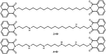

 求助内容:
求助内容: 应助结果提醒方式:


