Rituparna Sarkar, Daniel Darby, Sigolène Meilhac, Jean-Christophe Olivo-Marin
{"title":"3D cell morphology detection by association for embryo heart morphogenesis.","authors":"Rituparna Sarkar, Daniel Darby, Sigolène Meilhac, Jean-Christophe Olivo-Marin","doi":"10.1017/S2633903X22000022","DOIUrl":null,"url":null,"abstract":"<p><p>Advances in tissue engineering for cardiac regenerative medicine require cellular-level understanding of the mechanism of cardiac muscle growth during embryonic developmental stage. Computational methods to automatize cell segmentation in 3D and deliver accurate, quantitative morphology of cardiomyocytes, are imperative to provide insight into cell behavior underlying cardiac tissue growth. Detecting individual cells from volumetric images of dense tissue, poised with low signal-to-noise ratio and severe intensity in homogeneity, is a challenging task. In this article, we develop a robust segmentation tool capable of extracting cellular morphological parameters from 3D multifluorescence images of murine heart, captured via light-sheet microscopy. The proposed pipeline incorporates a neural network for 2D detection of nuclei and cell membranes. A graph-based global association employs the 2D nuclei detections to reconstruct 3D nuclei. A novel optimization embedding the network flow algorithm in an alternating direction method of multipliers is proposed to solve the global object association problem. The associated 3D nuclei serve as the initialization of an active mesh model to obtain the 3D segmentation of individual myocardial cells. The efficiency of our method over the state-of-the-art methods is observed via various qualitative and quantitative evaluation.</p>","PeriodicalId":72371,"journal":{"name":"Biological imaging","volume":" ","pages":"e2"},"PeriodicalIF":0.0000,"publicationDate":"2022-04-22","publicationTypes":"Journal Article","fieldsOfStudy":null,"isOpenAccess":false,"openAccessPdf":"https://www.ncbi.nlm.nih.gov/pmc/articles/PMC10951799/pdf/","citationCount":"0","resultStr":null,"platform":"Semanticscholar","paperid":null,"PeriodicalName":"Biological imaging","FirstCategoryId":"1085","ListUrlMain":"https://doi.org/10.1017/S2633903X22000022","RegionNum":0,"RegionCategory":null,"ArticlePicture":[],"TitleCN":null,"AbstractTextCN":null,"PMCID":null,"EPubDate":"2022/1/1 0:00:00","PubModel":"eCollection","JCR":"","JCRName":"","Score":null,"Total":0}
引用次数: 0
Abstract
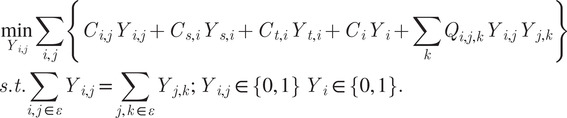
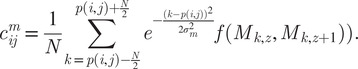
Advances in tissue engineering for cardiac regenerative medicine require cellular-level understanding of the mechanism of cardiac muscle growth during embryonic developmental stage. Computational methods to automatize cell segmentation in 3D and deliver accurate, quantitative morphology of cardiomyocytes, are imperative to provide insight into cell behavior underlying cardiac tissue growth. Detecting individual cells from volumetric images of dense tissue, poised with low signal-to-noise ratio and severe intensity in homogeneity, is a challenging task. In this article, we develop a robust segmentation tool capable of extracting cellular morphological parameters from 3D multifluorescence images of murine heart, captured via light-sheet microscopy. The proposed pipeline incorporates a neural network for 2D detection of nuclei and cell membranes. A graph-based global association employs the 2D nuclei detections to reconstruct 3D nuclei. A novel optimization embedding the network flow algorithm in an alternating direction method of multipliers is proposed to solve the global object association problem. The associated 3D nuclei serve as the initialization of an active mesh model to obtain the 3D segmentation of individual myocardial cells. The efficiency of our method over the state-of-the-art methods is observed via various qualitative and quantitative evaluation.


 求助内容:
求助内容: 应助结果提醒方式:


