Jessica Felton, Kunrong Cheng, Aaron C Shang, Shien Hu, Shannon M Larabee, Cinthia B Drachenberg, Jean-Pierre Raufman
{"title":"Two sides to colon cancer: mice mimic human anatomical region disparity in colon cancer development and progression.","authors":"Jessica Felton, Kunrong Cheng, Aaron C Shang, Shien Hu, Shannon M Larabee, Cinthia B Drachenberg, Jean-Pierre Raufman","doi":"10.20517/2394-4722.2018.39","DOIUrl":null,"url":null,"abstract":"<p><strong>Aim: </strong>Strong evidence reveals important differences between cancers in the proximal <i>vs</i>. distal colon. Animal models of metastatic colon cancer are available but with varying degrees of reproducibility and several important limitations. We explored whether there were regional differences in the location of murine colon cancers and assessed the utility of murine models to explore the biological basis for such differences.</p><p><strong>Methods: </strong>We re-analyzed data from our previous studies to assess the regional distribution of murine colon cancer. In survival surgery experiments, we injected HT-29 human colon cancer cells into the wall of the cecum or distal colon of Nu(NCr)-Foxn1<sup>nu</sup> or NOD.Cg-Prkdc<sup>scid</sup>Il2rg<sup>Tim1Wji</sup>/SzJ mice and compared the development of primary tumors and metastases.</p><p><strong>Results: </strong>Within 7-17 weeks after intramural cecal injection of HT-29 cells, eight mice failed to develop solid primary tumors or metastases. In contrast, within four weeks after cell injection into the distal colon, 13 mice developed metastases - 12 mice developed subcutaneous metastases; of these, four developed liver metastases and one developed both liver and lung metastases. One mouse developed liver metastases only. Histological examination confirmed these lesions were adenocarcinomas.</p><p><strong>Conclusion: </strong>Our findings reveal the preferential growth of murine colon neoplasia and invasive human orthotopic xenografts in the distal mouse colon. The new approach of injecting cells into the distal colon wall results in a pattern of colon cancer development that closely mimics the progression of metastatic colon cancer in humans. This novel model of colon neoplasia has great potential for exploring anatomical differences in colon cancer and testing novel therapeutics.</p>","PeriodicalId":15167,"journal":{"name":"Journal of Cancer Metastasis and Treatment","volume":"4 1","pages":""},"PeriodicalIF":1.0000,"publicationDate":"2018-01-01","publicationTypes":"Journal Article","fieldsOfStudy":null,"isOpenAccess":false,"openAccessPdf":"https://www.ncbi.nlm.nih.gov/pmc/articles/PMC6860924/pdf/","citationCount":"0","resultStr":null,"platform":"Semanticscholar","paperid":null,"PeriodicalName":"Journal of Cancer Metastasis and Treatment","FirstCategoryId":"3","ListUrlMain":"https://doi.org/10.20517/2394-4722.2018.39","RegionNum":0,"RegionCategory":null,"ArticlePicture":[],"TitleCN":null,"AbstractTextCN":null,"PMCID":null,"EPubDate":"2018/9/27 0:00:00","PubModel":"Epub","JCR":"Q4","JCRName":"ONCOLOGY","Score":null,"Total":0}
引用次数: 0
Abstract
Aim: Strong evidence reveals important differences between cancers in the proximal vs. distal colon. Animal models of metastatic colon cancer are available but with varying degrees of reproducibility and several important limitations. We explored whether there were regional differences in the location of murine colon cancers and assessed the utility of murine models to explore the biological basis for such differences.
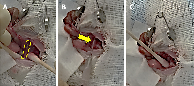
Methods: We re-analyzed data from our previous studies to assess the regional distribution of murine colon cancer. In survival surgery experiments, we injected HT-29 human colon cancer cells into the wall of the cecum or distal colon of Nu(NCr)-Foxn1nu or NOD.Cg-PrkdcscidIl2rgTim1Wji/SzJ mice and compared the development of primary tumors and metastases.
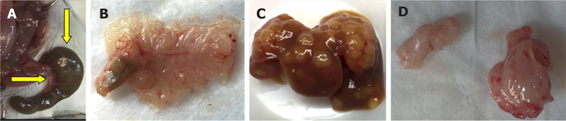
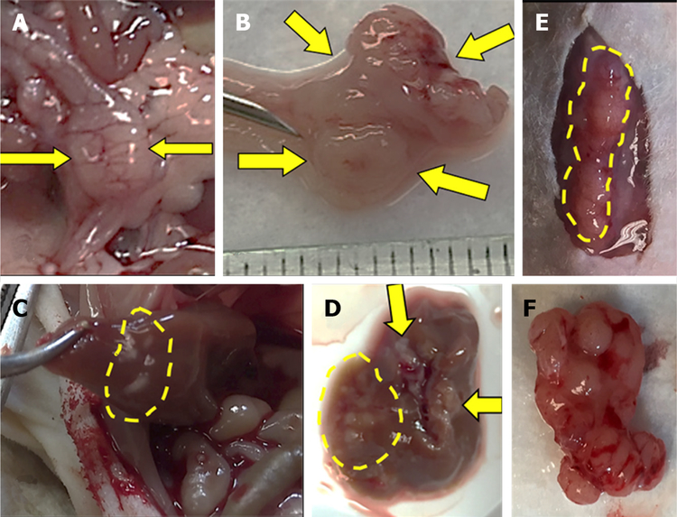
Results: Within 7-17 weeks after intramural cecal injection of HT-29 cells, eight mice failed to develop solid primary tumors or metastases. In contrast, within four weeks after cell injection into the distal colon, 13 mice developed metastases - 12 mice developed subcutaneous metastases; of these, four developed liver metastases and one developed both liver and lung metastases. One mouse developed liver metastases only. Histological examination confirmed these lesions were adenocarcinomas.
Conclusion: Our findings reveal the preferential growth of murine colon neoplasia and invasive human orthotopic xenografts in the distal mouse colon. The new approach of injecting cells into the distal colon wall results in a pattern of colon cancer development that closely mimics the progression of metastatic colon cancer in humans. This novel model of colon neoplasia has great potential for exploring anatomical differences in colon cancer and testing novel therapeutics.

 求助内容:
求助内容: 应助结果提醒方式:


