{"title":"Assessment of the developmental neurotoxicity of silver nanoparticles and silver ions with mouse embryonic stem cells in vitro","authors":"Nuoya Yin, Bowen Hu, Renjun Yang, Shaojun Liang, Shengxian Liang, Francesco Faiola","doi":"10.1002/jin2.49","DOIUrl":null,"url":null,"abstract":"<p>The wide applications of silver nanoparticles (AgNPs) have raised many concerns worldwide regarding their safety. The few previous studies on the developmental toxicity of AgNPs have been mostly dependent on animal experiments, which are time-consuming and costly. The rapid development of stem cell biology provides a new in vitro alternative to live animal assays for developmental toxicity studies. Here, we assessed the developmental neurotoxicity of AgNPs and Ag ions using a mouse embryonic stem cell (mESC) toxicology model. Our results showed that AgNP and Ag ion treatments did not affect mESC viability or cause accumulation of reactive oxygen species, at concentrations below 1 μg/mL. Conversely, AgNPs and Ag ions perturbed mESC global and neural progenitor cell-specific differentiation processes. In fact, both AgNPs and Ag ions induced the anomalous expression of neural ectoderm marker genes, such as <i>Sox1</i>, <i>Sox3</i>, <i>Map2</i>, <i>NeuroD</i>, <i>Nestin</i>, and <i>Pax6</i>, at concentrations lower than 0.1 μg/mL. Interestingly, AgNP effects manifested at earlier time points as compared with Ag ions. In addition, treatment with Ag ions generated neural progenitor cell abnormal morphology. Overall, our data proved that both AgNPs and Ag ions are toxicants, and their toxic effects are somehow different.</p>","PeriodicalId":91547,"journal":{"name":"Journal of interdisciplinary nanomedicine","volume":"3 3","pages":"133-145"},"PeriodicalIF":0.0000,"publicationDate":"2018-08-19","publicationTypes":"Journal Article","fieldsOfStudy":null,"isOpenAccess":false,"openAccessPdf":"https://sci-hub-pdf.com/10.1002/jin2.49","citationCount":"17","resultStr":null,"platform":"Semanticscholar","paperid":null,"PeriodicalName":"Journal of interdisciplinary nanomedicine","FirstCategoryId":"1085","ListUrlMain":"https://onlinelibrary.wiley.com/doi/10.1002/jin2.49","RegionNum":0,"RegionCategory":null,"ArticlePicture":[],"TitleCN":null,"AbstractTextCN":null,"PMCID":null,"EPubDate":"","PubModel":"","JCR":"","JCRName":"","Score":null,"Total":0}
引用次数: 17
Abstract
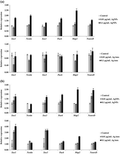
The wide applications of silver nanoparticles (AgNPs) have raised many concerns worldwide regarding their safety. The few previous studies on the developmental toxicity of AgNPs have been mostly dependent on animal experiments, which are time-consuming and costly. The rapid development of stem cell biology provides a new in vitro alternative to live animal assays for developmental toxicity studies. Here, we assessed the developmental neurotoxicity of AgNPs and Ag ions using a mouse embryonic stem cell (mESC) toxicology model. Our results showed that AgNP and Ag ion treatments did not affect mESC viability or cause accumulation of reactive oxygen species, at concentrations below 1 μg/mL. Conversely, AgNPs and Ag ions perturbed mESC global and neural progenitor cell-specific differentiation processes. In fact, both AgNPs and Ag ions induced the anomalous expression of neural ectoderm marker genes, such as Sox1, Sox3, Map2, NeuroD, Nestin, and Pax6, at concentrations lower than 0.1 μg/mL. Interestingly, AgNP effects manifested at earlier time points as compared with Ag ions. In addition, treatment with Ag ions generated neural progenitor cell abnormal morphology. Overall, our data proved that both AgNPs and Ag ions are toxicants, and their toxic effects are somehow different.

 求助内容:
求助内容: 应助结果提醒方式:


