{"title":"Mesocrystal aggregation of biological apatite nanocrystals","authors":"Abdulelah S. Alrebaish, Otto C. Wilson Jr","doi":"10.1002/mds3.10155","DOIUrl":null,"url":null,"abstract":"<p>A broader revelation of the mechanisms which contribute to the formation, growth, healing and remodelling of bone tissue is essential for advancing the design and development of biomaterials and devices which directly enhance bone health. Hydroxyapatite and associated calcium phosphate-based minerals play an essential role in bone tissue formation. Further insights into how biomineral crystals form, grow and integrate within bone tissue will provide key information to direct efforts in more comprehensive bone tissue engineering products and therapies. While previous studies proposed the aggregation of amorphous calcium phosphate clusters as a precursor to biological hydroxyapatite, the exact formation mechanism of the plate-like biological hydroxyapatite is still unclear. Here we report the analysis of high-resolution electron microscopy images of bone biomineral precipitated in a biological environment. We propose that 3 nm primary biologically synthesized (biosynthesized) hydroxyapatite (BHAp) single crystal units assemble and coalesce via an oriented aggregation mechanism to form larger (approximately 46 nm × 25 nm) plate-like biological hydroxyapatite mesocrystals. A better understanding of the biomineralization process can provide insights to improve the in vitro precipitation of bone biominerals with tailored properties and unique functionality. This will help to usher in the next generation of biobased biomaterials and devices to enhance the healing and remodelling of bone at the tissue, cell and subcellular level.</p>","PeriodicalId":87324,"journal":{"name":"Medical devices & sensors","volume":"4 1","pages":""},"PeriodicalIF":0.0000,"publicationDate":"2020-11-29","publicationTypes":"Journal Article","fieldsOfStudy":null,"isOpenAccess":false,"openAccessPdf":"https://sci-hub-pdf.com/10.1002/mds3.10155","citationCount":"3","resultStr":null,"platform":"Semanticscholar","paperid":null,"PeriodicalName":"Medical devices & sensors","FirstCategoryId":"1085","ListUrlMain":"https://onlinelibrary.wiley.com/doi/10.1002/mds3.10155","RegionNum":0,"RegionCategory":null,"ArticlePicture":[],"TitleCN":null,"AbstractTextCN":null,"PMCID":null,"EPubDate":"","PubModel":"","JCR":"","JCRName":"","Score":null,"Total":0}
引用次数: 3
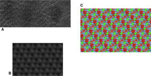
Abstract
A broader revelation of the mechanisms which contribute to the formation, growth, healing and remodelling of bone tissue is essential for advancing the design and development of biomaterials and devices which directly enhance bone health. Hydroxyapatite and associated calcium phosphate-based minerals play an essential role in bone tissue formation. Further insights into how biomineral crystals form, grow and integrate within bone tissue will provide key information to direct efforts in more comprehensive bone tissue engineering products and therapies. While previous studies proposed the aggregation of amorphous calcium phosphate clusters as a precursor to biological hydroxyapatite, the exact formation mechanism of the plate-like biological hydroxyapatite is still unclear. Here we report the analysis of high-resolution electron microscopy images of bone biomineral precipitated in a biological environment. We propose that 3 nm primary biologically synthesized (biosynthesized) hydroxyapatite (BHAp) single crystal units assemble and coalesce via an oriented aggregation mechanism to form larger (approximately 46 nm × 25 nm) plate-like biological hydroxyapatite mesocrystals. A better understanding of the biomineralization process can provide insights to improve the in vitro precipitation of bone biominerals with tailored properties and unique functionality. This will help to usher in the next generation of biobased biomaterials and devices to enhance the healing and remodelling of bone at the tissue, cell and subcellular level.

 求助内容:
求助内容: 应助结果提醒方式:


