Analysis of MRI radiomic features of hypoxic area in nasopharyngeal carcinoma patients
引用次数: 0
Abstract
Objective To analyze the magnetic resonance imaging (MRI) radiomic performance of hypoxic area in nasopharyngeal carcinoma patients, aiming to provide a reference for identification and analysis of hypoxic area. Methods The MRI-T1, MRI-T2, MRI-T1+ and PET/CT images of 32 patients initially diagnosed with nasopharyngeal carcinoma were retrospectively analyzed. The gross tumor volume (GTV) of nasopharynx was countoured and the hypoxic areas (GTV-H) were identified by 18F-FMISO-PET images. The non-hypoxic areas (GTV-NH) were defined as the rest of areas removed GTV-H from GTV. The radiomic features of GTV-H and GTV-NH were extracted and compared. Results The average volume of GTV-H and GTV-NH was (10.92±11.02) cm3 and (7.21±5.70) cm3, respectively. The maximum rate of change was 46% for intensity direct-global min (ID-GM) on MRI-T1(P 0.7 and Youden index>0.5). The average rate of change was 136% for long run emphasis (LRE), long run high gray level emphasis (LRHGLE) and long run low gray level emphasis (LRLGLE) on MRI-T2(P 0.7 and Youden index>0.5). The high change rates was greater than 90% on MRI-T1+ (P 0.7 and Youden index>0.5) for ID-GM, LRE, LRHGLE and LRLGLE. Conclusions The hypoxic area of tumor target can be reflected by MRI radiomics on T1/T2/T1+ . Quantifying and tracking the variations of these features can bring benefit to recognize the hypoxic area of nasopharyngeal carcinoma tumor target. Key words: Nasopharyngeal neoplasm hypoxic area; Radiomics; Hypoxic area identification鼻咽癌患者缺氧区MRI影像学特征分析
目的分析鼻咽癌患者缺氧区的磁共振成像(MRI)放射学表现,为缺氧区的识别和分析提供参考。方法回顾性分析32例初诊鼻咽癌患者的MRI-T1、MRI-T2、MRI-T1+及PET/CT影像。应用18F-FMISO-PET检测鼻咽部肿瘤总体积(GTV)及缺氧区(GTV- h)。非缺氧区(GTV- nh)定义为从GTV中去除GTV- h的其余区域。提取GTV-H和GTV-NH的放射学特征并进行比较。结果GTV-H和GTV-NH的平均体积分别为(10.92±11.02)cm3和(7.21±5.70)cm3。MRI-T1上强度直接全局最小值(ID-GM)的最大变化率为46% (P = 0.7,约登指数> = 0.5)。MRI-T2上长期灰度强调(LRE)、长期高灰度强调(LRHGLE)和长期低灰度强调(LRLGLE)的平均变化率为136% (P = 0.7,约登指数> = 0.5)。ID-GM、LRE、LRHGLE和LRLGLE的MRI-T1+高变化率均大于90% (P = 0.7,约登指数> = 0.5)。结论MRI放射组学可以在T1/T2/T1+上反映肿瘤靶区缺氧区域。量化和跟踪这些特征的变化,有助于识别鼻咽癌缺氧区肿瘤靶点。关键词:鼻咽肿瘤缺氧区;Radiomics;缺氧区识别
本文章由计算机程序翻译,如有差异,请以英文原文为准。
求助全文
约1分钟内获得全文
求助全文
来源期刊
自引率
0.00%
发文量
6375
期刊介绍:
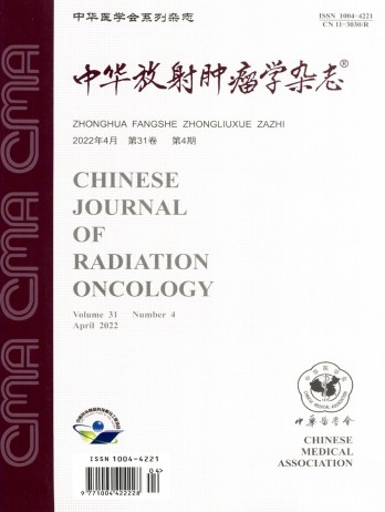
The Chinese Journal of Radiation Oncology is a national academic journal sponsored by the Chinese Medical Association. It was founded in 1992 and the title was written by Chen Minzhang, the former Minister of Health. Its predecessor was the Chinese Journal of Radiation Oncology, which was founded in 1987. The journal is an authoritative journal in the field of radiation oncology in my country. It focuses on clinical tumor radiotherapy, tumor radiation physics, tumor radiation biology, and thermal therapy. Its main readers are middle and senior clinical doctors and scientific researchers. It is now a monthly journal with a large 16-page format and 80 pages of text. For many years, it has adhered to the principle of combining theory with practice and combining improvement with popularization. It now has columns such as monographs, head and neck tumors (monographs), chest tumors (monographs), abdominal tumors (monographs), physics, technology, biology (monographs), reviews, and investigations and research.

 求助内容:
求助内容: 应助结果提醒方式:


