Gengxin Wang , Hongyi Yang , Juan Li , Jie Wen , Kai Zhong , Changlin Tian
{"title":"Overview and progress of X-nuclei magnetic resonance imaging in biomedical studies","authors":"Gengxin Wang , Hongyi Yang , Juan Li , Jie Wen , Kai Zhong , Changlin Tian","doi":"10.1016/j.mrl.2023.05.002","DOIUrl":null,"url":null,"abstract":"<div><p>Proton nuclear (<sup>1</sup>H) is the observed nucleus on which most magnetic resonance imaging (MRI) applications depend. Most traditional <sup>1</sup>H MRI can provide structural and functional information about organisms, while various non-proton nuclei (X-nuclei) MRI can provide more metabolic information. However, due to the relatively poor signal-to-noise ratio (SNR) of X-nuclei MRI, their applications are quite rare compared to <sup>1</sup>H. Benefit from the rapid developments of MRI hardware and software technologies, X-nuclei MRI has recently attracted increasing interests in biomedical research. This review firstly introduces some current methods to improve the SNR of X-nuclei MRI. Secondly, this review describes biomedical applications of X-nuclei MRI, especially focusing on the current use of X-nuclei (<sup>13</sup>C, <sup>17</sup>O, <sup>19</sup>F, <sup>23</sup>Na and <sup>31</sup>P) MRI to study related diseases in different organs, including the brain, liver, kidney, heart and bone. Finally, perspectives studies on X-nuclei imaging and its potential applications are described in biomedical research.</p></div>","PeriodicalId":93594,"journal":{"name":"Magnetic Resonance Letters","volume":"3 4","pages":"Pages 327-343"},"PeriodicalIF":0.0000,"publicationDate":"2023-11-01","publicationTypes":"Journal Article","fieldsOfStudy":null,"isOpenAccess":false,"openAccessPdf":"https://www.sciencedirect.com/science/article/pii/S2772516223000347/pdfft?md5=c189098f51e852f55ae6183d4e19ad08&pid=1-s2.0-S2772516223000347-main.pdf","citationCount":"1","resultStr":null,"platform":"Semanticscholar","paperid":null,"PeriodicalName":"Magnetic Resonance Letters","FirstCategoryId":"1085","ListUrlMain":"https://www.sciencedirect.com/science/article/pii/S2772516223000347","RegionNum":0,"RegionCategory":null,"ArticlePicture":[],"TitleCN":null,"AbstractTextCN":null,"PMCID":null,"EPubDate":"","PubModel":"","JCR":"","JCRName":"","Score":null,"Total":0}
引用次数: 1
Abstract
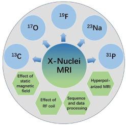
Proton nuclear (1H) is the observed nucleus on which most magnetic resonance imaging (MRI) applications depend. Most traditional 1H MRI can provide structural and functional information about organisms, while various non-proton nuclei (X-nuclei) MRI can provide more metabolic information. However, due to the relatively poor signal-to-noise ratio (SNR) of X-nuclei MRI, their applications are quite rare compared to 1H. Benefit from the rapid developments of MRI hardware and software technologies, X-nuclei MRI has recently attracted increasing interests in biomedical research. This review firstly introduces some current methods to improve the SNR of X-nuclei MRI. Secondly, this review describes biomedical applications of X-nuclei MRI, especially focusing on the current use of X-nuclei (13C, 17O, 19F, 23Na and 31P) MRI to study related diseases in different organs, including the brain, liver, kidney, heart and bone. Finally, perspectives studies on X-nuclei imaging and its potential applications are described in biomedical research.

 求助内容:
求助内容: 应助结果提醒方式:


