{"title":"A case of COVID-19 with pernio-like skin lesions and increased red blood cell distribution width","authors":"Satoko Minakawa MD, PhD, Yasushi Matsuzaki MD, PhD, Akari Terada MD, Shu Ogasawara PhD, Yuki Nishiya MD, PhD, Jin Irie MD, PhD, Yoshiya Ishizawa MD, PhD, Hiroyuki Hanada MD, PhD, Daisuke Sawamura MD, PhD","doi":"10.1002/cia2.12272","DOIUrl":null,"url":null,"abstract":"<p>Coronavirus disease 2019 (COVID-19) is related to cutaneous manifestations.<span><sup>1</sup></span> Previous reports indicate that pernio-like lesions are cutaneous manifestations of COVID-19.<span><sup>1, 2</sup></span> We report a case of pernio-like lesions related to COVID-19.</p><p>A 74-year-old Japanese woman with diabetes and hypertension was diagnosed with COVID-19. She was living with her daughter's family, who had also been diagnosed with COVID-19. On illness day 6, she presented to our emergency department with malaise and worsening shortness of breath (saturation percentage of oxygen in the artery blood 74%). The chest radiograph detected her bilateral interstitial pneumonia, and the treatment of artificial respiration started. Favipiravir, dexamethasone, remdesivir, and empiric antibiotic therapy were administrated because of a positive blood culture (<i>Corynebacterium striatum</i>, <i>Klebsiella</i> species). She was given lactulose, subcutaneous low molecular heparin, and continuous intravenous heparin infusion for prevent COVID-19-related thrombosis. On illness day 13, her breathing became worse. Extracorporeal membrane oxygenation was started. On illness day 25, erythema was seen on her body. On illness day 27, erythema and maculopapular exanthema appeared on her trunk (Figure 1A), legs (Figure 1B), and hands (Figure 1C). A hemorrhagic rash appeared on her hands (Figure 1D). Pernio-like lesions were seen on her fingers (Figure 1E,F) and toes. Laboratory inspection clarified a normal white blood cell count (5860/mm), normal liver and kidney function, decreased hemoglobin (10.1 g/dl; normal range: 10.6–14.4 g/dl), hematocrit (31.5%; normal range: 32.1%–42.7%), platelet count (45,000/mm; normal range: 13,800–309,000/mm), increased C-reactive protein (CRP, 10.56 mg/dl; normal range <0.3 mg/dl), lactate dehydrogenase (LD, 346 U/L; normal range: 119–229 U/L), fibrinogen-fibrin degradation product (FDP, 113.8 μg/ml; normal range <5.0 μg/ml), dimerized plasmin fragment D (D-dimer, 55.5 μg/ml; normal range <1.0 μg/ml), and red blood cell distribution width (RDW, 16.2%; normal range 11.5%–13.8%). Over the following days, gradual spontaneous improvement of skin lesions occurred. However, she became the cerebral infarction and died on illness day 44.</p><p>Previous studies found that 318 of 505 (63%) patients with dermatologic symptoms related to COVID-19 had pernio-like lesions.<span><sup>3</sup></span> Pernio-like lesions were sole symptoms in the 55% of patients.<span><sup>3</sup></span> Retiform purpura presented entirely in inpatients.<span><sup>1</sup></span> Mechanisms of COVID-19 coagulopathy have been proposed. In a hypercoagulable situation, the crosstalk between the inflammatory and hemostatic systems may advance for thrombosis in COVID-19.<span><sup>4</sup></span> Heparin resistance was observed in patients with COVID-19 infectious disease of the serious case.<span><sup>4</sup></span></p><p>Elevated RDW is associated with an increased risk for mortality from heart disease, pulmonary disease, sepsis, influenza, and cancer.<span><sup>5</sup></span> RDW related to the death rate in Cox proportional hazards models adjusted for age, D-dimer level, and common comorbidities such as diabetes and hypertension.<span><sup>5</sup></span> Patients whose RDW increased during hospitalization had higher mortality compared with those whose RDW did not change.<span><sup>5</sup></span></p><p>In our case, skin manifestations of sepsis with DIC or heparin-induced thrombocytopenia need to be considered. However, cutaneous manifestations, RDW, and levels of LD and D-dimer might be useful biomarkers to improve the efficiency of information input work related to symptoms of the determination of triage of patients with COVID-19.</p><p>The authors declare no conflicts of interest.</p><p>Approval of the research protocol: This study was approved by the Committee of Medical Ethics of Hirosaki University Graduate School of Medicine in Aomori, Japan. The patient was registrated to the trial was registered in the University Hospital Medical Information Network (UMIN) (Reservation No. UMIN000041301) by approved by the Committee of Medical Ethics of Hirosaki University Graduate School of Medicine in Aomori, Japan.</p><p>Informed Consent: Written informed consent was obtained from the patient's husband.</p><p>Registry and the Registration No. of the study/trial: 21,928.</p><p>Animal Studies: N/A.</p>","PeriodicalId":15543,"journal":{"name":"Journal of Cutaneous Immunology and Allergy","volume":"6 2","pages":"55-56"},"PeriodicalIF":0.9000,"publicationDate":"2022-08-15","publicationTypes":"Journal Article","fieldsOfStudy":null,"isOpenAccess":false,"openAccessPdf":"https://onlinelibrary.wiley.com/doi/epdf/10.1002/cia2.12272","citationCount":"0","resultStr":null,"platform":"Semanticscholar","paperid":null,"PeriodicalName":"Journal of Cutaneous Immunology and Allergy","FirstCategoryId":"1085","ListUrlMain":"https://onlinelibrary.wiley.com/doi/10.1002/cia2.12272","RegionNum":0,"RegionCategory":null,"ArticlePicture":[],"TitleCN":null,"AbstractTextCN":null,"PMCID":null,"EPubDate":"","PubModel":"","JCR":"Q4","JCRName":"ALLERGY","Score":null,"Total":0}
引用次数: 0
Abstract
Coronavirus disease 2019 (COVID-19) is related to cutaneous manifestations.1 Previous reports indicate that pernio-like lesions are cutaneous manifestations of COVID-19.1, 2 We report a case of pernio-like lesions related to COVID-19.
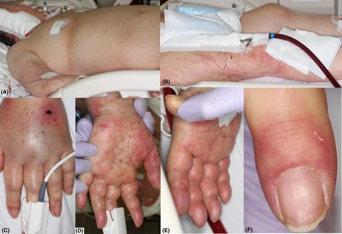
A 74-year-old Japanese woman with diabetes and hypertension was diagnosed with COVID-19. She was living with her daughter's family, who had also been diagnosed with COVID-19. On illness day 6, she presented to our emergency department with malaise and worsening shortness of breath (saturation percentage of oxygen in the artery blood 74%). The chest radiograph detected her bilateral interstitial pneumonia, and the treatment of artificial respiration started. Favipiravir, dexamethasone, remdesivir, and empiric antibiotic therapy were administrated because of a positive blood culture (Corynebacterium striatum, Klebsiella species). She was given lactulose, subcutaneous low molecular heparin, and continuous intravenous heparin infusion for prevent COVID-19-related thrombosis. On illness day 13, her breathing became worse. Extracorporeal membrane oxygenation was started. On illness day 25, erythema was seen on her body. On illness day 27, erythema and maculopapular exanthema appeared on her trunk (Figure 1A), legs (Figure 1B), and hands (Figure 1C). A hemorrhagic rash appeared on her hands (Figure 1D). Pernio-like lesions were seen on her fingers (Figure 1E,F) and toes. Laboratory inspection clarified a normal white blood cell count (5860/mm), normal liver and kidney function, decreased hemoglobin (10.1 g/dl; normal range: 10.6–14.4 g/dl), hematocrit (31.5%; normal range: 32.1%–42.7%), platelet count (45,000/mm; normal range: 13,800–309,000/mm), increased C-reactive protein (CRP, 10.56 mg/dl; normal range <0.3 mg/dl), lactate dehydrogenase (LD, 346 U/L; normal range: 119–229 U/L), fibrinogen-fibrin degradation product (FDP, 113.8 μg/ml; normal range <5.0 μg/ml), dimerized plasmin fragment D (D-dimer, 55.5 μg/ml; normal range <1.0 μg/ml), and red blood cell distribution width (RDW, 16.2%; normal range 11.5%–13.8%). Over the following days, gradual spontaneous improvement of skin lesions occurred. However, she became the cerebral infarction and died on illness day 44.
Previous studies found that 318 of 505 (63%) patients with dermatologic symptoms related to COVID-19 had pernio-like lesions.3 Pernio-like lesions were sole symptoms in the 55% of patients.3 Retiform purpura presented entirely in inpatients.1 Mechanisms of COVID-19 coagulopathy have been proposed. In a hypercoagulable situation, the crosstalk between the inflammatory and hemostatic systems may advance for thrombosis in COVID-19.4 Heparin resistance was observed in patients with COVID-19 infectious disease of the serious case.4
Elevated RDW is associated with an increased risk for mortality from heart disease, pulmonary disease, sepsis, influenza, and cancer.5 RDW related to the death rate in Cox proportional hazards models adjusted for age, D-dimer level, and common comorbidities such as diabetes and hypertension.5 Patients whose RDW increased during hospitalization had higher mortality compared with those whose RDW did not change.5
In our case, skin manifestations of sepsis with DIC or heparin-induced thrombocytopenia need to be considered. However, cutaneous manifestations, RDW, and levels of LD and D-dimer might be useful biomarkers to improve the efficiency of information input work related to symptoms of the determination of triage of patients with COVID-19.
The authors declare no conflicts of interest.
Approval of the research protocol: This study was approved by the Committee of Medical Ethics of Hirosaki University Graduate School of Medicine in Aomori, Japan. The patient was registrated to the trial was registered in the University Hospital Medical Information Network (UMIN) (Reservation No. UMIN000041301) by approved by the Committee of Medical Ethics of Hirosaki University Graduate School of Medicine in Aomori, Japan.
Informed Consent: Written informed consent was obtained from the patient's husband.
Registry and the Registration No. of the study/trial: 21,928.

 求助内容:
求助内容: 应助结果提醒方式:


