{"title":"Recurrent pregnancy-associated erythema annulare centrifugum in a single gestation period","authors":"Kie Imura MD, Yurika Masuda MD, Yuko Sano MD, Hiroaki Yagi MD, PhD","doi":"10.1002/cia2.12266","DOIUrl":null,"url":null,"abstract":"<p>Erythema annulare centrifugum (EAC) is reportedly related to many factors such as immunological disorders, infections, malignancies, foods, and drugs. Pregnancy-associated EAC is extremely rare, with only six cases reported in the literature.<span><sup>1-6</sup></span> To date, there have been no reports of EAC that disappeared and recurred during a single gestation period.</p><p>A 34-year-old woman visited our department in the 36th week of her first pregnancy with asymptomatic multiple annular erythematous lesions on the legs. On the examination, the erythematous areas were slightly raised and had slight scales (Figure 1A, B). She reported that the eruption had first appeared in week 10 and spontaneously disappeared within 2 months, then reappeared in week 28 and gradually enlarged centrifugally and increased in number. This was confirmed in serial photographs she had taken. Skin biopsy revealed hyperkeratosis, parakeratosis, mild spongiosis in the epidermis, and a superficial perivascular lymphohistiocytic infiltrate (Figure 1C). She was in good health and had not taken any medication or vaccination during the pregnancy. She had a good pregnancy course except that the fetus was in the breech position. The laboratory test results were normal, including complete blood count, renal and hepatic functions indices, immunoglobulins, antinuclear antibodies, anti-SS-A and anti-SS-B antibodies, and rheumatoid factors. She had no evidence of Sjögren syndrome. She delivered a healthy baby by elective cesarean section in week 38. Although all the erythematous lesions had been increasing in number and enlarging until the delivery, they started to disappear within 2 days after the delivery and wholly resolved within 1 month without any treatment. No recurrence was observed during 4 years of follow-up.</p><p>In all six reported cases of pregnancy-associated EAC, including ours, eruptions appeared during the first pregnancy. The onset of the lesions occurred in week 12 in two cases and weeks 26–33 in four.<span><sup>1-6</sup></span> The lesions improved in weeks 33–36 in two cases, and at 3 days to 1 month postpartum in four. These findings suggest that the change in maternal hormone levels during pregnancy is a crucial etiological factor.<span><sup>5</sup></span> The major possible causative candidates include estrogen, progesterone, and human chorionic gonadotropin (hCG).<span><sup>6</sup></span> The secretion of hCG differs from that of the other hormones and many other placental proteins. hCG is first detected in maternal serum within 9 days after conception. The hCG levels then rise in a logarithmic fashion, peaking at 8–10 weeks after the last menstrual period, followed by a decline to a nadir at 18 weeks, with subsequent levels remaining constant or slightly increasing in some individuals until delivery.<span><sup>7, 8</sup></span> The concentrations of estrogen and progesterone gradually increase and are maintained at high concentrations until delivery.<span><sup>8</sup></span> The fact that the initial lesions appeared during early pregnancy, spontaneously disappeared, and then recurred in a single gestation period in our case may suggest that hCG is the most promising causative candidate in our case. However, it is likely that unknown cofactors will exist because EAC never develops in the second pregnancy. EAC during pregnancy resolves when the hormones return to prepregnancy concentrations.</p><p><span>The authors declare no conflict of interest</span>.</p><p>Approval of research protocol: Not applicable.</p><p>Informed Consent: The patient provided informed consent to publish his photographs and case details.</p><p>Registry and the Registration No. of the study/trial: Not applicable.</p><p>Animal Studies: Not applicable.</p>","PeriodicalId":15543,"journal":{"name":"Journal of Cutaneous Immunology and Allergy","volume":"6 1","pages":"28-29"},"PeriodicalIF":0.9000,"publicationDate":"2022-07-06","publicationTypes":"Journal Article","fieldsOfStudy":null,"isOpenAccess":false,"openAccessPdf":"https://onlinelibrary.wiley.com/doi/epdf/10.1002/cia2.12266","citationCount":"0","resultStr":null,"platform":"Semanticscholar","paperid":null,"PeriodicalName":"Journal of Cutaneous Immunology and Allergy","FirstCategoryId":"1085","ListUrlMain":"https://onlinelibrary.wiley.com/doi/10.1002/cia2.12266","RegionNum":0,"RegionCategory":null,"ArticlePicture":[],"TitleCN":null,"AbstractTextCN":null,"PMCID":null,"EPubDate":"","PubModel":"","JCR":"Q4","JCRName":"ALLERGY","Score":null,"Total":0}
引用次数: 0
Abstract
Erythema annulare centrifugum (EAC) is reportedly related to many factors such as immunological disorders, infections, malignancies, foods, and drugs. Pregnancy-associated EAC is extremely rare, with only six cases reported in the literature.1-6 To date, there have been no reports of EAC that disappeared and recurred during a single gestation period.
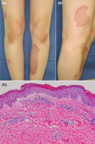
A 34-year-old woman visited our department in the 36th week of her first pregnancy with asymptomatic multiple annular erythematous lesions on the legs. On the examination, the erythematous areas were slightly raised and had slight scales (Figure 1A, B). She reported that the eruption had first appeared in week 10 and spontaneously disappeared within 2 months, then reappeared in week 28 and gradually enlarged centrifugally and increased in number. This was confirmed in serial photographs she had taken. Skin biopsy revealed hyperkeratosis, parakeratosis, mild spongiosis in the epidermis, and a superficial perivascular lymphohistiocytic infiltrate (Figure 1C). She was in good health and had not taken any medication or vaccination during the pregnancy. She had a good pregnancy course except that the fetus was in the breech position. The laboratory test results were normal, including complete blood count, renal and hepatic functions indices, immunoglobulins, antinuclear antibodies, anti-SS-A and anti-SS-B antibodies, and rheumatoid factors. She had no evidence of Sjögren syndrome. She delivered a healthy baby by elective cesarean section in week 38. Although all the erythematous lesions had been increasing in number and enlarging until the delivery, they started to disappear within 2 days after the delivery and wholly resolved within 1 month without any treatment. No recurrence was observed during 4 years of follow-up.
In all six reported cases of pregnancy-associated EAC, including ours, eruptions appeared during the first pregnancy. The onset of the lesions occurred in week 12 in two cases and weeks 26–33 in four.1-6 The lesions improved in weeks 33–36 in two cases, and at 3 days to 1 month postpartum in four. These findings suggest that the change in maternal hormone levels during pregnancy is a crucial etiological factor.5 The major possible causative candidates include estrogen, progesterone, and human chorionic gonadotropin (hCG).6 The secretion of hCG differs from that of the other hormones and many other placental proteins. hCG is first detected in maternal serum within 9 days after conception. The hCG levels then rise in a logarithmic fashion, peaking at 8–10 weeks after the last menstrual period, followed by a decline to a nadir at 18 weeks, with subsequent levels remaining constant or slightly increasing in some individuals until delivery.7, 8 The concentrations of estrogen and progesterone gradually increase and are maintained at high concentrations until delivery.8 The fact that the initial lesions appeared during early pregnancy, spontaneously disappeared, and then recurred in a single gestation period in our case may suggest that hCG is the most promising causative candidate in our case. However, it is likely that unknown cofactors will exist because EAC never develops in the second pregnancy. EAC during pregnancy resolves when the hormones return to prepregnancy concentrations.
The authors declare no conflict of interest.
Approval of research protocol: Not applicable.
Informed Consent: The patient provided informed consent to publish his photographs and case details.
Registry and the Registration No. of the study/trial: Not applicable.
据报道,环状离心性红斑(EAC)与许多因素有关,如免疫紊乱、感染、恶性肿瘤、食物和药物。妊娠相关EAC极为罕见,文献中仅报道了6例。1-6迄今为止,还没有关于EAC在单个妊娠期内消失和复发的报告。一位34岁的女性在她第一次怀孕的第36周来我科就诊,她腿部出现了无症状的多发性环状红斑。检查时,红斑区域轻微隆起,有轻微鳞屑(图1A、B)。她报告说,火山喷发最初出现在第10周,在2个月内自发消失,然后在第28周再次出现,并逐渐离心扩大,数量增加。她拍摄的一系列照片证实了这一点。皮肤活检显示表皮角化过度、角化不全、轻度海绵状血管病和浅表血管周围淋巴组织细胞浸润(图1C)。她健康状况良好,在怀孕期间没有服用任何药物或接种任何疫苗。除了胎儿处于臀位外,她的妊娠过程很顺利。实验室检测结果正常,包括全血细胞计数、肾和肝功能指数、免疫球蛋白、抗核抗体、抗SSA和抗SSB抗体以及类风湿因子。她没有干燥综合征的证据。她在第38周通过选择性剖宫产产术产下了一个健康的婴儿。尽管所有红斑病变在分娩前数量一直在增加和扩大,但在分娩后2天内开始消失,并在1个月内完全消退,无需任何治疗。在4年的随访中没有观察到复发。在所有六例报告的妊娠相关EAC病例中,包括我们的病例,皮疹出现在第一次妊娠期间。2例在第12周出现病变,4例在第26-33周出现病变。1-6 2例在33-36周出现病变改善,4例产后3天至1个月出现病变改善。这些发现表明,妊娠期间母体激素水平的变化是一个关键的病因。5可能的主要病因包括F I G U R E 1临床和组织病理学特征。(A,B)首次妊娠第36周出现无症状的多发性环状红斑病变。(C) 红斑病变的组织病理学表现为表皮角化过度、角化不全、轻度海绵状血管病和真皮浅表血管周淋巴组织细胞浸润(a)(B)

 求助内容:
求助内容: 应助结果提醒方式:


