Effects of dexmedetomidine on renal fibrosis in a mouse model of renal ischemia-reperfusion: the role of Akt

Q4 Medicine
Weimin Li, June Zhou, Yi-Hung Chen, Wanxian Lyu, Han-bing Wang, Zhihui Yang, J. Zhong
{"title":"Effects of dexmedetomidine on renal fibrosis in a mouse model of renal ischemia-reperfusion: the role of Akt","authors":"Weimin Li, June Zhou, Yi-Hung Chen, Wanxian Lyu, Han-bing Wang, Zhihui Yang, J. Zhong","doi":"10.3760/CMA.J.ISSN.0254-1416.2019.09.010","DOIUrl":null,"url":null,"abstract":"Objective \nTo evaluate the effect of dexmedetomidine on renal fibrosis in a mouse model of renal ischemia-reperfusion (I/R) and the role of serine-threonine kinase (Akt). \n \n \nMethods \nSixty male C57BL/6 mice, aged 8 weeks, weighing 20-25 g, were divided into 5 groups (n=12 each) using a random number table method: sham operation group (S group), renal I/R group (I/R group), renal I/R plus dexmedetomidine group (I/R + D group), renal I/R plus dexmedetomidine plus Akt agonist SC79 group (I/R + D + SC group), and renal I/R plus dexmedetomidine plus normal saline group (I/R+ D+ NS group). Renal I/R injury model was established by clamping the bilateral renal pedicle for 30 min followed by reperfusion.Dexmedetomidine was intraperitoneally injected at 30 min before surgery in I/R+ D, I/R+ D+ SC and I/R+ D+ NS groups.SC79 was intraperitoneally injected as a bolus of 0.04 mg/kg at 1 min of reperfusion, followed by an intraperitoneal injection of the same dose every 24 h until day 7.The serum blood urea nitrogen (BUN) and Scr concentrations were detected at 24 h of reperfusion.Renal tissues were taken, and the damage to the renal tubules was scored.Renal tissues were removed at 14 days of reperfusion to detect the degree of renal fibrosis and expression of collagen 1 (COL1), fibronectin (FN), and α-smooth actin (α-SMA) (by immunofluorescence and Western blot). The expression of phosphorylated Akt (p-Akt) in renal tissues was determined by Western blot at 24 h and 14 day of reperfusion. \n \n \nResults \nCompared with group S, the serum BUN and Scr concentrations, renal tubule damage score and degree of renal fibrosis were significantly increased, and the expression of COL1, FN, α-SMA and p-Akt was up-regulated in group I/R (P<0.05). Compared with I/R group, the serum BUN and Scr concentrations, renal tubular damage score and degree of renal fibrosis were significantly decreased, and the expression of COL1, FN, α-SMA and p-Akt was down-regulated in I/R+ D and I/R+ D+ NS groups (P<0.05). Compared with I/R+ D group, the serum BUN and Scr concentrations, renal tubule damage score and degree of renal fibrosis were significantly increased , and the expression of COL1, FN, α-SMA and p-Akt was up-regulated in I/R+ D+ SC group (P<0.05). \n \n \nConclusion \nDexmedetomidine can reduce the degree of renal fibrosis in a mouse model of renal I/R and the mechanism is related to inhibiting activation of Akt. \n \n \nKey words: \nDexmedetomidine; Fibrosis; Kidney; Reperfusion injury; Protein-serine-threonine kinases","PeriodicalId":10053,"journal":{"name":"中华麻醉学杂志","volume":"39 1","pages":"1062-1066"},"PeriodicalIF":0.0000,"publicationDate":"2019-09-20","publicationTypes":"Journal Article","fieldsOfStudy":null,"isOpenAccess":false,"openAccessPdf":"","citationCount":"0","resultStr":null,"platform":"Semanticscholar","paperid":null,"PeriodicalName":"中华麻醉学杂志","FirstCategoryId":"3","ListUrlMain":"https://doi.org/10.3760/CMA.J.ISSN.0254-1416.2019.09.010","RegionNum":0,"RegionCategory":null,"ArticlePicture":[],"TitleCN":null,"AbstractTextCN":null,"PMCID":null,"EPubDate":"","PubModel":"","JCR":"Q4","JCRName":"Medicine","Score":null,"Total":0}
引用次数: 0

Abstract

Objective To evaluate the effect of dexmedetomidine on renal fibrosis in a mouse model of renal ischemia-reperfusion (I/R) and the role of serine-threonine kinase (Akt). Methods Sixty male C57BL/6 mice, aged 8 weeks, weighing 20-25 g, were divided into 5 groups (n=12 each) using a random number table method: sham operation group (S group), renal I/R group (I/R group), renal I/R plus dexmedetomidine group (I/R + D group), renal I/R plus dexmedetomidine plus Akt agonist SC79 group (I/R + D + SC group), and renal I/R plus dexmedetomidine plus normal saline group (I/R+ D+ NS group). Renal I/R injury model was established by clamping the bilateral renal pedicle for 30 min followed by reperfusion.Dexmedetomidine was intraperitoneally injected at 30 min before surgery in I/R+ D, I/R+ D+ SC and I/R+ D+ NS groups.SC79 was intraperitoneally injected as a bolus of 0.04 mg/kg at 1 min of reperfusion, followed by an intraperitoneal injection of the same dose every 24 h until day 7.The serum blood urea nitrogen (BUN) and Scr concentrations were detected at 24 h of reperfusion.Renal tissues were taken, and the damage to the renal tubules was scored.Renal tissues were removed at 14 days of reperfusion to detect the degree of renal fibrosis and expression of collagen 1 (COL1), fibronectin (FN), and α-smooth actin (α-SMA) (by immunofluorescence and Western blot). The expression of phosphorylated Akt (p-Akt) in renal tissues was determined by Western blot at 24 h and 14 day of reperfusion. Results Compared with group S, the serum BUN and Scr concentrations, renal tubule damage score and degree of renal fibrosis were significantly increased, and the expression of COL1, FN, α-SMA and p-Akt was up-regulated in group I/R (P<0.05). Compared with I/R group, the serum BUN and Scr concentrations, renal tubular damage score and degree of renal fibrosis were significantly decreased, and the expression of COL1, FN, α-SMA and p-Akt was down-regulated in I/R+ D and I/R+ D+ NS groups (P<0.05). Compared with I/R+ D group, the serum BUN and Scr concentrations, renal tubule damage score and degree of renal fibrosis were significantly increased , and the expression of COL1, FN, α-SMA and p-Akt was up-regulated in I/R+ D+ SC group (P<0.05). Conclusion Dexmedetomidine can reduce the degree of renal fibrosis in a mouse model of renal I/R and the mechanism is related to inhibiting activation of Akt. Key words: Dexmedetomidine; Fibrosis; Kidney; Reperfusion injury; Protein-serine-threonine kinases
右美托咪定对肾缺血再灌注模型小鼠肾纤维化的影响:Akt的作用
目的评价右美托咪定对小鼠肾缺血再灌注(I/R)模型肾纤维化的影响及丝氨酸-苏氨酸激酶(Akt)的作用。方法60只雄性C57BL/6小鼠,年龄8周,体重20~25g,采用随机数表法分为5组(每组12只):假手术组(S组)、肾I/R组(I/R组)、肾脏I/R加右美托咪定组(I/R+D组)、,肾I/R加右美托咪定加生理盐水组(I/R+D+NS组)。夹闭双侧肾蒂30min后再灌注,建立肾I/R损伤模型。I/R+D组、I/R+D+SC组和I/R++D+NS组术前30min腹膜内注射右美托咪定。在再灌注1分钟腹膜内注射SC79,剂量为0.04 mg/kg,然后每24小时腹膜内注射相同剂量,直到第7天。在再灌注24小时检测血清尿素氮(BUN)和Scr浓度。取肾组织,对肾小管损伤进行评分。在再灌注14天时切除肾组织,以检测肾纤维化程度以及胶原1(COL1)、纤连蛋白(FN)和α-光滑肌动蛋白(α-SMA)的表达(通过免疫荧光和蛋白质印迹)。在再灌注24小时和14天,通过蛋白质印迹测定肾组织中磷酸化Akt(p-Akt)的表达。结果与S组相比,I/R组血清BUN和Scr浓度、肾小管损伤评分和肾纤维化程度显著升高,COL1、FN、α-SMA和p-Akt表达上调(p<0.05),I/R+D组和I/R+D+NS组COL1、FN、α-SMA和p-Akt表达下降(p<0.05),I/R+D+SC组α-SMA和p-Akt表达上调(p<0.05)。关键词:右美托咪定;纤维化;肾脏;再灌注损伤;蛋白质丝氨酸-苏氨酸激酶
本文章由计算机程序翻译,如有差异,请以英文原文为准。
求助全文
约1分钟内获得全文 求助全文
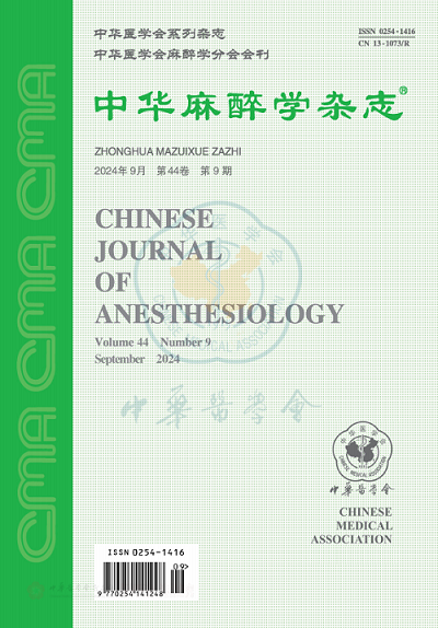
来源期刊
中华麻醉学杂志
中华麻醉学杂志 Medicine-Anesthesiology and Pain Medicine
CiteScore
0.10
自引率
0.00%
发文量
11211
期刊介绍:
×
引用
GB/T 7714-2015
复制
MLA
复制
APA
复制
导出至
BibTeX EndNote RefMan NoteFirst NoteExpress
×
提示
您的信息不完整,为了账户安全,请先补充。
现在去补充
×
提示
您因"违规操作"
具体请查看互助需知
我知道了
×
提示
确定
请完成安全验证×
copy
已复制链接
快去分享给好友吧!
我知道了
右上角分享
点击右上角分享
0
联系我们:info@booksci.cn Book学术提供免费学术资源搜索服务,方便国内外学者检索中英文文献。致力于提供最便捷和优质的服务体验。 Copyright © 2023 布克学术 All rights reserved.
京ICP备2023020795号-1
ghs 京公网安备 11010802042870号
Book学术文献互助
Book学术文献互助群
群 号:481959085
Book学术官方微信