Brain structural plasticity in rats subjected to early binocular enucleation characterized by high resolution anatomical magnetic resonance imaging and diffusion tensor imaging
{"title":"Brain structural plasticity in rats subjected to early binocular enucleation characterized by high resolution anatomical magnetic resonance imaging and diffusion tensor imaging","authors":"Xuxia Wang, Fuchun Lin, Yan Kang, Hao Lei","doi":"10.1016/j.mrl.2022.10.001","DOIUrl":null,"url":null,"abstract":"<div><p>Visual deprivation leads to structural neuroplasticity in the blind subjects, including gray matter (GM) and white matter (WM) atrophy and alterations in structural connectivity. The rat model of binocular enucleation (BE) is a frequently used animal model for studying brain plasticity induced by early blindness. Yet few neuroimaging studies have been performed on this model to investigate whether or not the BE rats have image phenotypes similar to or comparable to, those observed in the early blind subjects. The current study aimed to assess brain structural plasticity in BE rats using anatomical magnetic resonance imaging (MRI) and diffusion tensor imaging (DTI). The results demonstrated that early BE at postnatal day 4 (P4) caused almost complete degeneration of optic nerve (ON) and optic chiasma (OCH), atrophy in a number of visual and non-visual structures, including optic tract (OT), dorsal lateral geniculate nucleus (DLG) and corpus callosum (CC). The BE rats also exhibited impairments of WM microstructural integrity in the OT, and reduction of structural connectivity between the normal-appearing visual cortex (VC) and somatosensory/motor cortices at 4 months of age, likely as manifestations of deafferentation-induced maldevelopment. The structural neuroplasticity in BE rats observable to structural MRI parallels largely with what has been reported in blind subjects, suggesting that longitudinal neuroimaging studies on animal models of sensory deprivation can provide insights into how the brain changes its wiring and function during development/adaption in response to the lack of sensory stimuli.</p></div>","PeriodicalId":93594,"journal":{"name":"Magnetic Resonance Letters","volume":"3 1","pages":"Pages 14-21"},"PeriodicalIF":0.0000,"publicationDate":"2023-02-01","publicationTypes":"Journal Article","fieldsOfStudy":null,"isOpenAccess":false,"openAccessPdf":"","citationCount":"0","resultStr":null,"platform":"Semanticscholar","paperid":null,"PeriodicalName":"Magnetic Resonance Letters","FirstCategoryId":"1085","ListUrlMain":"https://www.sciencedirect.com/science/article/pii/S2772516222000511","RegionNum":0,"RegionCategory":null,"ArticlePicture":[],"TitleCN":null,"AbstractTextCN":null,"PMCID":null,"EPubDate":"","PubModel":"","JCR":"","JCRName":"","Score":null,"Total":0}
引用次数: 0
Abstract
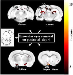
Visual deprivation leads to structural neuroplasticity in the blind subjects, including gray matter (GM) and white matter (WM) atrophy and alterations in structural connectivity. The rat model of binocular enucleation (BE) is a frequently used animal model for studying brain plasticity induced by early blindness. Yet few neuroimaging studies have been performed on this model to investigate whether or not the BE rats have image phenotypes similar to or comparable to, those observed in the early blind subjects. The current study aimed to assess brain structural plasticity in BE rats using anatomical magnetic resonance imaging (MRI) and diffusion tensor imaging (DTI). The results demonstrated that early BE at postnatal day 4 (P4) caused almost complete degeneration of optic nerve (ON) and optic chiasma (OCH), atrophy in a number of visual and non-visual structures, including optic tract (OT), dorsal lateral geniculate nucleus (DLG) and corpus callosum (CC). The BE rats also exhibited impairments of WM microstructural integrity in the OT, and reduction of structural connectivity between the normal-appearing visual cortex (VC) and somatosensory/motor cortices at 4 months of age, likely as manifestations of deafferentation-induced maldevelopment. The structural neuroplasticity in BE rats observable to structural MRI parallels largely with what has been reported in blind subjects, suggesting that longitudinal neuroimaging studies on animal models of sensory deprivation can provide insights into how the brain changes its wiring and function during development/adaption in response to the lack of sensory stimuli.

 求助内容:
求助内容: 应助结果提醒方式:


