Christa L LiBrizzi, Ashish Vankara, Christian F Meyer, Adam S Levin, Carol D Morris
{"title":"Bone Metastases in Patients with Leiomyosarcoma: A Retrospective Analysis of Survival and Surgical Management.","authors":"Christa L LiBrizzi, Ashish Vankara, Christian F Meyer, Adam S Levin, Carol D Morris","doi":"10.1155/2022/6806932","DOIUrl":null,"url":null,"abstract":"<p><strong>Background: </strong>Leiomyosarcomas (LMS) are malignancies with smooth muscle differentiation. Metastasis to the bone is not uncommon. The literature on the clinical course and management of such metastases is limited. Our study describes the clinical course of LMS to the bone, including survival rates, prognostic factors, and surgical management.</p><p><strong>Methods: </strong>We retrospectively reviewed 396 LMS patients presenting at an academic center between 1995 and 2020. We included LMS patients diagnosed with bone metastases and excluded patients with primary LMS of bone. We evaluated survival time with the Kaplan-Meier survival method and used Cox's proportional hazards regression analysis to determine factors associated with survival.</p><p><strong>Results: </strong>Forty-five patients with LMS (11%) had bone metastases. The most common LMS subtypes with bone metastases were uterine (<i>N</i> = 18, 40%) and retroperitoneal (<i>N</i> = 15, 33%). Bone metastasis was not an independent predictor of mortality by Cox regression analysis (HR 1.0, 95% CI: 0.67-1.5). Patients more frequently metastasized to the axial (<i>N</i> = 29, 64%) than to the appendicular (<i>N</i> = 5, 11%) skeleton. Bone was the first site of metastasis in 13 patients (29%). Patients presented with bone metastases at a median of 32.7 months (IQR: 5.2, 62.6) after initial LMS diagnosis. Twelve patients (27%) sustained a pathologic fracture. Twenty (44%) required surgical management, with 30 surgeries total. Three (15%) had a failure of reconstructive constructs. The median overall survival time was 69.7 months (IQR: 43.2, 124.5). There were no associations between the LMS subtype and survival. Pathologic fracture was an independent predictor of mortality by Cox regression analysis (HR 5.4, 95% CI: 1.8-16).</p><p><strong>Conclusion: </strong>The majority of patients with metastatic LMS to bone survive greater than 5 years and frequently require surgical intervention. Extended survival in this patient population should inform fixation and implant choice. No anatomic subtype was associated with risk for bone metastases. Pathologic fracture was associated with worse survival.</p>","PeriodicalId":21431,"journal":{"name":"Sarcoma","volume":" ","pages":"6806932"},"PeriodicalIF":0.0000,"publicationDate":"2022-05-06","publicationTypes":"Journal Article","fieldsOfStudy":null,"isOpenAccess":false,"openAccessPdf":"https://www.ncbi.nlm.nih.gov/pmc/articles/PMC9106492/pdf/","citationCount":"2","resultStr":null,"platform":"Semanticscholar","paperid":null,"PeriodicalName":"Sarcoma","FirstCategoryId":"1085","ListUrlMain":"https://doi.org/10.1155/2022/6806932","RegionNum":0,"RegionCategory":null,"ArticlePicture":[],"TitleCN":null,"AbstractTextCN":null,"PMCID":null,"EPubDate":"2022/1/1 0:00:00","PubModel":"eCollection","JCR":"Q2","JCRName":"Medicine","Score":null,"Total":0}
引用次数: 2
Abstract
Background: Leiomyosarcomas (LMS) are malignancies with smooth muscle differentiation. Metastasis to the bone is not uncommon. The literature on the clinical course and management of such metastases is limited. Our study describes the clinical course of LMS to the bone, including survival rates, prognostic factors, and surgical management.
Methods: We retrospectively reviewed 396 LMS patients presenting at an academic center between 1995 and 2020. We included LMS patients diagnosed with bone metastases and excluded patients with primary LMS of bone. We evaluated survival time with the Kaplan-Meier survival method and used Cox's proportional hazards regression analysis to determine factors associated with survival.
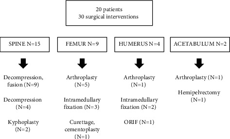
Results: Forty-five patients with LMS (11%) had bone metastases. The most common LMS subtypes with bone metastases were uterine (N = 18, 40%) and retroperitoneal (N = 15, 33%). Bone metastasis was not an independent predictor of mortality by Cox regression analysis (HR 1.0, 95% CI: 0.67-1.5). Patients more frequently metastasized to the axial (N = 29, 64%) than to the appendicular (N = 5, 11%) skeleton. Bone was the first site of metastasis in 13 patients (29%). Patients presented with bone metastases at a median of 32.7 months (IQR: 5.2, 62.6) after initial LMS diagnosis. Twelve patients (27%) sustained a pathologic fracture. Twenty (44%) required surgical management, with 30 surgeries total. Three (15%) had a failure of reconstructive constructs. The median overall survival time was 69.7 months (IQR: 43.2, 124.5). There were no associations between the LMS subtype and survival. Pathologic fracture was an independent predictor of mortality by Cox regression analysis (HR 5.4, 95% CI: 1.8-16).
Conclusion: The majority of patients with metastatic LMS to bone survive greater than 5 years and frequently require surgical intervention. Extended survival in this patient population should inform fixation and implant choice. No anatomic subtype was associated with risk for bone metastases. Pathologic fracture was associated with worse survival.
SarcomaMedicine-Radiology, Nuclear Medicine and Imaging
CiteScore
5.00
自引率
0.00%
发文量
15
审稿时长
14 weeks
期刊介绍:
Sarcoma is dedicated to publishing papers covering all aspects of connective tissue oncology research. It brings together work from scientists and clinicians carrying out a broad range of research in this field, including the basic sciences, molecular biology and pathology and the clinical sciences of epidemiology, surgery, radiotherapy and chemotherapy. High-quality papers concerning the entire range of bone and soft tissue sarcomas in both adults and children, including Kaposi"s sarcoma, are published as well as preclinical and animal studies. This journal provides a central forum for the description of advances in diagnosis, assessment and treatment of this rarely seen, but often mismanaged, group of patients.


 求助内容:
求助内容: 应助结果提醒方式:


