The impact of a magnetic field on the dose distribution using the Bebig60Co HDR sources
引用次数: 0
Abstract
Objective To evaluate the impact of an external magnetic field on the dose distribution and electronic disequilibrium region around a Bebig type 60Co HDR brachytherapy source and to judge the feasibility of applying MRI scanner during brachytherapy. Methods The source model was established based on the Monte Carlo package Geant4 software. The simulated geometries consisted of the 60Co source inside a water phantom of 10.0cm×10.0cm×10.0cm in size. The magnetic field strength of the 0T, 1.5T and 3.0T was considered, respectively. The voxels with a size of 0.2 mm, 0.5 mm and 0.5 mm were established along the x-, y-and z-axis. The influence of the magnetic field on the Kerma (kinetic energy released to matter) distribution and dose distribution within the 10.0mm region from the source center was evaluated. Furthermore, the ratio of the Kerma to dose as a function of the distance to the center source was acquired. Results The 1.5T magnetic field exerted no effect on the dose distribution adjacent to 60Co HDR brachytherapy source, whereas3.0T magnetic field caused significant increase in the dose distribution within r<6 mm from the source center. The dose distribution was increased by 40% at r=5.4 mm from the source center. The ratio of Kerma to dose was less than 1 within the region of 1.2 mm磁场对Bebig60Co HDR源剂量分布的影响
目的评价外磁场对贝比格型60Co HDR近距离放射治疗源周围剂量分布和电子不平衡区的影响,判断近距离放射治疗中应用MRI扫描仪的可行性。方法基于蒙特卡罗软件包Geant4软件建立源模型。模拟的几何形状包括60Co源在10.0cm×10.0cm×10.0cm大小的水影内。分别考虑0T、1.5T和3.0T的磁场强度。沿x、y、z轴分别建立尺寸为0.2 mm、0.5 mm和0.5 mm的体素。评价了磁场对源中心10.0mm范围内物质释放动能(Kerma)分布和剂量分布的影响。此外,还获得了Kerma与剂量的比值作为到中心源距离的函数。结果1.5T磁场对60Co HDR近距离放疗源附近的剂量分布没有影响,而3.0 t磁场对距离源中心r<6 mm范围内的剂量分布有显著增加。在距源中心r=5.4 mm处,剂量分布增加了40%。在1.2 mm
本文章由计算机程序翻译,如有差异,请以英文原文为准。
求助全文
约1分钟内获得全文
求助全文
来源期刊
自引率
0.00%
发文量
6375
期刊介绍:
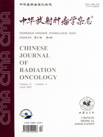
The Chinese Journal of Radiation Oncology is a national academic journal sponsored by the Chinese Medical Association. It was founded in 1992 and the title was written by Chen Minzhang, the former Minister of Health. Its predecessor was the Chinese Journal of Radiation Oncology, which was founded in 1987. The journal is an authoritative journal in the field of radiation oncology in my country. It focuses on clinical tumor radiotherapy, tumor radiation physics, tumor radiation biology, and thermal therapy. Its main readers are middle and senior clinical doctors and scientific researchers. It is now a monthly journal with a large 16-page format and 80 pages of text. For many years, it has adhered to the principle of combining theory with practice and combining improvement with popularization. It now has columns such as monographs, head and neck tumors (monographs), chest tumors (monographs), abdominal tumors (monographs), physics, technology, biology (monographs), reviews, and investigations and research.
联系我们:info@booksci.cn
Book学术提供免费学术资源搜索服务,方便国内外学者检索中英文文献。致力于提供最便捷和优质的服务体验。
Copyright © 2023 布克学术 All rights reserved.
京ICP备2023020795号-1
 京公网安备 11010802042870号
京ICP备2023020795号-1

Book学术文献互助群
群 号:604180095


 求助内容:
求助内容: 应助结果提醒方式:
