Domenico Mannatrizio, Laura Eusebi, Mamhmoud Yehia, Alessandra Filosa, Giuseppe Guglielmi
{"title":"Granulomatous prostatitis: mimicking locally advanced prostate adenocarcinoma.","authors":"Domenico Mannatrizio, Laura Eusebi, Mamhmoud Yehia, Alessandra Filosa, Giuseppe Guglielmi","doi":"10.23750/abm.v94i5.13751","DOIUrl":null,"url":null,"abstract":"<p><p>We report the case of a 63-year-old male who came to the urology clinic with an increasing value of the prostate specific antigen and an asymmetrical enlargement at the digital rectal examination. The man was subjected to an MRI of the prostate following which a convincing radiological diagnosis of prostate cancer was made. The patient was assigned a provisional stage of disease T3a N0. In order to confirm this diagnosis, a prostate biopsy was performed but the histological analysis reported non-specific granulomatous prostatitis (GP). It is an uncommon condition that both clinically and radiologically on TRUS and MRI usually mimics prostate cancer (PCa), representing a diagnostic challenge due to its non-specific symptoms and aspecific radiological findings. In this case report we discuss the magnetic resonance imaging features of this rare clinical condition in order to help radiologists in the timely diagnosis for a correct diagnostic framing.</p>","PeriodicalId":93849,"journal":{"name":"Acta bio-medica : Atenei Parmensis","volume":"94 5","pages":"e2023245"},"PeriodicalIF":0.0000,"publicationDate":"2023-10-17","publicationTypes":"Journal Article","fieldsOfStudy":null,"isOpenAccess":false,"openAccessPdf":"https://www.ncbi.nlm.nih.gov/pmc/articles/PMC10644919/pdf/","citationCount":"0","resultStr":null,"platform":"Semanticscholar","paperid":null,"PeriodicalName":"Acta bio-medica : Atenei Parmensis","FirstCategoryId":"1085","ListUrlMain":"https://doi.org/10.23750/abm.v94i5.13751","RegionNum":0,"RegionCategory":null,"ArticlePicture":[],"TitleCN":null,"AbstractTextCN":null,"PMCID":null,"EPubDate":"","PubModel":"","JCR":"","JCRName":"","Score":null,"Total":0}
引用次数: 0
Abstract
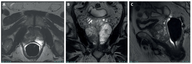
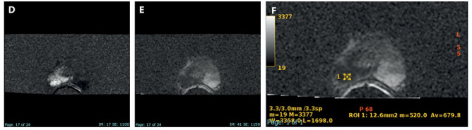
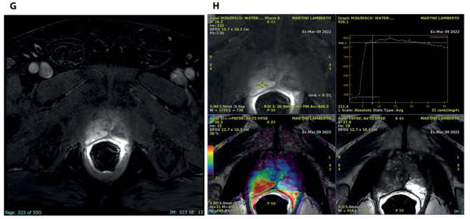
We report the case of a 63-year-old male who came to the urology clinic with an increasing value of the prostate specific antigen and an asymmetrical enlargement at the digital rectal examination. The man was subjected to an MRI of the prostate following which a convincing radiological diagnosis of prostate cancer was made. The patient was assigned a provisional stage of disease T3a N0. In order to confirm this diagnosis, a prostate biopsy was performed but the histological analysis reported non-specific granulomatous prostatitis (GP). It is an uncommon condition that both clinically and radiologically on TRUS and MRI usually mimics prostate cancer (PCa), representing a diagnostic challenge due to its non-specific symptoms and aspecific radiological findings. In this case report we discuss the magnetic resonance imaging features of this rare clinical condition in order to help radiologists in the timely diagnosis for a correct diagnostic framing.

 求助内容:
求助内容: 应助结果提醒方式:


