Magdi A Ali, Eman El-Abd, Mohamed Morsi, Mohamed M El Safwany, Mohamed Z El-Sayed
{"title":"The effect of hepatic steatosis on 18F-FDG uptake in PET-CT examinations of cancer Egyptian patients.","authors":"Magdi A Ali, Eman El-Abd, Mohamed Morsi, Mohamed M El Safwany, Mohamed Z El-Sayed","doi":"10.1186/s41824-023-00173-6","DOIUrl":null,"url":null,"abstract":"<p><strong>Background: </strong>Hepatic steatosis is the most common chronic hepatic disease. Imaging diagnosis of hepatic steatosis has been evaluated as an alternative to invasive histological diagnosis.</p><p><strong>Study aims: </strong>The study aimed to assess the effect of hepatic steatosis on Flourine-18 fluorodeoxyglucose (18F-FDG) uptakes in cancer patients.</p><p><strong>Patients and methods: </strong>Blood samples were collected from 50 cancer patients and analyzed to calculate fatty liver index and Hepatic steatosis index (HIS). Hepatic steatosis examined using high-resolution ultrasound and positron emission tomography-computed tomography (PET-CT). Linear attenuation coefficient, standardized-uptake value (SUV) mean (SUV mean), and SUV maximum (SUVmax) were measured. Accordingly, patients were divided equally into non-fatty liver, and fatty liver groups.</p><p><strong>Results: </strong>A significant increase in SUVmax and SUV mean was observed in the fatty liver group more than in the non-fatty liver group. HSI significantly increased in the fatty liver group compared to the non-fatty liver group. Liver tissue uptake FDG was significantly correlated with HSI values. SUV max significantly correlated with body mass index (BMI) in the non-fatty group only.</p><p><strong>Conclusion: </strong>Hepatic changes in cancer patients affect the liver metabolic activity and thus the 18 F-FDG uptake. Therefore, further corrections should be considered when the liver is used as a comparator for PET-CT scans of cancer patients.</p>","PeriodicalId":36160,"journal":{"name":"European Journal of Hybrid Imaging","volume":"7 1","pages":"19"},"PeriodicalIF":1.7000,"publicationDate":"2023-10-16","publicationTypes":"Journal Article","fieldsOfStudy":null,"isOpenAccess":false,"openAccessPdf":"https://www.ncbi.nlm.nih.gov/pmc/articles/PMC10577118/pdf/","citationCount":"0","resultStr":null,"platform":"Semanticscholar","paperid":null,"PeriodicalName":"European Journal of Hybrid Imaging","FirstCategoryId":"1085","ListUrlMain":"https://doi.org/10.1186/s41824-023-00173-6","RegionNum":0,"RegionCategory":null,"ArticlePicture":[],"TitleCN":null,"AbstractTextCN":null,"PMCID":null,"EPubDate":"","PubModel":"","JCR":"Q3","JCRName":"RADIOLOGY, NUCLEAR MEDICINE & MEDICAL IMAGING","Score":null,"Total":0}
引用次数: 0
Abstract
Background: Hepatic steatosis is the most common chronic hepatic disease. Imaging diagnosis of hepatic steatosis has been evaluated as an alternative to invasive histological diagnosis.
Study aims: The study aimed to assess the effect of hepatic steatosis on Flourine-18 fluorodeoxyglucose (18F-FDG) uptakes in cancer patients.
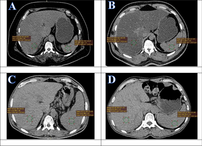
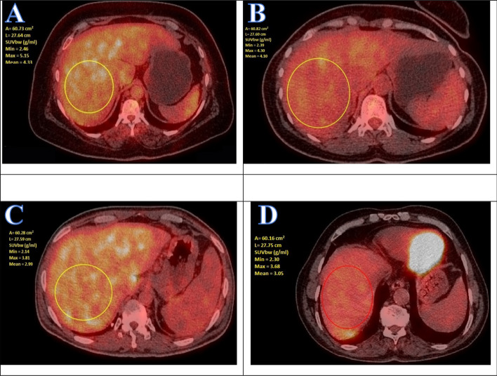
Patients and methods: Blood samples were collected from 50 cancer patients and analyzed to calculate fatty liver index and Hepatic steatosis index (HIS). Hepatic steatosis examined using high-resolution ultrasound and positron emission tomography-computed tomography (PET-CT). Linear attenuation coefficient, standardized-uptake value (SUV) mean (SUV mean), and SUV maximum (SUVmax) were measured. Accordingly, patients were divided equally into non-fatty liver, and fatty liver groups.
Results: A significant increase in SUVmax and SUV mean was observed in the fatty liver group more than in the non-fatty liver group. HSI significantly increased in the fatty liver group compared to the non-fatty liver group. Liver tissue uptake FDG was significantly correlated with HSI values. SUV max significantly correlated with body mass index (BMI) in the non-fatty group only.
Conclusion: Hepatic changes in cancer patients affect the liver metabolic activity and thus the 18 F-FDG uptake. Therefore, further corrections should be considered when the liver is used as a comparator for PET-CT scans of cancer patients.

 求助内容:
求助内容: 应助结果提醒方式:


